Novel Gd(OH)
3, GdOOH and Gd
2O
3Nanorods: Microwave-Assisted Hydrothermal
Synthesis and Optical Properties
Maciel Salomão de Almeidaa, Maria Aparecida Bezerra dos Santosa, Rosana de Fátima Gonçalvesb*,
Maria Rita de Cássia Santosa, Ana Paula de Azevedo Marquesb, Elson Longoc, Felipe de Almeida La
Portad, Ivo Mateus Pinattie, Maya Dayana Penha Silvae, Mario Junior Godinhoa,e
Received: March 27, 2016; Revised: June 28, 2016; Accepted: August 11, 2016
We present a study of the controlled synthesis and optical properties of single-crystals Gd(OH)3, GdOOH and Gd2O3 nanorods. In this work, Gd(OH)3 nanorods were synthesized by a simple and fast microwave-assisted hydrothermal method. This process combined with the thermal decomposition oxidation of Gd(OH)3 nanorods as precursors enabled the preparation of single-crystalline GdOOH and Gd2O3 structures with well-deined morphology at low temperatures. The crystal structure dependence
on the optical properties was investigated. We observed a green shift efect on the photoluminescence
(PL) emission spectra from Gd(OH)3 to Gd2O3 nanorods, which can be attributed to diferent types of
surface defects, as well as intrinsic properties that contribute signiicantly to the modiied PL behavior.
Keywords: Gd(OH)3, GdOOH, Gd2O3, nanorods
* e-mail: [email protected]
1. Introduction
Nowadays, rare earth hydroxide/oxide nanomaterials have received special attention because of their excellent physical and chemical properties, which provide a wide variety of
applications in the nanotechnology ield1-6. Among various
types of rare earth hydroxide/oxide materials, gadolinium hydroxide (Gd(OH)3), gadolinium oxyhydroxide (GdOOH) and gadolinium oxide (Gd2O3) exhibit important functional properties that depend on the structure. Gadolinium oxyhydroxide is a stable phase obtained by the thermal dehydration of gadolinium hydroxide and has a simple layered structure. So, these materials have been investigated as promising
candidates in the ield of high throughput luminescent
devices, catalysis and other functional devices based on their excellent electronic, optical and physicochemical responses arising from 4f electrons5-7.
Despite the recent improvements, few studies have been reported concerning the synthesis control of new rare earth hydroxide/oxide nanomaterials. Therefore, more studies leading to the development of new synthetic strategies that allow a superior control in the processing of such materials
are needed and this remains a highly challenging ield.
Some methods are reported in the literature to obtain
well deined Gd(OH)3, GdOOH and Gd2O3 nanostructures
such as electrochemical route, microwave, sol-gel synthesis, combustion, solid state reaction, and polyol method3-8.
However, most of these methods require long processing times due to slow reaction kinetics. Therefore, the microwave-assisted hydrothermal (MAH) method has shown numerous advantages such as the use of an environmentally friendly
solvent (water) and low processing temperatures (≤200 °C).
This method provides a simple and fast approach for the large-scale production of new emergent complex functional materials. In addition, it also enables the shape-controlled synthesis of micro- and/or nanomaterials at a lower cost, for their industrial exploitation with unprecedented capabilities in the new technologies9-14.
Herein, we report a facile and simple, low-temperature synthesis of single-crystalline Gd(OH)3, GdOOH and Gd2O3 nanorods obtained by the MAH method combined with thermal decomposition oxidation process. These
microcrystals were structurally characterized by diferent
techniques and the optical properties of the crystal structures obtained were investigated using ultraviolet-visible (UV-vis)
difuse relectance spectroscopy and photoluminescence
(PL) measurements at room temperature. Moreover, this
aUnidade Acadêmica Especial de Física e Química – Regional Catalão, Universidade Federal de Goiás
– UFG, 75.704–020, Catalão, GO, Brazil
bUniversidade Federal de São Paulo – UNIFESP, Rua Prof. Artur Riedel, 275, CEP 09972-270,
Diadema, SP, Brazil
cLaboratório Interdisciplinar de Eletroquímica e Cerâmica – LIEC, Instituto de Química – IQ,
Universidade Estadual Paulista, P.O. Box 355, 14801-907, Araraquara, SP, Brazil
dUniversidade Tecnológica Federal do Paraná – UTFP, 86036-370, Londrina, Brazil
eUniversidade Federal de São Carlos – UFSCar, Rod. Washington Luiz, Km. 235, CEP 13.565-905, São
paper discuss the electronic efects in order to establishes a
close correlation between the structure and properties for the single-crystalline Gd(OH)3, GdOOH and Gd2O3 nanorods.
2. Experimental
The Gd(OH)3 nanorods were prepared using a MAH method. Initially, a solution of Gd3+ was prepared by
dissolving gadolinium nitrate (99.9%, Aldrich) in distilled water to obtain a concentration of 0.1 mol L–1. Under
magnetic stirring, an ammonium hydroxide solution was
added dropwise to complete the precipitation (inal pH =
10), resulting in a white gel. In the second stage, the white gel is dispersed in water and the aqueous precursor solution
was transferred into a Telon autoclave, which was properly
sealed and placed inside a domestic MAH system (2.45 GHz, maximum power of 800 W). MAH processing was performed
at 130ºC under a constant pressure of approximately 3 bar
for 20 min. After the hydrothermal treatment, the dispersed powder was centrifuged and washed with distilled water until the residual ions in the solution were eliminated. The resulting product was dried at room temperature for 24 h.
GdOOH and Gd2O3 nanocrystals were obtained by a thermal decomposition oxidation process using the Gd(OH)3 nanocrystals prepared by the MAH synthesis as precursors.
In both cases, the heating rate was ixed at 3ºC min-1 using
a conventional furnace (ambient air), obtaining a white powder. The Gd(OH)3 was calcined at either 380 or 600ºC for 60 min, depending on the type of nanocrystals envisaged (GdOOH or Gd2O3, respectively).
XRD measurements were performed on a Shimadzu XRD
6100 with CuKα radiation (40 kV, 30 mA, λ = 0.15418 nm).
The difraction pattern was measured between 5º and 75º
with a step size of 2º s–1. The size and morphologies were
characterized by FE-SEM (FEG-VP Zeiss Supra 35) and high-resolution transmission electron microscopy (HRTEM, Jeol, JEM-2100). The thermogravimetric (TG) analyses were carried out with a Netzsch-409 STA simultaneous
thermogravimetric-diferential thermal analysis (TG-DTA) apparatus with a heating rate of 10ºC min−1 under lowing
air. DR spectra were produced using a Varian Cary 5G(USA) spectrophotometer in difuse relectance mode. PL spectra
were collected with a thermal Jarrel-Ash Mono Spec Monochromator and a Hamamatsu R446 photomultiplier.
A krypton ion laser (Coherent Innova) with an excitation
wavelength of 350.7 nm was used, with the laser nominal output kept at 200 mW. All measurements were performed at room temperature.
3. Results and discussion
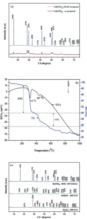
Figure 1(a) presents the XRD patterns of the as-prepared Gd(OH)3 powder and Gd(OH)3 powder after the MAH treatment. For the sample processed with the MAH method,
Figure 1: XRD patterns of (a) as-prepared Gd(OH)3 with and
without the use of MAH treatment (b) TG/DTA curves of the Gd(OH)3 prepared by the MAH method (c) XRD patterns of the Gd(OH)3, GdOOH and Gd2O3 nanocrystals.
all the difraction peaks can be perfectly indexed to the
hexagonal structure of the Gd(OH)3 powders, in agreement
with the respective Joint Committee on Powder Difraction Standard (JCPDS) cards nº 083-2037. No impurities were
the synthesis of highly crystalline products featuring a long-range-ordered single-phase (Figure 1(a)).
Figure 1(b) shows the TG/DTA curve for the Gd(OH)3
sample prepared by the MAH method at 130ºC for 20
min. Our measurements reveal the presence of two major
stages with rapid weight losses at about 312 and 437ºC
(8.9 and 4.4%, respectively), suggesting the existence of an intermediate phase during the thermal conversion process of Gd(OH)3 to Gd2O3. Interestingly, the total weight loss is in agreement with the theoretical value found for the complete dehydration of Gd(OH)3 to produce Gd2O3 (13.3%), as previously reported15,16. The thermal dehydration process of
the Gd(OH)3 and subsequent conversion to Gd2O3 observed in the Figure 1(b) is supposed to occur in two steps, as summarized by equations (1) and (2).
( )
Gd OH
Q
V
3GdOOH
+
H O
21
( )
GdOOH
Gd O
H O
2
2 3+
22
In this study, a two-step approach was used to synthesize the GdOOH and Gd2O3 powders at optimal conditions.
Based on these results, we chose two diferent temperatures (380 and 600ºC) to produce GdOOH and Gd2O3 powders,
respectively, using a thermal decomposition process of the Gd(OH)3 powder obtained previously by the MAH method.
As shown in Figure 1(c), all the difraction peaks can
be perfectly indexed to the hexagonal structure of Gd(OH)3
(JCPDS 083-2037), monoclinic structure of GdOOH (JCPDS
075-3267) and cubic structure of Gd2O3 (JCPDS 072-6362), respectively16-19. However, the peak widths for Gd
2O3 are
relatively broader than those observed in the XRD patterns of GdOOH and Gd(OH)3, suggesting a smaller crystallite size. The crystallite sizes were estimated using the Scherrer equation17, obtaining 34.9, 19.9 and 15.7 nm for Gd(OH)
3,
GdOOH and Gd2O3, respectively. Therefore, the average crystallite size decreases as a function of the dehydration process. This behavior is explained by the dehydration process during the phase change, as well as the synthesis method employed, which facilitates the preparation of materials with small particle sizes.
Figure 2 shows the representations of the unit cells for the hexagonal Gd(OH)3 structure with a P63/m space group, the monoclinic GdOOH structure with a P121/m1 space group, and the cubic Gd2O3 structure with an Ia-3 space group18-20. VESTA program was used for the construction
and visualization of these three models21. In this study, the
most important diference between the crystalline phases is
the coordination of the gadolinium ions in the ideal crystal structure. The hexagonal Gd(OH)3 structure has a nine-coordination complex with [Gd(OH)9] clusters, the monoclinic GdOOH structure has a seven-coordination complex with [GdO(OH)7] clusters, and the cubic Gd2O3 structure has a six-coordination complex with [GdO6] clusters. It is well known that lanthanide ions have a wide variety of coordination
Figure 2: Schematic representation of the unit cells corresponding to the hexagonal, monoclinic and cubic structure of the materials.
environments, and in general tend to form complex clusters with high coordination numbers, nine-coordination being particularly important in the structure of f-block elements22.
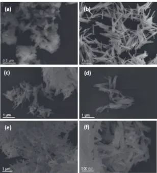
FE-SEM analysis was performed in order to determine the particle morphology in detail. Figures 3(a-f) present FE-SEM images of the Gd(OH)3, GdOOH and Gd2O3 nanorods. We can see that the as-prepared Gd(OH)3 sample consists of
particles with undeined shape (Figure 3(a)). On the other
hand, the Gd(OH)3 precursor treated by the MAH method
exhibits a controlled and well-deined morphology, where
nanorods prevail (Figure 3(b)). Similarly, GdOOH and Gd2O3 also exhibit a nanorods-like morphology, even after subsequent thermal treatment (Figures 3(b-f)).
Figure 3: FE-SEM images of (a) as-prepared Gd(OH)3 without the MAH treatment (b) Gd(OH)3 (c-d) GdOOH and (e-f) Gd2O3
nanorods.
It can be seen that Gd(OH)3 nanorods (Figure 3(b)) have a narrow size distribution with an average length of about 0.9 µm and diameter of about 12.4 nm, respectively. Figures 3 (c-d) exhibit the same morphology for the intermediate GdOOH compound formed with the thermal treatment
at 380 ºC. There was no morphological change after the
nanorods (average length of about 0.64 µm and diameter
of about 14.3 nm), conirming the crystallite size results
presented earlier. Moreover, similar results were found for the Gd2O3 nanorods (Figures 3(e-f)) formed after the thermal
treatment at 600 ºC, in which a reduction of the nanorods
size (average length of about 0.60 µm and diameter of about 13.2 nm) was also observed.
Then, we can verify that the synthetic strategy employed in this study allows the preparation of materials with high
purity and well-deined morphologies at low temperatures
and short synthesis times. Successful conditions for this synthesis using the MAH method are attributed to the rapid
and efective interaction between the electromagnetic radiation
and the permanent dipole moment of water molecules23,24. The
permanent dipoles of water are induced in solution and can produce a rapid heating of the system because they interact directly with microwaves. This kind of interaction is directly related to the water capacity for electromagnetic radiation
absorption and eiciency in converting electromagnetic
radiation to thermal energy25-28. Therefore, the reaction time
and temperature necessary for obtaining of such materials
in this work is signiicantly lower than those described in
the literature29-32 providing a signiicant improvement on
crystallinity and morphology.
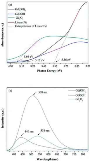
In this work, the Kubelka-Munk method was employed to calculate the Eg values. The direct Eg values were found to be 5.56, 5.12 and 5.04 eV for the Gd(OH)3, GdOOH and Gd2O3 nanorods, respectively (Figures 4 (a)). These values agree with those reported in the literature as compared to bulk band gap values33,34. Some small variations are attributed
to the fact that the Eg values depend on the method of preparation and the experimental conditions like the type of surfactant, temperature and processing time. These key factors can favor or inhibit the formation of structural defects, which can control the degree of structural order-disorder of the material and consequently, the number of intermediate energy levels within the band gap35.
However, it has been revealed that the Eg values are strongly dependent on the crystalline structure, and are very
sensitive to the efects of the structural order-disorder, as well
as the conditions of synthesis used in the preparation of these complex functional materials. The crystal growth process reported in this study might lead to both surface and bulk
modiications due to structural changes, which are directly
linked to the appearance of new intermediary energy levels within the forbidden band gap36. This has a major impact on
the physical and chemical properties of the nanorods, namely over its PL emission behavior (Figure 4(d)).
PL emission spectra of the Gd(OH)3, GdOOH and Gd2O3 nanorods, excited at 350 nm are shown in Figure 4(b). All the samples exhibit a broad emission band at room temperature, typical of a multiphonon process. As shown in Figure 4(b), the PL emission with a maximum at 508 nm corresponds to the Gd(OH)3 phase, while the other two peaks with lower
Figure 4: (a) Difuse relectance spectra of nanorods and (b) PL
at room temperature.
intensities centered at 440 and 538 nm are ascribed to the PL emissions of GdOOH and Gd2O3, respectively. Essentially, Gd3+ has a simpler emission spectrum in comparison to
the other trivalent rare-earth ions29. However, we observe
a green shift efect on the emission spectrum (Figure 4b)
when we compare the conversion of Gd(OH)3 on Gd2O3.
These emissions are attributed to diferent types of surface
defects of Schottky and Frenkel types35. According to Wang’s
proposal37, the green emission band may attribute to the
recombination of a delocalized electron close to the conduction band with a single charged state of surface oxygen vacancy. In oxide systems, typically oxygen vacancies are prevalent
and the presence of such defects contribute signiicantly to the modiied photoluminescence response37.
However, diferent density of oxygen vacancies might arouse diferences of PL intensities as shown in emission
pattern of Figure 4 (b). The PL emission can be a probe for the structural evaluation of materials9,36,38. Therefore, direct
studies can provide indirect evidences in support of them. More information about the nature of these defects may be elucidated in a future study, using quantum mechanical calculations and experimental techniques such as electron paramagnetic resonance (EPR), X-ray photoelectron spectroscopy (XPS), and X-ray absorption near-edge spectroscopy (XANES). Zhang and co-authors39 have demonstrated that
luminescence results can be employed as a simple probe for oxygen vacancies in TiO2 nanoparticles. In another work, studies of visible luminescence in TiO2 nanotubes and EPR spectrum provide strong evidence characteristic of single-electron-trapped oxygen vacancies40.
Indeed, it is well known that the crystal structure depends on the experimental conditions such as heat treatment, which may cause changes to its electronic structure, and thus,
has an important role in the PL proile9. We can observe
that the Gd(OH)3 compound presents the most intense PL
emission (Fig 4b). This efect can be attributed to defects
that cause structural disorder yielding intermediate levels in the band gap, which is essential for the PL phenomenon. An improved structural organization has been achieved with further thermal treatment during the annealing process. The resulting GdOOH and Gd2O3 compounds present lower PL intensities. These results corroborate with those obtained by UV measurements [Figure 4 (a)]. As shown before, the band gap values of the GdOOH and Gd2O3 samples were lower than those presented by Gd(OH)3. Studies on intermediate levels in band gap and structural order-disorder showed a decrease in the optical band gap according to the structural organization41. Our group has postulated that the PL intensity
in inorganic oxides is also associated with the thermal treatment history, structural order-disorder, and electronic levels in the band gap41,14.
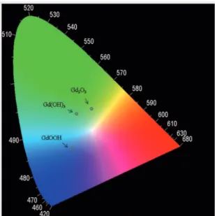
Figure 5 shows the Commission Internationale de L’Eclairage (CIE) diagram for the compounds with respective assignment of their colors. Speciically, the CIE chromaticity coordinates [x-axis, y-axis] are [x = 0.2801, y = 0.4052], [x = 0.2608, y = 0.2527], and [x = 0.3455, y =
0.4277] corresponding to emission color coordinates for the Gd(OH)3, GdOOH and Gd2O3, respectively. These results
clearly demonstrate the green shift efect and corroborate
the observed PL behavior.
Moreover, an earlier study has demonstrated the synthesis of rare-earth doped Gd2O3 nanoparticles42. Multicolor emissions
at selected wavelengths could be obtained by altering the doping concentration in the triple-doped sample. Based on
the CIE coordinates, it could be inferred that double-doped
Gd2O3 with the composition Tb = 0.05% and Dy = 1.95 mol%, when excited at 247 nm, emits white light, which is closest to the standard noon daylight. Hence, it can be inferred that by altering the doping content of the rare-earth ions in a suitable host lattice like Gd2O3, it is possible to achieve desired emission colors.
Figure 5: CIE chromaticity diagram for the nanorods.
4. Conclusions
In summary, the method employed is simple and fast to obtain highly crystalline nanorods-like oxides and with unique morphology once the reaction time necessary for the synthesis
of such materials is signiicantly lower than that used in the
most previous works. The results contribute to an insight of the crystal structure and chemical composition and its impact the on the optical properties of the Gd(OH)3, GdOOH and Gd2O3 obtained. So, shape, size, interaction between particles and synthesis technique play an important role for optical behavior of the studied oxides. Therefore, such properties enable the potential application of these complex functional materials as high-quality phosphors and optoelectronics devices.
5. Acknowledgments
The authors acknowledge the support of the Brazilian
agencies FAPESP-CDMF: 2013/07296-2, FAPESP 2013/07437-5, CAPES/PROCAD: 182441, CNPq: 554580/2010-1, CNPq:
485518/2013-9 and FAPEG. Special thanks by INTMF and LabMic/UFG.
6. References
1. Wang X, Sun XM, Yu D, Zou BS, Li Y. Rare earth compound nanotubes. Advanced Materials. 2003;15(17):1442-1445.
2. Wang X, Li Y. Rare-earth-compound nanowires, nanotubes, and fullerene-like nanoparticles: Synthesis, characterization, and properties. Chemistry - A European Journal. 2003;9(22):5627-5635.
4. Majeed S, Shivashankar SA. Microspherical, hierarchical structures of blue-green-emitting Dy:GdOOH and Dy:Gd2O3. Materials Letters. 2014;125:136-139.
5. Panda AB, Glaspell G, El-Shall MS. Microwave synthesis and optical properties of uniform nanorods and nanoplates of rare earth oxides. The Journal of Physical Chemistry C. 2007;111(5):1861-1864.
6. Xiao H, Li P, Jia F, Zhang L. General nonaqueous sol-gel synthesis of nanostructured Sm2O3, Gd2O3, Dy2O3, and Gd2O3:Eu3+ phosphor. The Journal of Physical Chemistry C.
2009;113(50):21034-21041.
7. Tamrakar RK, Bisen DP, Brahme N. Comparison of photoluminescence
properties of Gd2O3 phosphor synthesized by combustion and solid state reaction method. JournalofRadiation Researchand Applied Sciences. 2014;7(4):550-559.
8. Bedekar V, Dutta DP, Tyagi AK. White light emission from spin coated Gd2O3:Dy nano phosphors synthesized using polyol technique. Journal of Nanoscience and Nanotechnology. 2010;10(12):8234-8238.
9. La Porta FA, Andrés J, Li MS, Sambrano JR, Varela JA, Longo E. Zinc blende versus wurtzite ZnS nanoparticles: control of the phase and optical properties by tetrabutylammonium hydroxide.
Physical Chemistry Chemical Physics. 2014;16(37):20127-20137.
10. Ribeiro PC, Costa ACFM, Kiminami RHGA, Sasaki JM,
Lira HL. Synthesis of TiO2 by the Pechini method and photocatalytic degradation of methyl red. Materials Research. 2013;16(2):468-472.
11. Garcia-Contreras R, Scougall-Vilchis RJ, Contreras-Bulnes R,
Sakagami H, Morales-Luckie RA, Nakajima H. Mechanical, antibacterial and bond strength properties of nano-titanium-enriched glass ionomer cement. Journal of Applied Oral Science.
2015;23(3):321-328.
12. Gonçalves RF, Godinho MJ, Marques APA, Santos MRC,
Rosa ILV, Longo E, et al. Structure, morphology, and optical
properties of (Ca1−3x Eu2x )WO4 microcrystals. Electronic Materials Letters. 2015;11(2):193-197.
13. Prabaharan DMDM, Sadaiyandi K, Mahendran M, Sagadevan S. Structural, Optical, Morphological and Dielectric Properties
of Cerium Oxide Nanoparticles. Materials Research. 2016;19(2):478-482.
14. Gonçalves RF, Lima ARF, Godinho MJ, Moura AP, Espinosa J, Longo E, et al. Synthesis of Pr3+-doped CaTiO
3 using polymeric
precursor and microwave-assisted hydrothermal methods: a comparative study. Ceramics International. 2015;41(10 Part A):12841-12848.
15. Chang C, Zhang Q, Mao D. The hydrothermal preparation,
crystal structure and photoluminescent properties of GdOOH nanorods. Nanotechnology. 2006;17(8):1981-1993.
16. Jia G, Liu K, Zheng Y, Song Y, Yang M, You HP. Highly uniform Gd(OH)3 and Gd2O3:Eu3+ nanotubes: Facile synthesis and
luminescence properties. The Journal of Physical Chemistry C. 2009;113(15):6050-6055.
17. Guinier A. X-Ray Difraction in Crystals, Imperfect Crystals,
and Amorphous Bodies. San Francisco: W. H. Freeman; 1963.
18. Le Luyer C, García-Murillo A, Bernstein E, Mugnier J. Waveguide
Raman spectroscopy of sol–gel Gd2O3 thin ilms. Journal of Raman Spectroscopy. 2003;34(3):234-239.
19. Majeed S, Shivashankar SA. Novel spherical hierarchical structures of GdOOH and Eu:GdOOH: rapid microwave-assisted synthesis through self-assembly, thermal conversion to oxides, and optical studies. Journal of Materials Chemistry C.2014;2(16):2965-2974.
20. Zhang CC, Zhang ZM, Dai RC, Wang ZP, Ding ZJ. High
Pressure Luminescence and Raman Studies on the Phase Transition of Gd2O3:Eu3+ Nanorods. Journal of Nanoscience and Nanotechnology. 2011;11(11):9887-9891.
21. Momma K, Izumi F. VESTA 3 for three-dimensional visualization of crystal, volumetric and morphology data. JournalofApplied Crystallography. 2011;44:1272-1276.
22. Shriver DF, Atkins PW, Overton TL, Rourke JP, Weller MT,
Armstrong FA. Shriver & Atkins Inorganic Chemistry. New
York: W. H. Freeman; 2006. 848 p.
23. Dalmaschio CJ, Ribeiro C, Leite ER. Impact of the colloidal
state on the oriented attachment growth mechanism. Nanoscale. 2010;2(11):2336-2345.
24. Godinho M, Ribeiro C, Longo E, Leite ER. Inluence of
microwave heating on the growth of gadolinium-doped cerium oxide nanorods. Crystal Growth & Design. 2008;8(2):384-386.
25. Lee EJH, Ribeiro C, Longo E, Leite ER. Oriented attachment: An efective mechanism in the formation of anisotropic nanocrystals. The Journal of Physical Chemistry B. 2005;109(44):20842-20846.
26. Li C, Liu H, Yang J. A facile hydrothermal approach to the
synthesis of nanoscale rare earth hydroxides. Nanoelcale Research Letters. 2015;10:144.
27. Seo S, Yang H, Holloway PH. Controlled shape growth of Eu- or
Tb-doped luminescent Gd2O3 colloidal nanocrystals. Journal of Colloid and Interface Science. 2009;331(1):236-242.
28. Gonçalves RF, Moura AP, Godinho MJ, Longo E, Machado MAC, de Castro DA, et al. Crystal growth and photoluminescence of
europium-doped strontium titanate prepared by a microwave hydrothermal method. Ceramics International. 2015;41(3A):3549-3554.
29. Hazarika S, Paul N, Mohanta D. Rapid hydrothermal route to synthesize cubic-phase gadolinium oxide nanorods. Bulletin of Materials Science. 2014;37(4):789-796.
30. Kang JG, Min BK, Sohn Y. Synthesis and characterization of Gd(OH)3 and Gd2O3 nanorods. Ceramics International. 2015;41(1B):1243-1248.
31. Dhananjaya N, Nagabhushana H, Nagabhushana BM, Rudraswamy
B, Shivakumara C, Chakradhar RPS. Hydrothermal synthesis,
characterization and Raman studies of Eu3+ activated Gd 2O3
nanorods. Physica B: Condensed Matter. 2011;406(9):1639-1644.
32. Li S, Li Q, Wang K, Zhou M, Huang X, Liu J, et al.
Pressure-induced irreversible phase transition in the energetic material urea
nitrate: combined Raman scattering and X-ray difraction study. The Journal of Physical Chemistry C. 2013;117(1):152-159.
33. Chen F, Zhang XH, Hu XD, Zhang W, Zeng R, Liu PD, et
34. Sahoo NK, Thakur S, Senthikumar M, Bhattacharyya D, Das
NC. Reactive electron beam evaporation of gadolinium oxide optical thin ilms for ultraviolet and deep ultraviolet laser
wavelengths. Thin Solid Films. 2005;440(1-2):155-168.
35. Dhananjaya N, Nagabhushana H, Nagabhushana BM, Rudraswamy
B, Sharma SC, Sunitha DV, et al. Efect of diferent fuels on
structural, thermo and photoluminescent properties of Gd2O3 nanoparticles. Spectrochimica Acta Part A: Molecular and Biomolecular Spectroscopy. 2012;96:532-540.
36. Silva Junior E, La Porta FA, Liu MS, Andrés J, Varela JA, Longo E. A relationship between structural and electronic
order-disorder efects and optical properties in crystalline TiO2
nanomaterials. Dalton Transactions (Cambridge, England: 2003). 2015;44(7):3159-3175.
37. Hu CG, Liu H, Dong WT, Zhang YY, Bao G, Lao CS, et al.
La(OH)3 and La2O3 Nanobelts-Synthesis and Physical Properties.
Advanced Materials. 2007;19(3):470-474.
38. Longo VM, Cavalcante LS, Paris EC, Sczancoski JC, Pizani PS, Li MS, et al. Hierarchical assembly of CaMoO4 nano-octahedrons
and their photoluminescence properties. The Journal of Physical Chemistry C. 2011;115(13):5207-5219.
39. Zhang L, Wang S, Lu C. Detection of Oxygen Vacancies in
Oxides by Defect-Dependent Cataluminescence. Analytical Chemistry. 2015;87(14):7313-7320.
40. Qian L, Jin ZS, Zhang JW, Huang YB, Zhang ZJ, Du ZL. Study
of the visible-excitation luminescence of NTA-TiO2(AB) with single-electron-trapped oxygen vacancies. Applied Physics A: Materials Science & Processing. 2005;80(8):1801-1805.
41. Milanez J, de Figueiredo AT, de Lazaro S, Longo VM, Erlo R, Mastelaro VR, et al. The role of oxygen vacancy in the
photoluminescence property at room temperature of the CaTiO3. Journal of Applied Physics. 2009;106(4):043526.
42. Bedekar V, Dutta DP, Mohapatra M, Godbole SV, Ghildiyal R, Tyagi AK. Rare-earth doped gadolinia based phosphors for potential multicolor and white light emitting deep UV LEDs.