The quality control of glycoprotein
folding in the endoplasmic reticulum,
a trip from trypanosomes to mammals
Instituto de Investigaciones Bioquímicas, Fundación Campomar, Buenos Aires, Argentina
A.J. Parodi
Abstract
The present review deals with the stages of synthesis and processing of asparagine-linked oligosaccharides occurring in the lumen of the endoplasmic reticulum and their relationship to the acquisition by glycoproteins of their proper tertiary structures. Special emphasis is placed on reactions taking place in trypanosomatid protozoa since their study has allowed the detection of the transient glucosylation of glycoproteins catalyzed by UDP-Glc:glycoprotein glucosyltransferase and glucosidase II. The former enzyme has the unique property of covalently tagging improperly folded conformations by catalyzing the formation of protein-linked Glc1Man7GlcNAc2, Glc1Man8GlcNac2
and Glc1Man9GlcNAc2 from the unglucosylated proteins.
Glucosyl-transferase is a soluble protein of the endoplasmic reticulum that recognizes protein domains exposed in denatured but not in native conformations (probably hydrophobic amino acids) and the innermost N-acetylglucosamine unit that is hidden from macromolecular probes in most native glycoproteins. In vivo, the glucose units are removed by glucosidase II. The influence of oligosaccharides in glycoprotein folding is reviewed as well as the participation of endoplasmic reticu-lum chaperones (calnexin and calreticulin) that recognize monogluco-sylated species in the same process. A model for the quality control of glycoprotein folding in the endoplasmic reticulum, i.e., the mech-anism by which cells recognize the tertiary structure of glycoproteins and only allow transit to the Golgi apparatus of properly folded species, is discussed. The main elements of this control are calnexin and calreticulin as retaining components, the UDP-Glc:glycoprotein glucosyltransferase as a sensor of tertiary structures and glucosidase II as the releasing agent.
Correspondence A.J. Parodi
Instituto de Investigaciones Bioquímicas, Fundación Campomar Antonio Machado, 151
1405 Buenos Aires Argentina
Fax: (+ 54-1) 865-2246 E-mail: [email protected]
Presented as a PABMB (Pan-American Association for Biochemistry and Molecular Biology) Lecture at the XXVI Annual Meeting of the Sociedade Brasileira de Bioquímica e Biologia Molecular, Caxambu, MG, Brasil, May 3-6, 1997.
Research supported by the World Health Organization, the National Institutes of Health (USA) and the Howard Hughes Medical Institute.
Received December 10, 1997 Accepted January 5, 1998
Key words •Glycoproteins •N-glycosylation •Folding
•Endoplasmic reticulum •Glucosylation
Protein glycosylation and initial oligosaccharide-processing reactions
The initial biochemical steps in N-glyco-sylation (i.e., glycoN-glyco-sylation of asparagine units in proteins) have been known for some years but their truly biological meaning has
only been emerging in recent times (1). As was shown in 1972, N-glycosylation is initi-ated by the transfer of an oligosaccharide (Glc3Man9GlcNAc2 in most species, Figure
-P-P-dolichol involves first the transfer of GlcNAc-1-P from UDP-GlcNAc to dolichol-P, fol-lowed by transfer of an additional N-acetyl-glucosamine unit and five mannose resi-dues. The donor of the last ones is GDP-Man. The subsequent 4 mannose and 3 glu-cose residues are transferred from dolichol-P-Man and dolichol-P-Glc, respectively. The monophosphate derivatives are formed upon reaction of dolichol-P and GDP-Man or UDP-Glc (3).
Oligosaccharyltransferase is an enzyme bound to the membrane of the endoplasmic reticulum (ER) and composed of several subunits. Ribophorins I and II are two of them, this fact being compatible with trans-fer of the oligosaccharide to nascent polypep-tide chains in the lumen of the above men-tioned subcellular location (4). Cell-free as-says have shown that transfer of glucose-free oligosaccharides is about 10-20-fold slower than that of compounds having the full complement of three glucose units (5). This feature of oligosaccharyltransferase apparently explains why glycoproteins of Saccharomyces cerevisiae mutants unable to synthesize glucosylated derivatives of dolichol-P-P and that accumulate Man9Glc
NAc2-P-P-dolichol are heavily
underglyco-sylated (6). The presence of the consensus sequence Asn x Ser/Thr (where x may be any amino acid except proline) in the nascent peptide is necessary but not sufficient for N-glycosylation.
Processing of the oligosaccharide in the lumen of the ER starts as soon as it is trans-ferred to protein: the more external glucose unit is removed by glucosidase I, a mem-brane-bound α(1,2)glucosidase, whereas the remaining two glucoses are excised by glu-cosidase II, an α(1,3)glucosidase only loosely attached to the inner membrane of the ER (7,8). The more external, α(1,2)-linked glu-cose unit is removed cotranslationally whereas the half-life of the middle unit is longer and that of the innermost glucose unit
is even longer. The explanation for this last fact will be presented below. Another pro-cessing reaction also occurring in the ER lumen is the removal of one or two α (1,2)-linked peripheral mannose residues. Specif-ic ER α-mannosidases have been identified, one in S. cerevisiae and at least two in mam-malian cells (9-11). Cell-free experiments showed that both glucosylated or unglucosy-lated oligosaccharides may be substrates for mammalian ER α-mannosidases. At this stage Man7-9GlcNAc2 protein-linked
oli-gosaccharides are transported to the Golgi apparatus where further processing of the saccharide moieties may proceed.
Protein glycosylation and oligosaccharide processing in trypanosomatids
Trypanosomatids are parasitic protozoa of considerable medical and economic im-portance because they are the causative agents of chronic human and livestock diseases endemic in developing countries. Examples of the former are Chagas disease, present in most of Latin America and produced by Trypanosoma cruzi, sleeping sickness or African trypanosomiasis, caused by Trypa-nosoma brucei gambiense and Trypanoso-ma b. rhodesiense, and several forms of leishmaniasis (visceral, mucocutaneous and cutaneous), produced by species belonging to the genus Leishmania. On the other hand, T.b. brucei, Trypanosoma congolense and Trypanosoma vivax cause nagana in Africa, a disease affecting cattle, whereas Trypano-soma evansi is the agent of surra, a disease of camel and horses (12).
Trypanosomatids are the only wild type cells that transfer in vivo unglucosylated oli-gosaccharides in protein N-glycosylation, i.e., Man6GlcNAc2, Man7GlcNAc2 or Man9Glc
NAc2, depending on the species (Figure 1)
species transferring Man6GlcNAc2 or
Man7GlcNAc2 were found to be defective in
the dolichol-P-Man-dependent mannosyl-transferases responsible for the addition of the seventh, eighth and ninth mannose resi-dues or for the addition of the eighth and ninth mannose residues, respectively (15). It was found that glucosylated protein-linked oligosaccharides were transiently formed upon pulse chase-labeling trypanosomatid cells with [14C]glucose (16-20). Thus,
Glc1Man9GlcNAc2, Glc1Man8GlcNAc2 and
Glc1Man7GlcNAc2 were detected in
para-sites transferring Man9GlcNAc2, Glc1Man7
GlcNAc2 in those transferring Man7GlcNAc2
and Glc1Man6GlcNAc2 and Glc1Man5
GlcNAc2 in parasites where Man6GlcNAc2
is involved in N-glycosylation. In all cases the glucosylated compounds showed a tran-sient existence. A glucosidase II-like en-zyme was characterized in trypanosomatids but an activity with the specificity of glu-cosidase I was not found (21).
As unglucosylated oligosaccharides were transferred to proteins in trypanosomatids, the above mentioned results indicated that glucosylation of protein-linked compounds had occurred. Moreover, as the parasites were defective in the synthesis of dolichol-P-Glc, it was concluded that another sugar donor, probably UDP-Glc, was involved in the glucosylation reaction. In the case of T. cr uzi, Glc1Man9GlcNAc2, Glc1Man8Glc
NAc2 and Glc1Man7GlcNAc2 were
immedi-ately labeled in the glucose residues upon addition of [14C]glucose to the medium
con-taining intact cells. On the contrary, label in the mannose units of the two last compounds only appeared with a considerable delay af-ter it had appeared in the first one (17). This implied that Glc1Man8GlcNAc2 and Glc1
Man7GlcNAc2 were formed by glucosylation
of Man8GlcNAc2 and Man7Glc NAc2 and
not by demannosylation of Glc1Man9
GlcNAc2.
Transient glucosylation of glycoproteins in mammalian, plant and fungal cells
It was first thought that glucosylation of protein-linked high mannose-type com-pounds only occurred in trypanosomatids, but the process was later found to occur as well in mammalian, fungal and plant cells pulse-chased with [14C]glucose (initial
pro-cessing reactions occurring in mammals, plants and fungi and in T. cruzi are shown in Figure 2A and B, respectively) (22-24). In-cubation of rat liver microsomes with UDP-[14C]Glc led to the formation of
protein-linked Glc3Man9GlcNAc2, Glc1Man9Glc
NAc2, Glc1Man8GlcNAc2 and Glc1Man7Glc
NAc2. Addition of amphomycin, an
inhibi-tor of the synthesis of dolichol-P-Glc, to the incubation mixtures precluded formation of the first compound but not of the last three ones, thus indicating that they were formed by direct transfer of glucose units from the sugar nucleotide to unlabeled glycoproteins present in microsomes (23). Formation of the glucosylated compounds was not affected under conditions in which glucosidase I and II and α-mannosidase activities were inhib-ited, thus confirming that the glucosylated
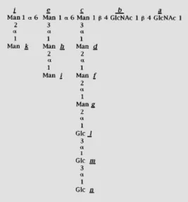
Figure 1 - Structure of oligosac-charides. The structure is that of the oligosaccharide transferred to proteins in wild type mamma-lian, plant and fungal cells. The lettering used for the identifica-tion of the individual monosac-charide residues (a, b, c, ...) fol-lows the same order as the addi-tion of the monosaccharides in the assembly of the oligosaccha-ride. The oligosaccharides with compositions Man9GlcNAc2, Man7GlcNAc2 and Man6Glc NAc2 transferred in different trypanosomatid species are de-void of residues l, m and n, or of
j, k, l, m, and n or of i, j, k, l, m
compounds were not formed by deglucosy-lation and demannosydeglucosy-lation of Glc3Man9Glc
NAc2. Moreover, upon fractionation of
mi-crosomes in sucrose gradients, maximal for-mation of the monoglucosylated compounds was detected in the rough ER-containing fractions and not in those containing Golgi apparatus-derived membranes (23). On the other hand, addition of thyroglobulin, a gly-coprotein containing high mannose-type oli-gosaccharides, to the incubation mixtures did not enhance formation of the monoglu-cosylated derivatives. This result stalled fur-ther research on the subject for several years. It is worth mentioning that the formation of Glc1Man8GlcNAc2 and Glc1Man7GlcNAc2
upon incubation of mammalian cells with [14C]glucose or [14C]galactose had been
ob-served previously by other researchers, but they had assumed without any experimental evidence that those compounds had been formed by successive deglucosylation and demannosylation of the transferred oligosac-charide (Glc3Man9GlcNAc2) (25,26). It was
the fact that direct glucosylation of high mannose type compounds was first detected in trypanosomatids that suggested the occur-rence of this pathway in mammalian cells.
The UDP-Glc:glycoprotein glucosyltransferase discriminates between misfolded and native glycoproteins or glycopeptides
Mainly by chance it was found that 8 M urea-denatured thyroglobulin was a good glucose acceptor when added to incubation mixtures containing UDP-[14C]Glc and rat
liver microsomes. The products formed were protein-linked Glc1Man9GlcNAc2, Glc1Man8
GlcNAc2 and Glc1Man7GlcNAc2 (27).
Evi-dence was obtained indicating that this reac-tion represented transfer of glucose units from UDP-Glc to the acceptor protein and that no dolichol monophosphate or diphos-phate derivatives were involved in it (27). Moreover, this result provided a convenient method for assaying the transferring enzyme (UDP-Glc:glycoprotein glucosyltransferase, GT), which was detected in microsomal membranes of mammalian, plant, fungal and trypanosomatid cells (27). Further work showed that GT was a soluble protein of the ER lumen and that the reaction products had the glucose unit linked to the same mannose with the same α(1,3) bond as in Glc1Man9
GlcNAc2-P-P-dolichol (Figure 1) (27,28).
As observed for thyroglobulin, other glyco-proteins (phytohemagglutinin, soybean ag-glutinin, ribonuclease B) were not gluco-sylated unless they had been previously de-natured (29). Moreover, glycopeptides ob-tained upon digestion of thyroglobulin with a nonspecific protease (Pronase) or of phy-tohemagglutinin or soybean agglutinin with trypsin were very poorly glucosylated by the enzyme (29).
The assay allowed purification to homo-geneity of the enzyme from rat liver and Schizosaccharomyces pombe (30,31). GT had an almost absolute calcium requirement for activity (maximal activity is attained at about 10 mM of the cation). The ER lumen is the major intracellular calcium reservoir. The concentration of the cation in such subcellu-lar location is about 3 mM, about three or-Figure 2 - Processing of
ders of magnitude higher than that in the cytosol. The luminal concentration of the cation is not constant and is apparently regu-lated by the action of an ion-motive ATPase that pumps calcium into the lumen and ino-sitol 1,4,5-triphosphate that releases it to the cytosol. The possibility exists, therefore, that variations in the intraluminal calcium con-centration may affect transient glucosylation of glycoproteins. The only donor substrate for the homogeneous rat liver or S. pombe GTs appeared to be UDP-Glc. Other sugar nucleotides such as ADP-Glc, TDP-Glc and UDP-Glc were not effective substrates. The problem posed by the fact that the donor substrate (UDP-Glc) is synthesized in the cytoplasm and acts in the interior of the ER was solved by the description of a transport system specific for UDP-Glc in the ER mem-brane of mammalian cells (32).
Not all oligosaccharides are transiently glucosylated in vivo by the UDP-Glc:glycoprotein
glucosyltransferase
Are the majority or only a minor fraction of glycoproteins transiently glucosylated in vivo? To answer this question in mammalian cells it would be necessary to have available compounds able to only inhibit removal of glucose units added by UDP-Glc:glycopro-tein glucosyltransferase and not those pres-ent in the transferred oligosaccharide. Avail-ability of such compounds is impossible be-cause, as mentioned above, Glc1Man9Glc
NAc2 formed by GT is structurally
indistin-guishable from that produced by deglucosy-lation of Glc3Man9GlcNAc2. Moreover,
known inhibitors of glucosidase II also in-hibit glucosidase I, and thence addition of them to intact mammalian cells produces an accumulation of Glc3Man9GlcNAc2 and
Glc2Man9GlcNAc2. The question can be
an-swered, nevertheless, for trypanosomatids since their glucosylated compounds are ex-clusively formed by action of UDP-Glc:
glycoprotein glucosyltransferase (Figure 2B). It was found that about 50% of all N-linked oligosaccharides were glucosylated in T. cruzi or Crithidia fasciculata cells incubated in the presence of castanospermine and/or 1-deoxynojirimycin, known inhibitors of glu-cosidase II (33,34). Analysis of individual glycoproteins indicated that all glycopro-teins are glucosylated in vivo but that the same oligosaccharide is glucosylated in some molecules but not in others (35). A possible explanation for this result will be given be-low.
Molecular basis for the selective glucosylation of misfolded glycoproteins by the
UDP-Glc:glycoprotein glucosyltransferase
native conformations. Interaction of those protein domains with GT was apparently necessary for the occurrence of the transfer reaction (29). Several hypotheses may be advanced concerning the nature of the pro-tein domains interacting with GT. They may be formed by a) specific amino acid se-quences common to all glycoproteins, b) certain specific amino acids that are sepa-rated in the primary sequence but that be-come close in the denatured conformations, and c) nonspecific amino acids but common three-dimensional structures shared by all denatured glycoproteins but generated by totally different amino acid sequences. The widespread distribution of transient gluco-sylation in nature indicates that very differ-ent glycoproteins are glucosylated in vivo, a fact that invalidates all the hypotheses ad-vanced above since it should not be reason-ably expected for any of the structural fea-tures alluded in them to be common to all glucosylated glycoproteins.
The interior of water-soluble proteins in their native states is predominantly com-posed of hydrophobic amino acids while the hydrophilic side chains are on the exterior where they interact with water. Denatured states have, in general, more hydrophobic side chains on the exterior than native ones. Thus, the effect of denaturation may be to provide a certain hydrophobic environment in the vicinity of the oligosaccharide and this environment may be recognized by GT. Fur-ther work showed that under physiological pH and salt concentration conditions GT very efficiently binds hydrophobic but not hydrophilic peptides and that binding of the former species can be inhibited by denatured but not native glycoproteins (36). On the other hand, a rather unexpected result was the fact that not only native but also dena-tured non-glycosylated proteins failed to in-hibit glucosylation of denatured glycopro-teins. Additional experiments showed that the GT recognized both protein domains exposed in denatured conformations and the
innermost N-acetylglucosamine unit of the oligosaccharide, i.e., the residue that is left linked to the protein moiety by endo-ß-N-acetylglucosaminidase H (36). In many na-tive glycoproteins the innermost N-acetyl-glucosamine interacts with neighboring amino acid residues and is not accessible to a macromolecular probe such as endo-ß-N-acetylglucosaminidase H. In these cases, cleavage of oligosaccharides by the endo-glycosidase requires denaturation of the gly-coprotein (37). It may be speculated that proper folding of most glycoproteins would hinder recognition of the innermost N-ace-tylglucosamine unit by GT and thus prevent glucosylation. Several experiments showed that both recognition elements, the protein domains (hydrophobic amino acids) and the oligosaccharides, had to be covalently linked. This is an important restriction. Since there are numerous unfolded, partially folded and misfolded proteins and glycoproteins in the ER lumen, if the protein domains recognized by the GT and the oligosaccharides were not required to be covalently linked it might be speculated that domains exposed in not prop-erly folded species would induce glucosyla-tion of glycoproteins already in their native conformations, provided that the innermost N-acetylglucosamine units are accessible to GT in the latter species.
GT as a sensor of glycoprotein tertiary structures
(38). A high mannose type glycopeptide was then linked to that position using N-succinimidyl 3-(2-pyridyldithio) propionate, a bifunctional reagent that reacts with amino and sulfhydryl groups. The neoglycoprotein thus formed (K70C-Glyc) had the same spe-cific nuclease activity as the unmodified pro-tein, thus suggesting that both species had the same tertiary structure (38,39). The neoglycoprotein had a very low glucose ac-ceptor capacity but addition of the nuclease inhibitor pdTp [3,5 diphosphothymidine], a stabilizer of the native nuclease conforma-tion and an inducer of proper folding in truncated species (see below), further dimin-ished the glucose acceptor capacity. The neoglycoprotein was nevertheless efficiently glucosylated when previously denatured with 8 M urea.
It has been reported that a truncated nu-clease lacking the last 14 amino acids at the C-terminal end is per se (i.e., without any denaturing treatment) in a compact but dis-ordered conformation, but that the enzyme can be induced to properly fold in the pres-ence of Ca2+ and pdTp or the substrate (DNA),
as judged by far UV circular dichroism (CD) and nuclear magnetic resonance spectra (39,40). This large fragment showed the same specific nuclease activity as the full length enzyme, thus indicating that both species had the same tertiary structure.
Two truncated neoglycoproteins were synthesized using this large fragment as a protein backbone, one with the oligosaccha-ride attached to a cysteine introduced at position 70 (1-135 K70C-Glyc), i.e., in the same position as in the full length species, and the other at position 124 (1-135 H124C-Glyc). In this case the cysteine was intro-duced in place of a histidine. Both truncated neoglycoproteins were efficiently glucosyl-ated when pdTp was omitted from the reac-tion mixture (no denaturing treatment was performed). This suggests that the position of the oligosaccharide is not an important factor for glucosylation. Upon addition of
pdTp, the glucose acceptor capacity of both truncated neoglycoproteins was reduced by about 60%, but without reaching the basal levels of the full length neoglycoprotein (K70C-Glyc) in the presence of pdTp (it is worth mentioning that Ca2+, the other
ele-ment required for inducing proper folding, is always present in reaction mixtures since it is required for GT activity).
In order to check that the incubation of the truncated neoglycoproteins with Ca2+
and pdTp had actually led to the transition to a native conformation, the far UV CD spec-tra of the truncated neoglycoprotein 1-135 K70C-Glyc and the full length wild type enzyme were compared. The truncated neoglycoprotein yielded a spectrum strik-ingly different from that of the wild type enzyme, but both spectra became superim-posable when Ca2+ and pdTp were added to
the former species. This indicated that both the truncated neoglycoprotein in the pres-ence of Ca2+ and pdTp and the wild type
enzyme had the same secondary structure. Nevertheless, the tertiary structures of K70C-Glyc (a full length neoglycoprotein) and of 1-135 K70C-Glyc (a truncated neoglycoprotein) were different: when sub-mitted to limited proteolysis in the presence of 1 mM pdTp and 10 mM CaCl2,
K70C-Glyc was barely cleaved even after a 60-min incubation with trypsin whereas the 1-135 K70C-Glyc was rapidly degraded. Moreover, the specific nuclease activities of both trun-cated neoglycoproteins (1-135 K70C-Glyc and 1-135 H124C-Glyc) were much lower than that of the full length neoglycoprotein (K70C-Glyc). The tertiary structures of the truncated neoglycoproteins in the presence of Ca2+ and pdTp or DNA were, therefore,
the latter had a residual glucose acceptor capacity in the presence of Ca2+ and pdTp
(36).
The excellence of GT as a sensor of the tertiary structure of glycoproteins was also revealed by the fact that the folding status of glycoproteins was paralleled by their ability to be glucosylated by the enzyme: soybean agglutinin was denatured with 6 M guani-dine hydrochloride, diluted and allowed to renature under controlled conditions. Rena-turation was monitored by measuring the fluorescent emission of tryptophan at 350 nm. The maximum wavelengths of tryp-tophan emission in soybean agglutinin are 328 and 350 nm in native and random coil conformations, respectively. The decrease in glucose acceptor capacity closely followed renaturation, thus confirming the conclusion reached above (36).
The primary sequence of UDP-Glc: glycoprotein glucosyltransferase: what it suggests
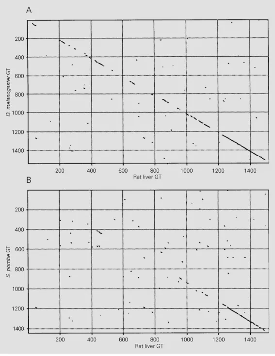
As mentioned above, GT is an excellent sensor of glycoprotein tertiary structures. The primary sequences of GT from two dif-ferent species (Drosophila melanogaster and S. pombe, GenBank accession numbers U20554 and U38417, respectively) are known (41,42). In addition, we have re-cently sequenced the enzyme from rat liver (Trombetta ES and Parodi AJ, unpublished results). The sequence of an open reading frame (gene F48E3.3; GenBank U28735) of the Caenorhabditis elegans genome prob-ably corresponds to the gene encoding GT due to its high identity with the other three sequences. The four sequences correspond to proteins of the same size (about 160 kDa). Moreover, retrieval sequences typical of ER soluble proteins are found at their C-termi-nal ends. All of them also have several N-glycosylation consensus sequences. This agrees with the fact that the mammalian and fungal enzymes interact with concanavalin
A. Comparison of the D. melanogaster and S. pombe GT sequences with that obtained for the rat liver enzyme is depicted in Figure 3A and B. All three enzymes show an ex-tremely high sequence homology at their C-terminal domains. A similar homology has been found between those domains and con-ceptual translations of expressed sequence tags from rice and Arabidopsis thaliana (GenBank D24933 and T23006, respec-tively). Since a limited but significant se-quence homology may be observed between the C-terminal domains of the fly, fungal and mammalian enzymes with several bacterial glycosyltransferases that use UDP-Glc or UDP-Gal as donor substrates (41) it may be speculated that the highly conserved GT C-terminal portions are responsible for UDP-Glc recognition. The same conserved en-zyme portions might also be responsible for the recognition of the other common sub-strate structure, that of the oligosaccharide acceptor.
structures, they may be constituted by quite different amino acid sequences. By further pursuing the analogy with hsp70 proteins, it may also be speculated that part of the en-ergy spent in the transfer of the glucose unit is also used to facilitate the release of the enzyme from the misfolded substrate, as is the case for chaperones upon ATP hydroly-sis.
The GT sequences show a certain homol-ogy with that of the S. cerevisiae Kre5 pro-tein. This is also a soluble ER protein similar in size to the GTs. The exact function of the Kre5 protein is unknown. Experimental evi-dence obtained both with cell-free extracts and with intact cells showed that S. cerevi-siae is the only eukaryote known to date to be devoid of GT activity (31). The
C-termi-D. melanogaster
GT
200
400
600
800
1000
1200
1400
200
600
800
1000
1200
1400
S. pombe
GT
200 400 600 800 1000 1200 1400
400
200 400 600 800 1000 1200 1400
Rat liver GT Rat liver GT A
B
Figure 3 - Homologies between GT primary sequences of D. melanogaster (A) and S. pombe
nal portions having the highest homology (over 66-70%) among rat liver, D. melano-gaster, C. elegans and S. pombe GTs only show a much reduced (about 25%) homol-ogy with the Kre5 protein. Another differ-ence is that certain Kre5 mutants appeared to be lethal whereas S. pombe mutant cells completely devoid of GT activity showed no discernible phenotype (42,44). As certain non-lethal Kre5 mutants were affected in cell wall glucan biosynthesis it may be specu-lated that Kre5 is a glucosyltransferase in-volved in the formation of that polysaccha-ride (44). Alternatively, the possibility exists that Kre5 is a GT that has lost its glucosyl-transferase activity but has conserved the postulated chaperone activity.
The influence of saccharides on glycoprotein folding
Glycoproteins, as all proteins entering the so called secretory pathway followed by secreted proteins, lysosomal enzymes and by proteins resident on several cellular mem-branes, acquire their tertiary structure (and in some cases also their quaternary one) in the ER. Glycoproteins that fail to fold prop-erly are retained in that subcellular location and are degraded in the proteasome. This implies the occurrence of a very stringent quality control of folding in the ER.
Except for a few exceptions, saccharide moieties have no influence on the tertiary structure of mature glycoproteins. On the other hand, their influence on folding varies for different species. Three approaches have been followed to study the effect of oli-gosaccharide on the folding process. The first takes advantage of the fact that tunicamycin, an N-acetylglucosamine ana-log, inhibits the synthesis of dolichol-P-P-GlcNAc, i.e., the first step in the synthesis of Glc3Man9GlcNac2-P-P-dolichol. Addition of
this drug to intact cells drastically inhibits protein glycosylation. The second approach uses cell lines defective in steps leading to
the formation of dolichol-P-P derivatives. Finally, a third approach consists of abolish-ing or creatabolish-ing new glycosylation sites in glycoproteins by site-directed mutagenesis (45).
In most cases glycoproteins show an ab-solute requirement for oligosaccharides for proper folding. There are, however, numer-ous exceptions to this rule (45). In some cases folding becomes temperature depend-ent whereas in others a fraction of the pro-teins folds properly and is transported to the Golgi apparatus, whereas the rest is degraded (46-49). Finally in some cases the saccha-rides are not required for attaining the cor-rect tertiary structure (48,50-52). Small dif-ferences in the amino acid primary sequence are often responsible for determining whether or not the oligosaccharide is required for folding. For instance, only one G protein of two strains of vesicular stomatitis virus re-quires the presence of the oligosaccharide for proper folding. The saccharide-depend-ent species can be converted into an inde-pendent one by a single point mutation (53). Site-directed mutagenesis of glycopro-teins having several N-linked oligosaccha-rides has very often shown that no single oligosaccharide is essential for folding and that several glycosylation consensus se-quences must be suppressed to affect fold-ing. On the other hand, the consequences of suppressing glycosylation sites may be re-versed by creating new ones at entirely dif-ferent locations (54-57).
Endoplasmic reticulum chaperones
sequence Arg-Lys-x-Arg-Arg-x which is probably an ER retention sequence for trans-membrane type I proteins (59). Calreticulin (molecular mass 46 kDa) is a soluble ho-molog of calnexin that ends with the known retention sequence for soluble proteins of the endoplasmic reticulum Lys-Asp-Glu-Leu (60). Both calnexin and calreticulin have calcium-binding motifs.
What is particularly interesting about cal-nexin and calreticulin is that both exclu-sively bind glycoproteins (61,62). Moreover, inhibition of glucosidases I and II by the addition of castanospermine or 1-deoxynoji-rimycin to cells yielded glycoproteins that were not precipitated with anticalnexin or anticalreticulin antibodies under native con-ditions. Evidence was presented indicating that both calnexin and calreticulin behaved as lectin-like proteins that recognized mono-glucosylated oligosaccharides. Further ex-periments showed that binding of calnexin and calreticulin to glycoproteins depended exclusively on the presence of monogluco-sylated oligosaccarides and was independ-ent from the tertiary structure of the protein moieties (63,64). Although mammalian cells have both calnexin and calreticulin, only the first one has been detected in S. cerevisiae and S. pombe and only calreticulin was found to be present in trypanosomatid protozoa (65; Labriola C and Parodi AJ, unpublished results).
The quality control of glycoprotein folding
As mentioned above, a very stringent quality control of folding is required to pre-vent passage of misfolded proteins from the ER to the Golgi cisternae. A model for such quality control applicable to glycoproteins has been recently proposed (45,62,66). Ac-cording to it, the transferred oligosaccharide (Glc3Man9GlcNAc2) would be first
degluco-sylated to Glc1Man9GlcNAc2 or Man9Glc
NAc2. The high mannose-type
oligosaccha-rides thus formed would then shuttle be-tween monoglucosylated and unglucosylated structures, their formation being catalyzed by the GT and glucosidase II.
Calnexin and calreticulin would bind the monoglucosylated structures in denatured conformations and thus retain glycoproteins in the ER as long as the protein moieties are not properly folded, that is, as long as glyco-proteins are reglucosylated by the GT. On attaining the correct native conformations, glycoproteins would become substrates for glucosidase II but not for GT and thus the element recognized by the calnexin/calretic-ulin anchor would be eliminated. Glycopro-teins would then be able to be transported to the Golgi apparatus. It was found that the interaction of calnexin/calreticulin with monoglucosylated oligosaccharides not only retains improperly folded glycoproteins in the ER but also facilitates folding of bound species probably by preventing their aggre-gation and thus allowing interaction with other chaperones such as BiP (67). It was also found that for individual glycoproteins not all the molecules interact with calnexin/ calreticulin (67). This finding correlates with the known fact that folding is an asynchro-nous process, i.e., different molecules of the same species might follow different folding pathways. All molecules end up with the same native conformation although at differ-ent rates. It may be assumed that glycopro-tein molecules that rapidly attain their proper tertiary structures would not interact with calnexin/calreticulin. This would also ex-plain the fact, mentioned above, that the same oligosaccharide in individual T. cruzi glycoproteins appeared to be glucosylated by GT in some molecules but not in others (35).
cannot be tested, however, in most eukary-otic cells because, as mentioned above, glu-cosidase II is responsible for removal of both α(1,3)-linked glucose units and known in-hibitors of glucosidase II also inhibit glu-cosidase I. Addition of those drugs to cells leads to the accumulation of oligosaccha-rides having two or three glucoses that are not recognized by calnexin or calreticulin. The prediction can be tested, however, in trypanosomatid protozoa because, as shown in Figure 1A and B, the only glucose present in their glycoproteins is that added by the
GT. It was found that addition of 1-deoxyno-jirimycin (an inhibitor of glucosidase II) to T. cruzi cells delayed the exit of glycopro-teins from the ER (35).
What started as the study of the initial steps in the processing of N-linked oligosac-charides in trypanosomatid protozoa ended up as a model for the quality control of glycoprotein folding in the ER. This con-firms the ultimate principle, one of Murphys laws: by definition when you are investigating the unknown, you do not know what you will find.
References
1. Kornfeld R & Kornfeld S (1985). Assem-bly of asparagine-linked oligosaccharides.
Annual Review of Biochemistry, 54: 631-664.
2. Parodi AJ, Behrens NH, Leloir LF & Carminatti HC (1972). The role of poly-prenol-bound saccharides as intermedi-ates in glycoprotein synthesis in liver. Pro-ceedings of the National Academy of Sci-ences, USA, 69: 3268-3272.
3. Hubbard SC & Ivatt RJ (1981). Synthesis and processing of asparagine-linked oli-gosaccharides. Annual Review of Bio-chemistry, 50: 555-583.
4. Kelleher DJ, Kreibich G & Gilmore R (1992). Oligosaccharyltransferase activity is associated with a protein complex com-posed of ribophorins I and II and a 48 kDa protein. Cell, 69: 55-65.
5. Turco S, Stetson B & Robbins PW (1977). Comparative rates of transfer of lipid-linked oligosaccharides to endogenous glycoprotein acceptors in vitro. Proceed-ings of the National Academy of Sci-ences, USA, 74: 4411-4414.
6. Ballou L, Gopal P, Krummel B, Tammi M & Ballou CE (1986). A mutation that pre-vents glucosylation of the lipid-linked oli-gosaccharide precursor leads to under-glycosylation of secreted yeast invertase.
Proceedings of the National Academy of Sciences, USA, 83: 3081-3085.
7. Hettkamp H, Legler G & Bause E (1984). Purification by affinity chromatography of glucosidase I, an endoplasmic reticulum hydrolase involved in the processing of asparagine-linked oligosaccharides. Euro-pean Journal of Biochemistry, 142: 85-90.
8. Brada D & Dubach UC (1984). Isolation of a homogeneous glucosidase II from pig kidney microsomes. European Journal of Biochemistry, 141: 149-156.
9. Bischoff J, Liscum L & Kornfeld R (1986). The use of 1-deoxymannojirimycin to evaluate the role of various α -mannosi-dases in oligosaccharide processing in in-tact cells. Journal of Biological Chemistry, 261: 4766-4774.
10. Weng S & Spiro RG (1993). Demonstra-tion that a kifunensin-resistant α -mannosi-dase with a unique processing action on
N-linked oligosaccharides occurs in rat liver endoplasmic reticulum and various cultured cells. Journal of Biological Chem-istry, 268: 25656-25663.
11. Jelinek-Kelly S & Herscovics A (1988). Gly-coprotein biosynthesis in Saccharomyces cerevisiae. Purification of the α -mannosi-dase which removes one specific man-nose residue from Man9GlcNAc. Journal of Biological Chemistry, 263: 14757-14763.
12. Vickerham K (1976). The diversity of the kinetoplastid flagellates. In: Lumsden WHR & Evans DA (Editors), Biology of Kinetoplastida. Academic Press, New York. 13. Parodi AJ (1993). N-glycosylation in trypa-nosomatid protozoa. Glycobiology, 3: 193-199.
14. Parodi AJ (1993). Biosynthesis of protein-linked oligosaccharides in trypanosomatid flagellates. Parasitology Today, 9: 373-377.
15. de la Canal L & Parodi AJ (1987). Synthe-sis of dolichol derivatives in trypanosoma-tids. Characterization of enzymatic pat-terns. Journal of Biological Chemistry,
262: 11128-11133.
16. Parodi AJ & Cazzulo JJ (1982). Protein glycosylation in Trypanosoma cruzi. II. Par-tial characterization of protein-bound oli-gosaccharides labeled in vivo. Journal of Biological Chemistry, 257: 7641-7645. 17. Parodi AJ, Lederkremer GZ & Mendelzon
DH (1983). Protein glycosylation in Try-panosoma cruzi. The mechanism of gly-cosylation and structure of protein-bound oligosaccharides. Journal of Biological Chemistry, 258: 5589-5595.
18. Parodi AJ, Martín-Barrientos J & Engel JC (1984). Glycoprotein assembly in Leish-mania mexicana. Biochemical and Bio-physical Research Communications, 118: 1-7.
19. Mendelzon DH & Parodi AJ (1986). N-linked, high mannose-type oligosaccha-rides in the protozoa Crithidia fasciculata
and Crithidia harmosa contain galactofura-nose residues. Journal of Biological Chemistry, 261: 2129-2133.
20. Mendelzon DH, Previato JO & Parodi AJ (1986). Characterization of protein-linked oligosaccharides in trypanosomatid flagel-lates. Molecular and Biochemical Parasi-tology, 18: 355-367.
21. Bosch M, Trombetta S, Engstrom U & Parodi AJ (1988). Characterization of dolichol diphosphate oligosaccharide:protein oligosaccharyltransferase and of glycopro-tein processing glucosidases occurring in trypanosomatids. Journal of Biological Chemistry, 263: 17360-17365.
possible recognition signal in the process-ing of glycoproteins. Journal of Biological Chemistry, 258: 8260-8265.
23. Parodi AJ, Mendelzon DH, Lederkremer GZ & Martín-Barrientos J (1984). Evidence that transient glucosylation of protein-linked Man9GlcNAc2, Man8GlcNAc2 and Man7GlcNAc2 occurs in rat liver and Pha-seolus vulgaris cells. Journal of Biological Chemistry, 259: 6351-6357.
24. Lederkremer GZ & Parodi AJ (1986). Pro-cessing of asparagine-linked saccharides in Mucor rouxii.Biochimica et Biophysica Acta, 884: 363-369.
25. Kornfeld S, Li E & Tabas I (1978). The synthesis of complex-type oligosaccha-rides. II. Characterization of the process-ing intermediates in the synthesis of the complex units of the vesicular stomatitis virus G protein. Journal of Biological Chemistry, 253: 7771-7778.
26. Godelaine D, Spiro MJ & Spiro RG (1981). Processing of the carbohydrate units of thyroglobulin. Journal of Biological Chem-istry, 256: 10161-10168.
27. Trombetta S, Bosch M & Parodi AJ (1989). Glucosylation of glycoproteins by mam-malian, plant, fungal and trypanosomatid protozoa microsomal proteins. Biochem-istry, 28: 8108-8116.
28. Trombetta S, Gañán S & Parodi AJ (1991). The UDP-Glc:glycoprotein glucosyltrans-ferase is a soluble protein of the endo-plasmic reticulum. Glycobiology, 1: 155-161. 29. Sousa M, Ferrero-García MA & Parodi AJ (1992). Recognition of the oligosaccha-ride and protein moieties of glycoproteins by the UDP-Glc:glycoprotein glucosyl-transferase. Biochemistry, 31: 97-105. 30. Trombetta S & Parodi AJ (1992).
Purifica-tion to apparent homogeneity and partial characterization of rat liver UDP-Glc:gly-coprotein glucosyltransferase. Journal of Biological Chemistry, 267: 9236-9240. 31. Fernández F, Trombetta S, Hellman U &
Parodi AJ (1994). Purification to homoge-neity of UDP-Glc:glycoprotein glucosyl-transferase from Schizosaccharomyces pombe and apparent absence of the en-zyme from Saccharomyces cerevisiae.
Journal of Biological Chemistry, 269: 30701-30706.
32. Pérez M & Hirschberg C (1986). Topogra-phy of glycosylation reactions in the rough endoplasmic reticulum membrane. Jour-nal of Biological Chemistry, 261: 6822-6830.
33. Gañán S, Cazzulo JJ & Parodi AJ (1991). A major proportion of N-glycoproteins are transiently glucosylated in the endoplas-mic reticulum. Biochemistry, 30:
3098-3104.
34. Gotz G, Gañán S & Parodi AJ (1991). Glu-cosylation of glycoproteins in Crithidia fasciculata. Molecular and Biochemical Parasitology, 45: 265-274.
35. Labriola C, Cazzulo JJ & Parodi AJ (1995). Retention of glucose residues added by the UDP-Glc:glycoprotein glucosyltrans-ferase delays exit of glycoproteins from the endoplasmic reticulum. Journal of Cell Biology, 130: 771-779.
36. Sousa M & Parodi AJ (1995). The molecu-lar basis for the recognition of misfolded glycoproteins by the UDP-Glc:glycopro-tein glucosyltransferase. EMBO Journal, 14: 4196-4203.
37. Trimble RB & Maley F (1984). Optimizing hydrolysis of N-linked high mannose oli-gosaccharides by endo-ß-N-acetylglu-cosaminidase H. Analytical Biochemistry, 141: 515-522.
38. Ermacora MR, Ledman DW, Hellinga HW, Hsu GW & Fox RO (1994). Mapping staph-ylococcal nuclease conformation using an EDTA-Fe derivative attached to geneti-cally engineered cysteine residues. Bio-chemistry, 33: 13625-13641.
39. Shortle DA & Meeker A (1989). Residual structure in large fragments of staphylo-coccal nuclease: effects of amino acid substitutions. Biochemistry, 28: 936-944. 40. Flanagan JM, Kataoka M, Shortle D & Engenman DM (1992). Truncated staphy-lococcal nuclease is compact but disor-dered. Proceedings of the National Acad-emy of Sciences, USA, 89: 748-752. 41. Parker CG, Fessler LI, Nelson RE &
Fessler JH (1995). Drosophila UDP-Glc:gly-coprotein glucosyltransferase: sequence and characterization of an enzyme that distinguishes between denatured and na-tive proteins. EMBO Journal, 14: 1294-1303.
42. Fernández F, Jannatipour M, Hellman U, Rokeach L & Parodi AJ (1996). A new stress protein: synthesis of Schizosaccha-romyces pombe UDP-Glc:glycoprotein glucosyltransferase mRNA is induced un-der stress conditions but the enzyme is not essential for cell viability. EMBO Jour-nal, 15: 705-713.
43. Hendrick JP & Hartl FU (1993). Molecular chaperone functions of heat-shock pro-teins. Annual Review of Biochemistry, 62: 349-384.
44. Meaden P, Hill K, Wagner J, Slipetz D, Sommer SS & Bussey H (1990). The yeast KRE5 gene encodes a probable endoplas-mic reticulum protein required for (1-6)-ß-D-glucan synthesis and normal cell growth. Molecular and Cellular Biology,
10: 3013-3019.
45. Helenius A (1994). How N-linked oligosac-charides affect glycoprotein folding in the endoplasmic reticulum. Molecular Biology of the Cell, 5: 253-265.
46. Machamer CE & Rose JK (1988). Vesicu-lar stomatitis virus G proteins with altered glycosylation sites display temperature-sensitive intracellular transport and are subject to aberrant intermolecular disul-fide bonding. Journal of Biological Chem-istry, 263: 5955-5960.
47. Roberts PC, Garten W & Klenk HD (1993). Role of conserved glycosylation sites in maturation and transport of influenza A virus hemagglutinin. Journal of Virology, 67: 3048-3060.
48. Hickman J & Kornfeld S (1978). Effect of tunicamycin on IgM, IgA and IgG secre-tion by mouse plasmacytoma cells. Jour-nal of Immunology, 121: 990-996. 49. Miyazaki JL, Appella EHZ, Forman J &
Ozato K (1986). Expression and function of a nonglycosylated major histocompat-ibility class I antigen. Journal of Experi-mental Medicine, 163: 856-871. 50. Olden K, Pratt RM & Yamada KM (1978).
Role of carbohydrates in protein secretion and turnover: effect of tunicamycin on the major cell surface glycoprotein of chick embryo fibroblasts. Cell, 13: 461-473.
51. Shackelford DA & Strominger JL (1983). Analysis of the oligosaccharides on the HLA-DR and DC-1B cell antigens. Journal of Immunology, 130: 274-282.
52. Landolfi N, Rich R & Cook RG (1985). Differential glycosylation requirements for the cell surface expression of class I mol-ecules. Journal of Immunology, 134: 423-430.
53. Pitta AM, Rose JK & Machamer CE (1989). A single amino acid substitution eliminates the stringent carbohydrate re-quirement for intracellular transport of a viral glycoprotein. Journal of Virology, 63: 906-913.
54. Williams AM & Enns CA (1993). A region of the C-terminal portion of the human transferrin receptor contains an aspar-agine-linked glycosylation site critical for receptor structure and function. Journal of Biological Chemistry, 268: 12780-12786.
55. Shipley JM, Grubb JH & Sly WS (1993). The role of glycosylation and phosphory-lation in the expression of active human ß-glucuronidase. Journal of Biological Chemistry, 268: 12193-12198.
ex-pression of the vesicular stomatitis virus G protein at the plasma membrane. Jour-nal of Biological Chemistry, 263: 5948-5954.
57. Machamer CE, Florkiewicz RZ & Rose JK (1985). A single N-linked oligosaccharide at either of the two normal sites is suffi-cient for transport of vesicular stomatitis virus G protein to the cell surface. Molec-ular and CellMolec-ular Biology, 5: 3074-3083. 58. Degen E & Williams DB (1991).
Participa-tion of a novel 88-kDa protein in the bio-genesis of murine class I histocompatibil-ity molecules. Journal of Cell Biology, 112: 1099-1115.
59. David V, Hochstenbach F, Rajagopalan S & Brenner MB (1993). Interaction with newly synthesized and retained proteins in the endoplasmic reticulum suggests a chaperone function for human integral membrane protein IP90 (calnexin). Jour-nal of Biological Chemistry, 268:
9585-9592.
60. Michalak M, Milner RE, Burns K & Opas M (1992). Calreticulin. Biochemical Jour-nal, 285: 681-692.
61. Ou WJ, Cameron PH, Thomas DY & Bergeron JJM (1993). Association of fold-ing intermediates of glycoproteins with calnexin during protein maturation. Na-ture, 364: 771-776.
62. Hammond C, Braakman I & Helenius A (1994). Role of N-linked oligosaccharide recognition, glucose trimming, and cal-nexin in glycoprotein folding and quality control. Proceedings of the National A-cademy of Sciences, USA, 91: 913-917. 63. Rodan AR, Simons JF, Trombetta ES &
Helenius A (1996). N-linked oligosaccha-rides are necessary and sufficient for as-sociation of glycosylated forms of bovine RNase with calnexin and calreticulin.
EMBO Journal, 15: 6921-6930.
64. Zapun A, Petrescu SM, Rudd PM, Dwek
RW, Thomas DY & Bergeron JJM (1997). Conformation independent binding of monoglucosylated ribonuclease B to calnexin. Cell, 88: 29-38.
65. Josi M, Pogue GP, Duncan RC, Lee NS, Singh NK, Atreya CD, Dwyer DM & Nakhasi HL (1996). Isolation and charac-terization of Leishmania donovani calre-ticulin gene and its conservation of the RNA binding activity. Molecular and Bio-chemical Parasitology, 81: 53-64. 66. Helenius A, Trombetta ES, Hebert DN &
Simons JF (1997). Calnexin, calreticulin and the folding of proteins. Trends in Cell Biology, 7: 193-200.