Thyro id pe ro xidase activity is
inhibite d by am ino acids
Laboratório de Fisiologia Endócrina, Instituto de Biofísica Carlos Chagas Filho, Universidade Federal do Rio de Janeiro, Rio de Janeiro, RJ, Brasil
D.P. Carvalho, A.C.F. Ferreira, S.M. Coelho, J.M. Moraes, M.A.S. Camacho and D. Rosenthal
Abstract
Normal in vitro thyroid peroxidase (TPO) iodide oxidation activity was completely inhibited by a hydrolyzed TPO preparation (0.15 mg/ ml) or hydrolyzed bovine serum albumin (BSA, 0.2 mg/ml). A pancre-atic hydrolysate of casein (trypticase peptone, 0.1 mg/ml) and some amino acids (cysteine, tryptophan and methionine, 50 µM each) also inhibited the TPO iodide oxidation reaction completely, whereas casamino acids (0.1 mg/ml), and tyrosine, phenylalanine and histidine (50 µM each) inhibited the TPO reaction by 54% or less. A pancreatic digest of gelatin (0.1 mg/ml) or any other amino acid (50 µM) tested did not significantly decrease TPO activity. The amino acids that impair iodide oxidation also inhibit the TPO albumin iodination activity. The inhibitory amino acids contain side chains with either sulfur atoms (cysteine and methionine) or aromatic rings (tyrosine, tryptophan, histidine and phenylalanine). Among the amino acids tested, only cysteine affected the TPO guaiacol oxidation reaction, producing a transient inhibition at 25 or 50 µM. The iodide oxidation inhibitory activity of cysteine, methionine and tryptophan was re-versed by increasing iodide concentrations from 12 to 18 mM, while no such effect was observed when the cofactor (H2O2) concentration was increased. The inhibitory substances might interfere with the enzyme activity by competing with its normal substrates for their binding sites, binding to the free substrates or reducing their oxidized form.
Co rre spo nde nce
D. Rosenthal
Laboratório de Fisiologia Endócrina Instituto de Biofísica, UFRJ 21949-900 Rio de Janeiro, RJ Brasil
Fax: + 55-21-280-8193 E-mail: doris@ biof.ufrj.br
Research supported by FINEP, CEPG/UFRJ, FAPERJ and CNPq.
Received June 17, 1999 Accepted January 21, 2000
Ke y wo rds ·Thyroid
·Thyroperoxidase
·Amino acids
·Iodide oxidation
·Lactoperoxidase
Intro ductio n
Thyroid peroxidase (TPO) is a heme-containing, membrane bound, glycoprotein enzyme that plays a key role in the biosyn-thesis of thyroid hormones (1,2). The oxida-tion of iodide, the iodinaoxida-tion of thyroglobu-lin and the coupthyroglobu-ling of iodotyrosyl residues of thyroglobulin are catalyzed by TPO (2,3). TPO activity has been analyzed in various
por-cine thyroid tissues. The presence of a poorly characterized TPO inhibitor has been previ-ously described in some sporadic and dyshormonogenetic goiters due to absent or defective TPO (6-9). These findings are con-sistent with the fact that the iodide oxidation activity might be affected by factors that do not influence the guaiacol oxidation activity, as described by Hosoya et al. (5).
As reported here, the enzymatic hydroly-sates of TPO preparations and of bovine serum albumin (BSA), as well as some amino acids and peptides can act as TPO iodide oxidation inhibitors, suggesting that a pep-tide or even some amino acids might inter-fere with the in vivo TPO iodide oxidation and iodination reactions. However, with the exception of cysteine, the inhibitory amino acids do not inhibit the TPO guaiacol oxida-tion reacoxida-tion.
Mate rial and Me tho ds
Human thyroid tissue samples from dif-fuse toxic goiters (DTG) and from the para-nodular tissue of a para-nodular goiter with nor-mal TPO activity (NT) were obtained by thyroidectomy, immediately frozen and stored at -20oC until further processing. The
thyroid tissue was cleaned of fibrous tissue and calcified or hemorrhagic areas on an ice-cooled glass plate. Thyroid peroxidase was extracted as previously described (4,9). Briefly, the thyroid tissue samples were cleaned, homogenized in 50 mM Tris-HCl buffer, pH 7.2, containing 1 mM KI, and centrifuged (100,000 g, 4o
C, 60 min). The pellet was suspended in 2 ml digitonin (1%, w/v) and incubated at 4o
C for 48 h. The digitonin-treated suspension was centrifuged (100,000 g, 4o
C, 60 min), and the superna-tant containing solubilized TPO was used in the activity and inhibitory assays. Protein concentration was measured by the method of Warburg and Christian (10).
DTG-TPO and NT-TPO iodide oxida-tion assays were performed using 12 mM KI
in 50 mM phosphate buffer, pH 7.4, and glucose-glucose oxidase as the hydrogen peroxide (H2O2) generating system, as
previ-ously described (4,9). For the guaiacol oxi-dation assay a minor modification of the method described by Hosoya et al. (5,9) was used. Briefly, the reaction mixture contained 33 mM guaiacol (Aldrich Chemical Co., Milwaukee, WI, USA, a kind gift from Dr. Alvin Taurog, South-Western Medical School, UT, Dallas, TX, USA) in 50 mM phosphate buffer, pH 7.4, and glucose-glu-cose oxidase as the H2O2 generating system.
The increase in absorbance at 353 nm for the iodide oxidation assay (DA353) or at 470 nm
for the guaiacol oxidation assay (DA470) was
monitored using a U-3300 (Hitachi) double beam spectrophotometer. TPO activity was estimated from the DA353/min or DA470/min
rate determined from the linear portion of the reaction curve. One unit of iodide (or guaiacol) oxidation activity is defined as
DA353/min = 1.0 (or DA470/min = 1.0).
Spe-cific activity was defined as units of iodide (or guaiacol) oxidation activity per gram of protein in the enzyme preparation.
The TPO iodination activity was deter-mined as previously described, using BSA as iodine acceptor, and trichloroacetic acid (TCA) precipitation of the iodine bound to 660 µg BSA (11). The assay mixture con-tained TPO, 660 µg BSA, 30 nmol 125
I-iodide, and glucose-glucose oxidase as the H2O2 generating system and was made up to
a final volume of 1.0 ml with 50 mM sodium phosphate buffer, pH 7.4. The reaction was carried out at 37o
C for different periods of time, BSA was then precipitated with 10% TCA and the fraction of total iodine bound to protein was determined.
Protein preparations were hydrolyzed with 1 mg/ml pronase (Protease type XIV, Sigma Chemical Co., St. Louis, MO, USA) for 90 min, at 37o
C. The hydrolysis was stopped by boiling for 30 min; non-hydro-lyzed preparations incubated at 100o
pronase solution (100o
C for 30 min) were used as controls.
For the iodide oxidation inhibition as-says, the same iodide oxidation activity (DA353/min) of either active DTG-TPO or
lactoperoxidase (Sigma) was assayed with and without the addition of a) different con-centrations of propylthiouracyl (PTU) or methimazole (MMI); b) 0.4 mg of hydro-lyzed BSA (Sigma) or its control; c) 0.3 mg of hydrolyzed NT-TPO preparation or its control; d) 0.2 mg of pancreatic digest of casein (trypticase peptone, BBL Microbiol-ogy Systems, Cockeysville, MD, USA); e) 0.2 mg of pancreatic digest of gelatin (gelysate peptone, BBL); f) 0.2 mg of casamino acids (Difco, Detroit, MI, USA), or g) the L-amino acids (different concentrations/assay) from the Aldrich Library of Chemical Standards (Aldrich), except alanine, aspartic acid, gly-cine and serine.
The possibility of inhibition caused by competition with the substrate (iodide) or with the enzyme cofactor (H2O2) was
evalu-ated by increasing H2O2 generation (11 mM
glucose + 20 µl 0.1% glucose oxidase; origi-nally 5.5 mM glucose + 10 µl 0.1% glucose oxidase) or iodide concentration. Further-more, to determine if the inhibitory amino acids were able to scavenge H2O2, 4.0 µM
H2O2 (Merck S.A., Rio de Janeiro, RJ,
Bra-zil) was incubated in the presence or absence of the amino acid concentrations necessary to inhibit TPO iodide oxidation activity by 50 and 100%. Aliquots of 100 µl were trans-ferred to a tube and 1 ml of 0.2 M sodium phosphate buffer, pH 7.8, containing scopo-letin (5 µM) and horseradish peroxidase (5 µg/ml) was added. Fluorescence was meas-ured with a Hitachi (F4000) spectrofluorom-eter (excitation = 360 nm, emission = 460 nm), as previously described (12). The fluo-rescence measurements were plotted against H2O2 concentrations.
To determine the kinetic parameters of TPO iodide oxidation inhibition, the con-centration necessary to inhibit 50% of the
original TPO activity (IC50) of the inhibitory
amino acids was assayed in the presence of a given TPO activity and variable iodide con-centrations. Results are the mean of at least three different experiments.
Since amino acids were less potent in-hibitors of TPO iodination activity, for the iodination inhibition assays the active DTG-TPO was assayed with and without the addi-tion of 50 µM of the iodide oxidaaddi-tion inhib-itory amino acids or of some of the non-inhibitory ones. Results are the mean of at least three different experiments and are ex-pressed as percentage of DTG-TPO activity at 5, 10, 20 or 30 min of incubation.
Re sults
The iodide oxidation specific activity of DTG-TPO (1169 U/g protein) and NT-TPO (431 U/g protein) was within our previously reported ranges for DTG and normal tissue TPO activities (4). The concentrations of PTU and MMI necessary to produce 50% inhibition of TPO-mediated thyroglobulin iodination have been reported to be 19.5 and 10 µM, respectively (1). Under our experi-mental conditions we found similar differ-ences in the IC50 values for the inhibitory
effects of PTU (10 µM) and MMI (4 µM) on the TPO iodide oxidation reaction. Thus, our TPO assay system can be compared with those reported by other authors, at least for the well-known antithyroid drugs PTU and MMI.
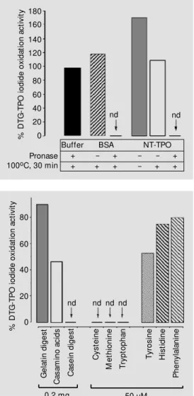
The DTG-TPO iodide oxidation activity was inhibited by hydrolyzed NT-TPO or hydrolyzed BSA, whereas no inhibitory ac-tivity was present before proteolysis. In fact, the addition of non-hydrolyzed NT-TPO be-fore boiling produced an increase of 71% over the original DTG-TPO activity. Dena-tured (boiled) BSA or NT-TPO preparations did not affect the original DTG-TPO activity (Figure 1). TPO activity was also unaffected by the addition of denatured pronase.
DTG-partial inhibition (47%), and phenylalanine (50 µM) and histidine (50 µM) produced a maximal inhibition of 20-25% even at higher concentrations (100 µM). The concentra-tions of inhibitory amino acids necessary to inhibit 50% of the original TPO iodide oxi-dation activity (IC50) were 12.5 µM cysteine,
13 µM methionine and 9.7 µM tryptophan. Other amino acids tested did not alter the enzyme iodide oxidation activity. The iodide oxidation activity of lactoperoxidase (LPO) was inhibited by the same amino acids that inhibited the TPO-catalyzed reaction, and to a similar extent (data not shown).
The guaiacol oxidation reaction was not significantly influenced by the amino acids, except for a very marked time lag produced by cysteine (Figure 3B). This time lag de-creased when cysteine concentration was reduced (25 µM) or the concentration of the enzyme was doubled (data not shown). In contrast to cysteine, cystine did not signifi-cantly alter the iodide oxidation or the guai-acol oxidation TPO activity (Figure 3A and B).
The increase in H2O2 in the reaction
mix-ture did not change the proportion of TPO iodide oxidation inhibition by amino acids. Furthermore, none of the inhibitory amino acids were able to scavenge H2O2 in vitro.
However, when the amount of iodide was increased to 18 mM, the inhibition produced by casamino acids, tyrosine, phenylalanine or histidine was decreased, resulting in a 10-25% increment in the iodide oxidation activ-ity. The same increment in iodide did not change the strong inhibition promoted by 50 µM cysteine, tryptophan and methionine. Nevertheless, this strong inhibition was de-pendent on the amino acid concentration in the assay. Kinetic iodide oxidation studies showed that the inhibition produced by cys-teine (12.5 µM), methionine (13 µM) or tryptophan (9.7 µM) was reversible, since the original inhibition is reversed at higher iodide concentrations, and the enzyme K0.5
for iodide was significantly increased in the
% D T G -T P O i o d id e o x id a ti o n a c ti v it y 80 60 40 20 0 G e la ti n d ig e s t C a s a m in o a c id s C a s e in d ig e s t C y s te in e M e th io n in e T ry p to p h a n T y ro s in e H is ti d in e P h e n y la la n in e
nd nd nd nd Figure 2 - Iodide oxidation
inhibi-tory assays. The same iodide oxi-dation activity (DA353 nm/min =
0.1) of active diffuse toxic goiter-thyroid peroxidase (DTG-TPO) w as assayed w ith or w ithout 0.2 mg of pancreatic gelatin digest, 0.2 mg of casamino acids, 0.2 mg of pancreatic casein digest, or 50 µM L-amino acids. nd = Activity not detectable. Figure 1 - Effect of hydrolyzed proteins on diffuse toxic goiter-thyroid peroxidase (DTG-TPO) iodide oxidation activity. The TPO iodide oxidation activity w as measured in the presence of non-hydrolyzed (-) or pronase-hy-drolyzed (+) bovine serum albu-min (BSA, 0.4 mg) or normal TPO (NT, 0.3 mg) preparations. The amount of solubilized TPO producing a fixed iodide oxida-tion activity (DA353 nm/min = 0.1)
w as assayed for each experi-mental condition. nd = Activity not detectable. % D T G -T P O i o d id e o x id a ti o n a c ti v it y 180 160 140 120 100 80 60 40 20 0 nd nd
Buffer BSA NT-TPO + - + - - + + + + - + + Pronase
100oC, 30 min
TPO was determined in the presence of several peptides or amino acids, and the extent of inhibition of the original TPO activity is shown in Figure 2. The pancreatic digest of gelatin did not significantly change TPO activity, and inhibited the original DTG-TPO activity by no more than 10%. In con-trast, the pancreatic digest of casein com-pletely abolished DTG-TPO activity. Nev-ertheless, a mixture of amino acids derived from acid hydrolysis of casein (casamino acids) inhibited TPO activity only partially (54% inhibition). Among the tested amino acids, only cysteine (Figure 3A), methio-nine and tryptophan completely inhibited TPO iodide oxidation at the concentration of 50 µM. Tyrosine (50 µM) caused only
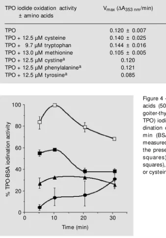
presence of these amino acids, indicating a competitive inhibition of the reaction, at least at the concentrations tested (Table 1).
DTG-TPO iodination activity was also inhibited by all the amino acids that inhib-ited the TPO iodide oxidation reaction, but the inhibitory patterns were different. In the iodide oxidation reaction tryptophan and cysteine were the most potent inhibitors, with equivalent increases in K0.5 (Table 1);
however, cysteine was more potent than tryp-tophan in inhibiting the iodination reaction (Figure 4). Tyrosine also inhibited the iodi-nation reaction, but to a lesser extent, while 50 µM phenylalanine, histidine, proline or cystine did not inhibit the iodination activity.
D iscussio n
The presence of endogenous dialyzable inhibitors of TPO iodination activity in the soluble fraction of bovine and ovine thyroid gland homogenates was first reported in stud-ies carried out in the sixtstud-ies (13,14), which suggested that endogenous sulfhydryl-con-taining compounds such as glutathione were responsible, at least in part, for iodination inhibition. It has also been shown that ascor-bic acid, cysteine, epinephrine, norepineph-rine, serotonin, NADH, and NADPH can inhibit tyrosine iodination (13). More recent studies have shown that some exogenous substances such as dietary flavonoids and sulfamethazine may impair TPO activity, at least in vitro (15-17).
As shown here, enzymatic proteolysis of BSA, and even of a TPO preparation, led to the release of TPO inhibitors. Furthermore, a pancreatic digest of casein (but not of gela-tin) and a mixture of amino acids derived from acid hydrolysis of casein were able to significantly impair TPO activity in vitro, suggesting that some amino acids, the component(s) of these mixtures, could be responsible for TPO inhibition.
Since the pancreatic digest of gelatin was unable to alter the TPO iodide oxidation
Table 1 - Kinetic parameters of thyroid peroxidase (TPO) iodide oxidation in the presence of some amino acids.
Results are reported as mean ± SEM . an = 1. * P<0.05; * * P<0.001 vs TPO (paired
Student t-test).
TPO iodide oxidation activity Vmax (DA353 nm/min) K0.5 (mM iodide)
± amino acids
TPO 0.120 ± 0.007 7.70 ± 0.55 TPO + 12.5 µM cysteine 0.140 ± 0.025 18.81 ± 1.12* * TPO + 9.7 µM tryptophan 0.144 ± 0.016 18.92 ± 2.82* * TPO + 13.0 µM methionine 0.105 ± 0.005 12.37 ± 0.77* TPO + 12.5 µM cystinea 0.120 9.12
TPO + 12.5 µM phenylalaninea 0.121 8.80
TPO + 12.5 µM tyrosinea 0.085 7.71
Figure 3 - Diffuse toxic goiter-thyroid peroxidase (DTG-TPO) in-hibitory assays. Iodide oxidation (A) or guaiacol oxidation (B) ac-tivities of a) TPO, b) DTG-TPO + cystine, and c) DTG-DTG-TPO + cysteine. DTG-TPO iodide oxi-dation or guaiacol oxioxi-dation as-says w ere performed in the ab-sence or preab-sence of cystine (50 µM ) or cysteine (50 µM ).
A
b
s
o
rb
a
n
c
e
(
3
5
3
n
m
)
0.5
0
0 100 200 Ab 0 100 200
s
o
rb
a
n
c
e
(
4
7
0
n
m
)
0.5
0 a
b c
a
b c
A B
Time (s) Time (s)
%
T
P
O
-B
S
A
i
o
d
in
a
ti
o
n
a
c
ti
v
it
y
100
80
60
40
20
0
0 10 20 30
Time (min)
Figure 4 - Effect of some amino acids (50 µM ) on diffuse toxic goiter-thyroid peroxidase (DTG-TPO) iodination activity. The io-dination of bovine serum albu-m in (BSA) by DTG-TPO w as measured in the absence or in the presence of tyrosine (open squares), t rypt ophan (f illed squares), methionine (triangles) or cysteine (circles).
tryptophan). However, the extent of TPO inhibition is quite different among the inhib-itory amino acids.
The present findings show that amino acids other than cysteine can inhibit the TPO iodide oxidation and iodination activities. However, cysteine was the most potent in-hibitor of the iodination reaction and was the only amino acid that inhibited both iodide and guaiacol oxidation activities, although the guaiacol oxidation inhibition was self-limited, and the lag period was reduced when the cysteine concentration was halved. A transient inhibition of the TPO iodination reaction has also been described for thio-ureylene drugs, depending on their concen-tration (1). Furthermore, the degree of iodide oxidation inhibition produced by cysteine, methionine and tryptophan seems to be de-pendent on iodide concentration, as also shown by Taurog (1) for the inhibitory ef-fects of thioureylene drugs. So, it would seem that the mechanism of TPO inhibition by these amino acids resembles that of thio-ureylene drugs as proposed by Taurog (1). As suggested by this author, since the oxi-dized form of iodide might act to oxidize the thioureylene drugs, as well as to iodinate tyrosine residues, the thioureylenes may com-pete with the tyrosyl residues for the oxi-dized iodide and so inhibit the iodination reaction. Some authors have also demon-strated that several mechanisms can be re-sponsible for the inhibition of TPO- and
LPO-catalyzed reactions, such as suicide in-activation of the enzyme, rapid equilibrium binding, and alternate substrate competition for the iodinating intermediate (15).
Thus, it is tempting to speculate that, at least under the conditions of the guaiacol oxidation assay, cysteine might be oxidized, just as proposed for the thioureylenes, into a non-inhibitory form such as cystine. Guai-acol oxidation was not inhibited by tryp-tophan and methionine, indicating that these amino acids do not interact with the oxidized form of guaiacol. However, tryptophan and methionine, like cysteine, seem to be com-petitive inhibitors of the iodide oxidation reaction, and therefore may react with the oxidized form of iodide, as we propose for cysteine, and/or compete reversibly with the iodide ion for its site on TPO.
In conclusion, our findings indicate that the inhibition of the in vitro peroxidase ac-tivity of some amino acids may be produced by their interaction with the oxidized form of iodide and/or with the iodide site on the TPO molecule. Further studies are needed to de-fine a possible physiological role for amino acids in thyroid gland regulation.
Ackno wle dgm e nts
We would like to thank Dr. Alvin Taurog for providing a generous sample of the old Aldrich guaiacol, and Dr. Luiz Francisco Macedo for critical advice.
Re fe re nce s
1. Taurog A (1996). Hormone synthesis. In: Braverman LE & Utiger RD (Editors),
Werner and Ingbar’s The Thyroid. A Fun-dam ental and Clinical Text. 7th edn. Lippincott-Raven, Philadelphia, PA, 47-84. 2. DeGroot LJ, Larsen PR & Hennemann G (Editors) (1996). Thyroid hormone synthe-sis and secretion. In: The Thyroid and its Diseases. 6th edn. Churchill Livingstone, New York, NY, 33-60.
3. Dème D, Pommier J & Nunez J (1976). Kinetics of thyroglobulin iodination and
hormone synthesis catalysed by thyroid peroxidase. Role of iodine in the coupling reaction. European Journal of Biochemis-try, 70: 435-440.
4. M oura EG, Rosent hal D & Carvalho-Guimarães DP (1989). Thyroid peroxidase activity in human nodular goiters. Brazil-ian Journal of M edical and Biological Re-search, 22: 31-39.
5. Hosoya T, Sato I, Hiyama Y, Yoshimura H, Niimi H & Taturani O (1985). An improved assay method for thyroid peroxidase
ap-plicable for a few milligrams of abnormal human thyroid tissues. Journal of Bio-chemistry, 98: 637-647.
6. Pommier J, Dominici R, Bougneres P, Rahmoun B & Nunez J (1977). A dialysa-ble inhibitor bound to thyroglobulins from four simple goiters and from tw o goiters w ith iodine organification defect. Journal of M olecular M edicine, 2: 169-177. 7. M edeiros-Neto GA, Okamura K, Cavaliere
peroxi-dase defect. Clinical Endocrinology, 17: 1-14.
8. Rosenthal D, Carvalho-Guimarães DP, Knobel M & M edeiros-Neto GA (1990). Dyshormonogenetic goiter: presence of an inhibitor of normal human thyroid per-oxidase. Journal of Endocrinological In-vestigation, 13: 901-904.
9. Carvalho DP, Rego KGM & Rosenthal D (1994). Thyroid peroxidase in dyshormo-nogenetic goiters w ith organification and thyroglobulin defects. Thyroid, 4: 421-426. 10. W arburg O & Christ ian W (1941). Isolierung und Kristallisation des Gärungs-ferments Enolase. Biochemische Zeit-schrift, 310: 384-421.
11. Carvalho-Guimarães DP, Ramos CF & Rosenthal D (1989). A technical improve-ment for the thyroid peroxidase
iodina-tion assay. Brazilian Journal of M edical and Biological Research, 22: 821-823. 12. Carvalho DP, Dupuy C, Gorin Y, Legue O,
Pommier J, Haye B & Virion A (1996). The Ca2+- and reduced nicotinamide adenine
dinucleotide phosphate-dependent hydro-gen peroxide hydro-generating system is in-duced by thyrotropin in porcine thyroid cells. Endocrinology, 137: 1007-1012. 13. Schussler GC, Ingbar SH & Eveleth P
(1961). The role of intermediary carbohy-drate metabolism in regulating organic io-dinations in the thyroid gland. Journal of Clinical Investigation, 40: 1394-1412. 14. Klebanoff SJ, Yip C & Kessler D (1962).
The iodination of tyrosine by beef thyroid preparations. Biochimica et Biophysica Acta, 58: 563-574.
15. Doerge DR & Decker CJ (1994). Inhibition
of peroxidase-cat alyzed react ions by arylamines: mechanism for the anti-thy-roid action of sulfamethazine. Chemical Research in Toxicology, 7: 164-169. 16. Doerge DR & Divi RL (1995). Porphyrin p
-cation and protein radicals in peroxidase catalysis and inhibition by anti-thyroid chemicals. Xenobiotica, 25: 761-767. 17. Divi RL & Doerge DR (1996). Inhibition of
thyroid peroxidase by dietary flavonoids.
Chemical Research in Toxicology, 9: 16-23.