O R I G I N A L R E S E A R C H
Phthaloyl amino acids as anti-inflammatory
and immunomodulatory prototypes
Ana Cristina Lima Leite•Fa´bio Fernandes Barbosa •Marcos Verı´ssimo de Oliveira Cardoso•
Diogo R. M. Moreira•Lucas Cunha D. Coeˆlho•Elany Barbosa da Silva•
Gevanio Bezerra de Oliveira Filho•Valdeˆnia Maria Oliveira de Souza•
Vale´ria Reˆgo A. Pereira•Luiza de C. Reis• Paulo Michel Pinheiro Ferreira•
Claudia Pessoa•Almir Gonc¸alves Wanderley•Fernanda Virgı´nia B. Mota •
Teresinha G. da Silva
Received: 7 May 2013 / Accepted: 15 August 2013 / Published online: 12 September 2013 ÓSpringer Science+Business Media New York 2013
Abstract A series of phthalimide analogs were synthesized by derivatization of phthalic anhydride, a highly toxic sub-stance, using a ‘‘one pot’’ condensation reaction toa-amino acids. All phthaloyl amino acid derivatives presented anti-oral inflammatory activity, but compounds2eand2gwere found to possess the best activities comparable to thalidomide. Most of the compounds effectively suppressed nitric oxide pro-duction in murine cells stimulated with lipopolysaccharide.N -phthaloyl amino acids did not exhibit any significant cyto-toxicity in vitro when tested against tumor cells as well as a spleen cell culture of BALB/c mice. Compounds2a,2g, and 2h were able to inhibit TNF-a and IL-1b production by macrophages. At the same concentration, thalidomide did not exhibit significant inhibitory activity.
Keywords N-phthaloyl amino acids
Anti-inflammatoryImmunomodulatory
Introduction
It is well known that the free radicals formed during inflammation play an important role in killing the micro-organism and activating leukocytes and macrophages. Overproduction of these radicals is associated with a wide range of pathological conditions (Ariel and Serhan,2007; Nathan, 2002). Thus, it is important to develop anti-inflammatory and immunomodulatory drugs that could regulate the overproduction of these undesirable species (Moormannet al.,2001).
Phthalimides are well-known plant growth regulators (Butula et al., 1975; Hoffmann and Smith, 1949; Koch, 1971), bacteriostatic agents (Kant and Saksena, 2003; Midtvedt, 1963), and fungicides (Kennedy et al., 1975). Thalidomide, a multi-target drug, is the best-known phthalimide and is a hypnotic/sedative drug with teratogenic effects. Despite these effects, thalidomide has never com-pletely vanished as a therapeutic substance. It was found to have a powerful anti-inflammatory effect due to its ability to inhibit the production of the cytokine tumor necrosis factor alpha (TNF-a), a potent stimulator of inflammation, cellular A. C. L. Leite (&)F. F. BarbosaM. V. O. Cardoso
D. R. M. MoreiraL. C. D. CoeˆlhoE. B. da Silva G. B. O. FilhoV. M. O. de Souza
Departamento de Cieˆncias Farmaceˆuticas, Centro de Cieˆncias da Sau´de, Universidade Federal de Pernambuco, Recife,
PE 50740-520, Brazil
e-mail: [email protected]
V. R. A. PereiraL. de C. Reis
Departamento de Imunologia, Centro de Pesquisas Ageu Magalha˜es-FIOCRUZ, Recife, PE, Brazil
P. M. P. Ferreira
Departamento de Cieˆncias Biolo´gicas, Universidade Federal do Piauı´, Picos, Piauı´ 64607-670, Brazil
C. Pessoa
Departamento de Fisiologia e Farmacologia, Centro de Cieˆncias da Sau´de, Universidade Federal do Ceara´, Fortaleza,
CE 60430-270, Brazil
A. G. Wanderley
Departamento de Fisiologia e Farmacologia, Centro de Cieˆncias da Sau´de, Universidade Federal de Pernambuco, Recife, PE 50740-520, Brazil
F. V. B. MotaT. G. da Silva
Departamento de Antibio´ticos, Universidade Federal de Pernambuco, Recife, PE 50740-520, Brazil
necrosis, and tissue damage in general (Ferna´ndez-Martı´nez et al.,2001). The anticancer activity of thalidomide is based on the inhibition of the growth of new vessels in the process of angiogenesis (Lepper et al., 2004). Thalidomide is being increasingly used in the clinical management of a wide spectrum of immunologically mediated infectious diseases and cancers such as erythema nodosum leprosum, multiple myeloma, renal and intestinal carcinomas, Behc¸et’s, Crohn’s, and a number of dermatologic diseases, as well as rheumatoid arthritis and wasting syndrome in AIDS (Frankset al.,2004; Gockelet al.,2004; Kerr and Ship, 2003; Lu et al.,2003; Sayarliogluet al.,2004; Srinivasan and Lichtenstein,2004; Zhuet al.,2003). It is also effective for mycobacterial infec-tion in the central nervous system, for example, tuberculous meningitis caused byMycobacterium bovisorMycobacterium bacillus(Tsenovaet al.,1999).
Based on the broad spectra of thalidomide properties, the phthalimide pharmacophore has been the target of research with varying intentions. The method by which phthalimide reduces TNF-a production is associated with the induction of the degradation of TNF-a m-RNA (Kim et al.,2004; Lentzsch et al.,2002; Orzeszko et al., 2003; Yogeeswariet al.,2003).N-substituted phthalimides are of high interest because they have been reported to possess hypolipidemic activity (Srivastavaet al.,2001) as well as anti-inflammatory and immunomodulatory properties (Ha-shimoto, 2002; Lima et al., 2002; Sena et al., 2003). Phthalimides also have a number of applications in syn-thetic chemistry (Casimir et al., 2000; Kukolja and Lammert, 1975). In fact, a number of reports have described the synthesis ofN-phthaloyl amino acids fusing free amino acids with phthalic anhydride (Billman and Harting, 1948; Zeng et al., 2004). To the best of our knowledge, no biological activity tests have been per-formed for this structural core; therefore, we were inter-ested in carrying out the pharmacological evaluations of these small compounds. This communication reports the results of the in vivo anti-inflammatory tests and immu-nomodulatory profile. In addition, the compounds were evaluated for their cytotoxicity against human tumor cell lines and a spleen cell culture from BALB/c mice.
Results and discussion
Chemistry
The compounds were synthesized according to described procedures by fusing free amino acids with phthalic anhydride (Fig. 1) (Zeng et al., 2004). It is worth men-tioning that only the desiredS-enantiomers were obtained in all cases, as verified by the addition of the chiral shift reagent Eu(hfc)3in the1H-NMR spectra.
Biological activities
Compounds 2a–g were screened for anti-inflammatory activity in an air pouches model assay with a single dose of 100 mg/kg orally administered to mice and evaluated for their ability to inhibit leukocyte migration from the blood circulation into air pouches (Table1).
To determine the selectivity and immunomodulatory action, the effect of the derivatives on the proliferation of spleen cells from BALB/c mice (a method that is effective for the evaluation of specific T lymphocyte cytotoxicity) and their effects on NO-induced production in murine spleen cells were tested using thalidomide (Thl) as a reference.
Compounds 2a and2b showed a weak anti-inflamma-tory profile, whereas 2c, 2d, and 2h presented moderate activity. The derivatives 2e, 2f, and 2g (an isoleucine, glutamic acid, and phenylalanine derivative, respectively) were the most potent anti-inflammatory agents. It is
Table 1 Anti-inflammatory activity orally of phthalimides2a–hand thalidomide (Thl)
Treated (100 mg/kg) No. of PMNL/mL (9106) Inhibition %
Vehicle 5.4±0.1* –
2a 2.8±0.2* 47.7
2b 3.3±0.2* 38.4
2c 2.5±0.1* 54.2
2d 2.3±0.3* 57.8
2e 1.8±0.2* 67.5
2f 2.1±0.4 61.7
2g 1.9±0.1* 64.2
2h 3.0±0.2* 43.8
Thl 1.5±0.2* 72.1
The data represent the mean±the standard error of 6 animals PMNLpolymorphonuclear leukocytes
*P\0.05. Significance was determined with one way ANOVA
followed by Bonferroni’s post hoc test when compared with control group
interesting to note that the amino acid derivative 2f (a synthetic precursor of Thl) presented a 15 % decrease in anti-inflammatory activity relative to Thl.
Previous work has shown that the inflammatory effects of carrageenan in the air pouch are due to an influx of predominantly neutrophilic leukocytes (mainly PMNL) from the blood circulation, and the reduction in PMNL suggests the inhibition of prostaglandin (PG) production (Jacobset al.,1981). Therefore, the air pouch model is an attractive method for the direct measurement of PG, TNF-a, and NO, among other inflammatory mediators.
The data shown in Table2provide evidence that these phthalimides are efficient at reducing lipopolysaccharide (LPS)-induced nitric oxide production at non-cytotoxic levels and at stimulating the proliferation of spleen cells without affecting cell viability at the dose assayed. These results indicate that these compounds are promising anti-inflammatory agents, especially compounds 2e and 2g. Compound2b(an alanine derivative) did not provide sig-nificant anti-inflammatory activity, but it was able to reduce the production of NO at non-cytotoxic levels.
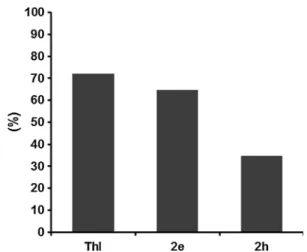
To better understand the inhibitory effects on NO for the amino acid2h, the in vivo biological aspects were inves-tigated using the air pouches model and the NO content of the fluid was measured by the Griess reaction. In the fluid samples from the air pouch, 2h and Thl (at a dose of 10 mg/kg) significantly decreased the concentration of LPS-induced nitric oxide production compared to the control group (Table2).
Thereafter, compounds2eand2hwere selected to test the dose-dependent response of the anti-inflammatory activity. When compound2ewas given at an oral dose of
10 mg/kg, it displayed a 64.7 % inhibition of inflamma-tion, which is comparable to thalidomide (76.3 % inhibi-tion). Compound 2h (a tryptophyl derivative), which presented poor anti-inflammatory activity orally, was very potent in inhibiting the production of NO, and it did not show cytotoxicity for mammalian cells at a high dose (50lg/mL) (Fig. 2).
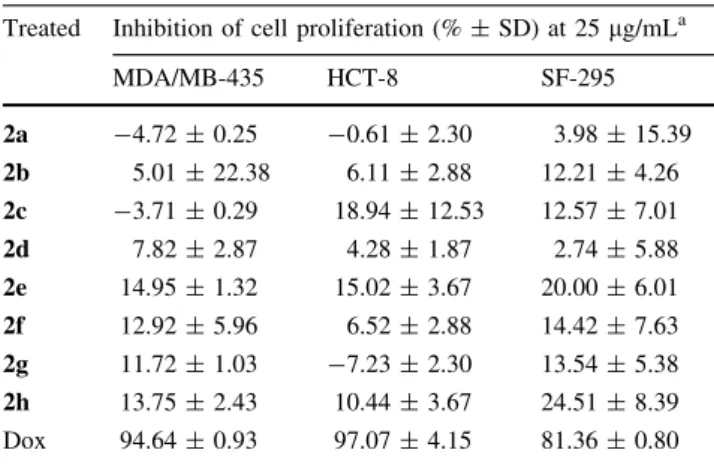
Furthermore, the compounds were evaluated in vitro against a 4-cell-line panel consisting of MDA/MB-435 (human breast), HCT-8 (human colon carcinoma), and SF-295 (human glioblastoma) using a previously described MTT assay (Mosmann, 1983). In this protocol, each cell line is inoculated and preincubated on a microtiter plate. Test agents were then added at a single concentration (25lg/mL), and the culture was incubated for 48 h. End-point determinations are made with MTT. The results for each test agent were reported as the percent of growth of the treated cells compared to the untreated control cells. Compounds that reduce the growth of any one of the cell lines to 32 % or less are considered cytotoxic (de Moreira et al., 2007). According to these criteria, the derivatives were considered non-cytotoxic (Table3).
To evaluate the levels of TNF-a and IL-1b, human macrophages were incubated with 50lmol of compounds 2a–h over a 24-h time course. These cells were then stimulated with LPS. The purpose of this test was to evaluate the biological capacity of the molecules found to inhibit the synthesis of these extremely important cytokines in inflammatory processes and cancers. The data presented in Fig.3a show that compounds2aand2hboth inhibit the secretion of TNF-a by macrophages at 50lmol/mL. In addition, as shown in Fig.3b, compounds2a,2g,and 2h inhibit IL-1b production at the same concentration Table 2 Inhibitory effects on LPS-induced nitric oxide production
and cytotoxic effect on spleen cell BALB/c mice
Treated?LPSa NO-levels (lg/mL)±SD* Cytotoxic effects
at 25lg/mL at 50lg/mL Conc. (lg/mL)b % Inhibitionc
Thl 3.6±0.0* 3.6±0.0* [100 None
2a 4.2±0.9 3.6±2.3 50 25.60
2b 2.1±0.3* 1.5±0.6* [100 None
2c 3.6±0.0* 4.4±1.2 50 22.20
2d 6.4±0.0 2.7±0.6* 25 24.69
2e 3.1±0.0* 2.3±0.0* 12.5 26.06
2f 2.9±0.9* 3.4±0.3* 50 08.75
2g 2.3±0.0* 0.7±0.0* 25 21.52
2h 1.3±0.9* 2.1±1.4* 50 27.62
NO-induced production by stimulating with lipopolysaccharide (LPS-1.0lg/
mL) and measured by Griess’s method [36]
*P\0.05 compared with control (only LPS stimulus)
aNO-induced levels induced by LPS 7.5±1.2lg/mL
b
The highest nontoxic concentration on spleen cell of BALB/c mice
c% of viable cells
(50lmol/mL). Thalidomide did not provide significant inhibitory activity.
Compound 2a (a phthaloyl-glycine derivative) had presented weak anti-inflammatory activity, but it exhibited an immunomodulatory profile when tested for NO, TNF-a, and IL-1bproduction. Compound 2g(a phthaloyl-phenyl-alanine derivative) showed oral anti-inflammatory activity in the air pouch model, and its immunomodulatory profile showed moderate activity for NO production and strong inhibitory activity for IL-1b production. The phthaloyl-tryptophyl derivative, 2h, exhibited immunomodulatory activity toward NO, TNF-a, and IL-1bproduction but poor anti-inflammatory activity with oral administration.
Conclusion
In summary, N-phthaloyl amino acids were readily syn-thesized and evaluated for their anti-inflammatory activity and cytotoxicity, as well as their NO, TNF-a, and IL-1b production inhibition. These amino acids possess simple structures and could be easily structurally improved. In this series, derivatives from the amino acids, isoleucine and phenylalanine, showed the best oral anti-inflammatory activity in an air pouch model. Moreover, the stimulation of lymphocyte proliferation and the inhibition of NO pro-duction suggest that the compounds could be considered as potential immunomodulatory agents. The tryptophyl derivative presented the best immunomodulatory activity of the series. Investigation of immunomodulation and toxicological profiles are subjects for further studies. Experimental
General
Melting points were determined on an electrothermal capillary melting point apparatus and were uncorrected.1H and13C NMR spectra were recorded on a Varian Unit Plus instrument (300 MHz for protons and 75.5 MHz for car-bon). All chemical shifts are reported in ppm relative to TMS as an internal standard. IR spectra were recorded with a Brucker model IFS66 FT-IR spectrophotometer using KBr pellets. All chemicals were purchased from Aldrich, Vetec, or Fluka and were used without additional purification.
Chemistry
All reagents were used as purchased from commercial sources (Sigma-Aldrich, Acros Organics, Vetec, or Fluka). The progress of the reactions was monitored by thin-layer chromatography (TLC) analysis (Merck, silica gel 60 F254 in aluminum foil). The chemical identity was confirmed by NMR and IR spectroscopy and accurate mass (HRMS). IR was performed in KBr pellets. For NMR spectroscopy, we used either a Varian UnityPlus 300 MHz 1H (300 MHz) and 13C (75.5 MHz) or a Bruker AMX-300 MHz 1H (300 MHz) and 13C (75.5 MHz) instrument. DMSO-d6and
D2O were purchased from CIL or Sigma-Aldrich.
Chemi-cal shifts are reported in ppm, and multiplicities are given as s (singlet), d (doublet), t (triplet), q (quartet), and m (multiplet). NH signals were localized in each spectrum after the addition of a few drops of D2O. Mass
spectrom-etry experiments were performed on an LC–IT–TOF (Shimadzu). Unless otherwise specified, ESI was carried out in the positive ion mode. Typical conditions were as follows: capillary voltage of 3 kV, cone voltage of 30 V, Table 3 Antiproliferative activity against human cancer cells
Treated Inhibition of cell proliferation (%±SD) at 25lg/mLa
MDA/MB-435 HCT-8 SF-295
2a -4.72±0.25 -0.61±2.30 3.98±15.39 2b 5.01±22.38 6.11±2.88 12.21±4.26 2c -3.71±0.29 18.94±12.53 12.57±7.01 2d 7.82±2.87 4.28±1.87 2.74±5.88 2e 14.95±1.32 15.02±3.67 20.00±6.01 2f 12.92±5.96 6.52±2.88 14.42±7.63 2g 11.72±1.03 -7.23±2.30 13.54±5.38 2h 13.75±2.43 10.44±3.67 24.51±8.39 Dox 94.64±0.93 97.07±4.15 81.36±0.80
a Standard deviation is given in parentheses
Fig. 3 a Effect of the amino acyl phthalimides and Thl in on the production of TNF-a in mouse macrophages (29106 cells/mL) stimulated with LPS (2lg/mL). TNF-awas measured after 24 h of incubation with compounds (50lM) by sandwich ELISA (eBio-science kit). Data are the mean±S.D obtained in triplicate.bEffect of phthalimides and Thl on the production of interleukin-1b(IL-1b) in mouse macrophages (29106) stimulated with LPS (2lg/mL-1).
and a peak scan between 50 and 1,000 m/z. The com-pounds were previously synthesized by Zeng et al. (Zeng et al.,2004).
Synthesis
A mixture of 0.5 g (3.38 mmol) of phthalic anhydride 1 and the respective L-amino acid (3.38 mmol) in TEA or 4-DMAP (0.5 mL) and DMF (three drops) was heated by microwave irradiation for 2 min. The crude mixture was then washed with n-hexane (30 mL), filtered through a Buchner funnel and recrystallized with 0.1 M KHSO4aq.
or chloroform (40 mL), and dried over anhydrous Na2SO4.
The compounds2fand2hwere obtained by heating at 180
8C in an oil bath for 10 min.
(S)-N-Phthaloylglycine (2a) yield: 96 %. M.p. (°C):187–191. Rf:0.5 (8:2-chloroform/methanol).1H-NMR (300 MHz, ppm):d 4.15 (s, 2H, CH2); 7.50–7.70 (m, 4H,
Ar); 10.58 (s, 1H, CO2H). 13C-NMR (75.5 MHz, ppm):d
39.1 (CH2); 122.4 (C2 and C5); 132.6 (C1 and C6); 135.8
(C3 and C4); 165.9 (C=O); 174.9 (CO2H). IR (KBr,
cm-1):3212 (mOH); 1775 (mC=O); 1715 (mC=O); 1401 (mC–N–C); 1374 (mC–N–C).
(S)-N-Phthaloylalanine (2b) yield: 70 %. M.p. (°C):135. Rf:0.46 (8:2-chloroform:methanol). 1H-NMR (300 MHz, ppm):d1.70 (d,J=7.5 Hz, 3H, CH3); 5.02 (q,
J=7.5 Hz, 1H, CH); 7.70–7.85 (m, 4H, Ar); 10.02 (s, 1H,
CO2H). 13C-NMR (75.5 MHz, ppm):d 16.9 (CH3); 51.7
(CHa); 122.4 (C2 and C5); 132.7 (C1 and C6); 135.8 (C3 and C4); 165.6 (C=O); 171.9 (CO2H). IR (KBr,
cm-1):3205 (mOH); 1752 (mC=O); 1740 (mC=O); 1469
(mC–N–C); 1397 (mC–N–C).
(S)-N-Phthaloylbetaalanine (2c) yield: 90 %. M. p. (°C):133–135. Rf:0.5(8:2-chloroform:methanol). 1H-NMR (300 MHz, ppm):d2.50 (t,J=15.0 Hz, 2H, CH2); 3.77 (t,
J=15.0 Hz, 2H, CH2), 7.67–7.51 (m, 4H, Ar), 8.07 (s,
1H, CO2H). 13
C-NMR (75.5 MHz, ppm):d 29.5 (CH2);
33.1 (CHa); 122.4 (C2 and C5); 132.6 (C1 and C6); 135.8 (C3 and C4); 165.9 (C=O); 175.9 (CO2H). IR (KBr,
cm-1):3170 (mOH); 1771 (mC=O); 1727 (mC=O); 1466
(mC–N–C); 1411 (mC–N–C).
(S)-N-Phthaloylvaline (2d) yield: 50 %. M.p. (°C):79–81. Rf:0.60 (8:2-chloroform:methanol). 1H-NMR (300 MHz, ppm):d 1.9 (d, J=4.0 Hz, 6H, CH3);
2.70–2.79 (m, 1H, CH); 4.98 (d, J=5.0 Hz, 1H, CHa);
8.11 (d,J=7 Hz, Ar); 8.16 (d,J=7.0 Hz, Ar); 9.14 (s,
1H, OH).13C-NMR (75.5 MHz, ppm):d18.9 (CH3); 29.5
(CH); 56.5 (CHa); 122.4 (C2 and C5); 130.7 (C1 and C6); 135.9 (C3 and C4); 165.5 (C=O); 172.9 (CO2H). IR
(KBr, cm-1):3234 (mCO2H); 1763 (mC=O); 1691
(mC=O); 1469 (mC–N–C); 1401 (mC–N–C).
(S)-N-Phthaloylisoleusine (2e) yield: 74 %. M.p. (°C):110–112. Rf:0.5 (8:2-chloroform:methanol).1H-NMR
(300 MHz, ppm):d 0.87 (d, J=7.2 Hz, 3H); 1.22 (t,
J=6.3 Hz, 3H); 1.50–1.49 (m, 2H); 2.54–2.52 (m, 1H);
4.70 (d,J=8.4 Hz, 1H); 7.87–7.72 (m, 4H); 9.45 (s, 1H, OH). 13C-NMR (75.5 MHz, ppm):d 10.6 (CH3); 16.6
(CH3); 25.4 (CH2); 33.9 (CH); 56.5 (CHa); 122.9 (C4 and
C9); 131.3 (C5 and C8); 133.8 (C6 and C7); 167.5 (C=O); 170.3 (CO2H). IR (KBr, cm
-1
):3250 (mOH); 1763 (mC=O); 1703 (mC=O); 1460 (mC–N–C); 1394
(mC–N–C).
(S)-N-Phthaloylglutamine (2f) yield=31.9 %. M.p. (°C):204–209. Rf=0.58 (8:2-chloroform:methanol). 1
H-NMR (300 MHz, ppm):d 2.48–2.63 (m, 1H, CH2);
2.11–2.05 (m, 1H, CH2); 2.96–2.84 (m, 2H, CH2);
5.13–5.19 (m, 1H, CHa); 7.87–7.90 (m, 4H, Ar); 7.80 (m, 4H, Ar); 11.15 (s, 1H, CO2H); 11.33 (s, 1H, CO2H); The
11.15 and 11.33 signals disappear after adding D2O. 13
C-NMR (75.5 MHz, ppm):d 22.044 (CH2); 30.995 (CH2);
49.025 (CHa); 122.967 (C2 and C5); 131.261 (C1 and C6); 134.927 (C3 and C4); 167.217 (C =O); 169.960 (CO2H);
172.873 (CO2H). IR (KBr, cm
-1
): 3201 (mOH); 1775 (mC=O); 1740 (mC=O); 1465 (mC–N–C).
(S)-N-Phthaloylphenylalanine (2 g) yield=87.4 %. M.p. (°C):167–170. Rf:0.54 (8:2-chloroform:methanol).
1
H-NMR (300 MHz, ppm):d 3.60 (d, J=9.0 Hz, 2H,
CH2); 5.258 (t,J=7.0 Hz, 1H, CHa); 7.15–7.196 (m, 5H,
Ar); 7.65–7.68 (m, 2H, Ar); 7.76–7.790 (m, 2H, Ar); 9.01 (s, 1H, OH). 13C-NMR (75.5 MHz, ppm):d 34.0 (CH2);
52.9 (CHa); 123.4 (C20and C50); 126.6 (C5 and C9); 128.3 (C6); 128.7 (C8); 130.7 (C70); 135.0 (C30 and C40); 137.3 (C4); 167.2 (C=O); 170.2 (CO2H). IR (KBr, cm-1):3274
(mCO2H); 1752 (mC=O); 1704 (mC=O); 1450 (mC–N–C);
1398 (mC–N–C).
(S)-N-Phthaloyltryptophan (2 h) yield =96 %. M.p.
(°C):94–96. Rf=0.48 (8:2-chloroform:methanol). 1
H-NMR (300 MHz, ppm):d3.58 (d,J=7.00 Hz, 2H, CH2);
5.15 (t,J=7.00 Hz, 1H, CHa); 6.89 (t,J=8.00 Hz, 4H,
Ar); 7.03 (m, 4H, Ar); 7.27 (d,J=9.00 Hz, 4H, Ar); 7.49
(d,J=9.00 Hz, 4H, Ar); 7.79 (s, 4H, Ar); 10.762 (s, 1H, NH); The 10.762 signal disappear after adding D2O. 13
C-NMR (75.5 MHz, ppm):d 24.140 (CH2); 52.680 (CHa);
109.758 (C7); 111.517 (C4); 117.960 (C10); 118.475 (C9); 121.049 (C8); 123.399 (C20 and C50); 126.964 (C11); 130.920 (C10 and C60); 134.923 (C30 and C40); 168.545 (C=O); 170.461 (CO2H). IR (KBr, cm
-1
): 3406 (mNH); 3258 (mOH); 1771 (mC=O); 1715 (mC=O); 1464 (mC–
N–C).
Biological evaluation
Culture of normal, tumor cells, and cytotoxicity assays
prior to sampling, aged between 18 and 35 years old) was collected, and peripheral blood mononuclear cells (PBMCs) were isolated by a standard method of density gradient centrifugation over Ficoll-Hypaque. All studies were performed in accordance with Brazilian research guidelines (Law 196/96, National Council of Health) and with the Declaration of Helsinki.
Culture of colon (HCT-8)
Glioblastoma (SF-295) and melanoma (MDA/MB-435) cancer lines and PBMC were grown in RPMI 1640 medium supplemented with 20 % fetal bovine serum, 2 mM glu-tamine, 100 U/mL penicillin, and 100lg/mL streptomycin at 37°C with 5 % CO2. The cytotoxicity of the compounds against human cancer cells was determined by the MTT assay (Mosmann, 1983), which analyzes the ability of living cells to reduce the yellow dye 3-(4,5-dimethyl-2-thiazolyl)-2,5-diphenyl-2H-tetrazolium bromide (MTT) to a purple formazan product. Briefly, cells were plated in 96-well plates (0.3-0.79105cells/well) and incubated to
allow cell adhesion. Twenty-four hours later, extracts were added to each well (0.04–100 lg/mL). After 72 h of incubation, the supernatant was replaced by fresh medium containing 10 % MTT, the formazan product was dissolved in DMSO, and the absorbance was measured at 595 nm. Quantification of cell proliferation was determined spec-trophotometrically using a multiplate reader (DTX 880 Multimode Detector, Beckman Coulter). Control groups (negative and positive) received the same amount of DMSO (0.1 %). Doxorubicin (Dox) (0.02–8.6lM) was used as a positive control.
Preparation of splenocytes
Mouse splenocytes were obtained by Pereira et al. (Her-nandeset al.,2010). After killing the animal with CO2gas,
the spleen of each mouse was removed aseptically and placed in a Falcon tube containing RPMI 1640 with fetal calf serum (complete medium). In a vertical flow, each spleen was transferred to a Petri dish where they were soaked. The cell suspensions obtained were transferred to Falcon tubes containing approximately 10 mL of incom-plete medium by spleen. Spleen homogenates were over-laid onto a Ficoll-PaqueTM PLUS layer with the density adjusted to 1.076 g/mL and centrifuged at 1,0009g at room temperature for 25 min. The interface cell layer containing immune cells was recovered by a Pasteur pip-ette, washed twice in PBS, and centrifuged two times at 5009g for 10 min. Cells were counted in a Neubauer chamber, and cell viability was determined by the trypan blue exclusion method. Cells were used only when viability was greater than 98 %.
In vitro cytotoxicity assays
The cytotoxicity of compounds2a–hwas determined using BALB/c mice splenocytes (69105cells/well) cultured in
96-well plates in RPMI 1640 media (Sigma Chemical Co., St. Louis, MO) supplemented with 10 % fetal bovine serum (FCS; Cultilab, Campinas, SP, Brazil) and 50lg/ mL of gentamycin (Novafarma, Ana´polis, GO, Brazil). Each compound was evaluated at six concentrations (100, 50, 25, 10, 5, and 1lg/mL) in triplicate in two independent assays. Cultures were incubated in the presence of [3 H]-thymidine (Amersham Biosciences) (1lCi/well) for 24 h at 378C and 5 % CO2. After this period, the content of the
plate was harvested to determine the [3H]-thymidine ([3H]TdR) incorporation using a beta-radiation counter (b-matrix 9600, Packard). The toxicity of compounds 2a–h was determined by comparing the percentage of [3 H]-thy-midine incorporation (as an indicator of cell viability) of lectin-treated wells compared to untreated wells. Saponine (0.05 %), concanavalin A (Con A), and phytohemaggluti-nin (PHA) were used as positive controls. Non-cytotoxic concentrations were defined as those causing a reduction in [3H]-thymidine incorporation below 30 % in relation to untreated controls.
Carrageenan-induced air pouch
The anti-inflammatory activity of the compounds was tes-ted by the formation of air pouches on the dorsal cervical region of mice (25–30 g) by subcutaneous injection of 2.5 mL of sterile air on day 0 followed by a second injection of 2.5 mL of sterile air 3 days later. On the 6th day, the mice received vehicle or the test compounds orally. One hour after drug administration, inflammation was induced by injecting 1 mL of carrageenan suspension (1 % in saline solution) into the air pouch. After 6 h, the mice were killed and the lumen of the air pouch was lavaged by intrapouch injection of 3 mL of PBS containing 50 ng/mL heparin and subsequent aspiration of the fluid. Total numbers of PMNL cell infiltration were determined with a Neubauer hemocytometer.
In vitro nitrite analysis
TNF-aand IL-1blevels
Mice peritoneal macrophages were placed into 96-well plates at a cell density of 29106cells/mL and incubated for
2 h at 37°C and 5 % CO2. Cells at a concentration of 29106were suspended in RPMI 1640 with 5 % FBS, 100
UI/mL of penicillin, 100lg/mL of streptomycin, and 50 mM 2-mercaptoethanol. One hundred microliters of the suspension and 100lL of the samples were incubated with 2lg/mL LPS (positive control) or with the test compounds in different concentrations. After 24 h, the supernatants were removed and kept at-80°C until the evaluation of cytokine levels (TNF-a and IL-1b). The doses of cytokines in the exudates were assayed by sandwich ELISA using mono-clonal antibodies specific to the detection of the cytokine. Acknowledgments We would like to thank the Brazilian National Research Council (CNPq), FACEPE, and FIOCRUZ for financial support. MVOC received a CNPq doctoral scholarship and currently receives a FACEPE researcher scholarship. We also thank the Department of Fundamental Chemistry-UFPE for recording the1 H-NMR and IR spectra of all compounds.
Conflict of interest The authors state no conflict of interest.
References
Ariel A, Serhan CN (2007) Resolvins and protectins in the termination program of acute inflammation. Trends Immunol 28:176–183. doi:10.1016/j.it.2007.02.007
Billman JH, Harting WF (1948) Amino acids; phthalyl derivatives. J Am Chem Soc 70:1473. doi:10.1021/ja01184a051
Butula LJ, Kujundzic´ N, Malnar M, Vukusic´ I (1975) Mitodepressive effect of someN-substituted phthalimides onLepidium sativum. Die Pharm 30:754
Casimir JR, Didierjean C, Aubry A et al (2000) Stereoselective alkylation ofN-boc-protected-5-substitutedd-lactams: synthesis ofa,d-disubstitutedd-amino acids. Org Lett 2:895–897. doi:10. 1021/ol9913136
de Moreira DRM, Lima Leite AC, Pinheiro Ferreira PM et al (2007) Synthesis and antitumour evaluation of peptidyl-like derivatives containing the 1,3-benzodioxole system. Eur J Med Chem 42:351–357. doi:10.1016/j.ejmech.2006.10.007
Ding A, Nathan C, Stuehr D (1988) Release of reactive nitrogen intermediates and reactive oxygen intermediates from mouse peritoneal macrophages. Comparison of activating cytokines and evidence. J Immunol 141:2407–2412
Ferna´ndez-Martı´nez E, Morales-Rı´os MS, Pe´rez-A´ lvarez V, Muriel P (2001) Effects of thalidomide and 3-phthalimido-3-(3,4-dime-thoxyphenyl)-propanamide on bile duct obstruction-induced cirrhosis in the rat. Drug Dev Res 54:209–218. doi:10.1002/ ddr.10022
Franks ME, Macpherson GR, Figg WD (2004) Thalidomide. Lancet 363:1802–1811. doi:10.1016/S0140-6736(04)16308-3
Gockel HR, Lu¨gering A, Heidemann J, et al. (2004) Thalidomide induces apoptosis in human monocytes by using a cytochrome c-dependent pathway. J Immunol (Baltimore Md: 1950) 172:5103–9
Hashimoto Y (2002) Structural development of biological response modifiers based on thalidomide. Bioorg Med Chem 10:461–479
Hernandes MZ, Rabello MM, Leite ACL et al (2010) Studies toward the structural optimization of novel thiazolylhydrazone-based potent antitrypanosomal agents. Bioorg Med Chem 18:7826–7835. doi:10.1016/j.bmc.2010.09.056
Hoffmann OL, Smith AE (1949) A new group of plant growth regulators. Science (New York, NY) 109:588. doi: 10.1126/ science.109.2841.588
Jacobs RS, White S, Wilson L (1981) Selective compounds derived from marine organisms: effects on cell division in fertilized sea urchin eggs. Fed Proc 40:26–29
Kant P, Saksena RK (2003) Synthesis and antimicrobial activity of some new 2-phenyl-3-p-(20-methyl-30-aryl-40-oxo-thiazolin-20 -yl) phenyl quinazolin-4-ones and 2-phenyl-3-p-(10 -aryl-3-phtha-limido-40-methylazetidin-20-one-20-yl) phenyl-quinazolin-4-ones. Indian J Heterocycl Chem 12:315–318
Kennedy GL, Arnold DW, Keplinger ML (1975) Mutagenicity studies with captan, captofol, folpet and thalidomide. Food and cosmetics toxicology 13:55–61
Kerr AR, Ship JA (2003) Management strategies for HIV-associated aphthous stomatitis. Am J Clin Dermatol 4:669–680. doi:10. 2165/00128071-200304100-00002
Kim YS, Kim JS, Jung HC, Song IS (2004) The effects of thalidomide on the stimulation of NF-kappaB activity and TNF-alpha production by lipopolysaccharide in a human colonic epithelial cell line. Mol Cells 17:210–216
Koch H (1971) Phytopharmakologische Untersuchung von Thalido-mid, seinen Metaboliten and einigen strukturverwandten Ver-bindungen. Sci Pharm 39:209–247
Kukolja S, Lammert SR (1975) Azetidinone antibiotics. XIV. Removal of a phthaloyl protective group from acid and base sensitive compounds. J Am Chem Soc 97:5582–5583. doi:10. 1021/ja00852a044
Lentzsch S, Rogers MS, LeBlanc R et al (2002) S-3-Amino-phthalimido-glutarimide inhibits angiogenesis and growth of B-cell neoplasias in mice. Cancer Res 62:2300–2305
Lepper ER, Ng SSW, Gu¨tschow M et al (2004) Comparative molecular field analysis and comparative molecular similarity indices analysis of thalidomide analogues as angiogenesis inhibitors. J Med Chem 47:2219–2227. doi:10.1021/jm0304820 Lima LM, Castro P, Machado AL et al (2002) Synthesis and anti-inflammatory activity of phthalimide derivatives, designed as new thalidomide analogues. Bioorg Med Chem 10:3067–3073 Lu KQ, Brenneman S, Burns R et al (2003) Thalidomide inhibits
UVB-induced mouse keratinocyte apoptosis by both TNF-alpha-dependent and TNF-alpha-inTNF-alpha-dependent pathways. Photodermatol Photoimmunol Photomed 19:272–280
Midtvedt T (1963) The effect of thalidomide on the growth curve of a riboflavin dependent microbe. Acta Pathol Microbiol Scandi-navica 58:355–362
Moormann AE, Metz S, Toth MV et al (2001) Selective heterocyclic amidine inhibitors of human inducible nitric oxide synthase. Bioorg Med Chem Lett 11:2651–2653
Mosmann T (1983) Rapid colorimetric assay for cellular growth and survival: application to proliferation and cytotoxicity assays. J Immunol Methods 65:55–63
Nathan C (2002) Points of control in inflammation. Nature 420:846–852. doi:10.1038/nature01320
Orzeszko A, Vilpo J, Vilpo L, Kamin´ska B (2003) Synthesis and anticancer activity of 50-phthaloylnucleosides. Pharmazie 58: 169–172
Sayarlioglu M, Kotan MC, Topcu N et al (2004) Treatment of recurrent perforating intestinal ulcers with thalidomide in Behc¸et’s disease. Ann Pharmacother 38:808–811. doi:10.1345/aph.1D524 Sena VLM, Srivastava RM, Silva RO, Lima VLM (2003) Synthesis
Srinivasan R, Lichtenstein GR (2004) Recent developments in the pharmacological treatment of Crohn’s disease. Expert Opin Investig Drugs 13:373–391. doi:10.1517/13543784.13.4. 373
Srivastava RM, Oliveira FJ, Da Silva LP et al (2001) Synthesis and hypolipidemic activity of N-phthalimidomethyl tetra-O-acyl-alpha-D-mannopyranosides. Carbohydr Res 332:335–340 Tsenova L, Bergtold A, Freedman VH et al (1999) Tumor necrosis
factor alpha is a determinant of pathogenesis and disease progression in mycobacterial infection in the central nervous system. Proc Natl Acad Sci USA 96:5657–5662
Yogeeswari P, Sriram D, Saraswat V et al (2003) Synthesis and anticonvulsant and neurotoxicity evaluation of N4-phthalimido phenyl (thio) semicarbazides. Eur J Pharmaceut Sci Off J Eur Fed Pharmaceut Sci 20:341–346
Zeng Q, Liu Z, Li B, Wang F (2004) Mild and effective N-phthaloylation of amino acids. Amino Acids 27:183–186. doi:10. 1007/s00726-004-0109-1