Mo nito ring human cyto me galo virus
viral lo ad in pe riphe ral blo o d le uko
-cyte s o f re nal transplant re cipie nts by
a simple limiting dilutio n-PCR assay
Laboratórios de 1Virologia and 2Genética do Câncer,
Instituto Ludwig de Pesquisa sobre o Câncer, São Paulo, SP, Brasil
3Departamento de Ciências Biológicas, Faculdade de Medicina do Triângulo Mineiro,
Uberaba, MG, Brasil
4Hemagen S/A, São Paulo, SP, Brasil
5Departamento de Patologia Clínica e Hemoterapia, Hospital A.C. Camargo,
São Paulo, SP, Brasil
6Hospital Felício Rocho, and 7Laboratório São Marcos, Belo Horizonte, MG, Brasil 8Unidade de Transplante Renal, Hospital das Clínicas, Universidade de São Paulo,
São Paulo, SP, Brasil
9Departamento de Anatomia Patológica e Medicina Legal, Faculdade de Medicina,
Universidade Federal de Minas Gerais, Belo Horizonte, MG, Brasil O .L. Caballero1,3,
M.C.S.L. Costa4,
A. Trevisan1, R.M. O liveira5,
E.A. Viotti6, E.R.F. Távora6,
S.S. Vilaça6, E. Sabagga8,
F.J. de-Paula8, P.F. Távora7,
G. Brasileiro-Filho9,
L.L. Villa1 and
A.J.G. Simpson2
Abstract
To assess the clinical relevance of a semi-quantitative measurement of human cytomegalovirus (HCMV) DNA in renal transplant recipients within the typical clinical context of a developing country where virtually 100% of both receptors and donors are seropositive for this virus, we have undertaken HCMV DNA quantification using a simple, semi-quantitative, limiting dilution polymerase chain reaction (PCR). We evaluated this assay prospectively in 52 renal transplant patients from whom a total of 495 serial blood samples were collected. The samples scored HCMV positive by qualitative PCR had the levels of HCMV DNA determined by end-point dilution-PCR. All patients were HCMV DNA positive during the monitoring period and a diagnosis of symptomatic infection was made for 4 of 52 patients. In symptomatic patients the geometric mean of the highest level of HCMV DNAemia was 152,000 copies per 106 leukocytes, while for the asymptomatic group this value was 12,050. Symptomatic patients showed high, protracted HCMV DNA levels, whereas asymptomatic patients demonstrated intermittent low or moderate levels. Using a cut-off value of 100,000 copies per 106 leukocytes, the limiting dilution assay had sensitivity of 100%, specificity of 92%, a positive predictive value of 43% and a negative predictive value of 100% for HCMV disease. In this patient group, there was universal HCMV infection but relatively infrequent symptomatic HCMV disease. The two patient groups were readily distinguished by monitoring with the limiting dilution assay, an extremely simple technology immediately applicable in any clinical laboratory with PCR capability.
Co rre spo nde nce A.J.G. Simpson
Laboratório de Genética do Câncer Instituto Ludwig de Pesquisa sobre o Câncer
Rua Prof. Antonio Prudente, 109 4º andar
01509-010 São Paulo, SP Brasil
Fax: + 55-11-270-7001 E-mail: asimpson@ node1.com.br Research partially supported by the PADCT program, administered by FINEP. A. Trevisan is the recipient of a FAPESP fellowship, and M.C.S.L. Costa received a salary from Hemagen S/A.
Received January 25, 1999 Accepted September 21, 1999
Ke y words
Intro ductio n
Human cytomegalovirus (HCMV) infec-tion is a major cause of morbidity in immu-nosuppressed transplant recipients but, for the most part, it is a clinically silent infection in normal individuals (1). In developing coun-tries approximately 100% of individuals have been infected with HCMV. Thus, in such locations it may be assumed that virtually all transplant donors and recipients will be HCMV positive since individuals generally maintain the virus in a latent form for life. This situation is clinically relevant since re-activation of an existing infection during immunosuppression is less likely to lead to symptomatic disease than a new infection (1,2). Thus, transplant programs in develop-ing countries face a situation where detect-able HCMV infection immediately follow-ing organ transplant is expected to be ex-tremely frequent, but where symptomatic disease may be relatively rare. In this situa-tion, diagnostic methodologies that permit silent infections to be distinguished from those that are likely to cause disease are of great importance. Furthermore, given the lim-ited resources available for health care in most developing countries, both the sparing use of prophylactic chemotherapy and the simplicity and cost of the diagnostic assay are important considerations. Thus, the de-velopment of cost effective methods that might allow the rational use of preemptive chemotherapy in such situations is a priority. The quantification of HCMV infection using the polymerase chain reaction (PCR) appears to be a powerful option for distin-guishing transplant patients with low level HCMV infections from those with clinically relevant infections requiring treatment. Sev-eral PCR-based methods for the assessment of HCMV viral load have been reported, including semi-quantitative analysis, such as end-point dilution, or quantitative com-petitive PCR. Irrespective of the use of semi-quantitative or semi-quantitative methods, there is
evidence that the quantitation of HCMV ge-nomes permits the differentiation between clinically relevant symptomatic HCMV in-fections and clinically unimportant CMV DNAemia (3-6). Competitive methods are proving to be the most accurate for quantifi-cation by PCR, but they are technically diffi-cult to perform and to use routinely in clini-cal laboratories. In contrast, semi-quantita-tive methods are more easily applicable to laboratories in which PCR is regularly per-formed.
In this study we have assessed the use of a semi-quantitative PCR assay for HCMV DNA in renal transplant recipients in Brazil, a developing country where virtually 100% of both receptors and donors are seroposi-tive for CMV. We have used a technical approach that is extremely simple and cost effective, allowing immediate implementa-tion in any laboratory where basic PCR tech-niques are established.
Mate rial and Me thods
Subjects and specimens
Fifty-two consecutive patients submitted to renal transplantation at Hospital Felício Rocho, Belo Horizonte, Brazil, or at Hospi-tal das Clínicas, University of São Paulo, Brazil, from May to July 1996 were included in this study. Blood samples (5 ml) contain-ing EDTA as anti-coagulant were drawn weekly after renal transplantation for a pe-riod of up to 12 weeks. A total of 495 samples were collected within 100 days of transplan-tation. The number of peripheral blood leu-kocyte (PBL) preparations obtained per pa-tients ranged from 2 to 16, with a mean of 9 samples per patient.
Se rology te sting
Cryopreserved (-20oC) sera from all
stan-dard indirect ELISA tests.
Clinical e valuation
HCMV infection was defined as the de-tection of HCMV DNA in blood and consid-ered asymptomatic when it occurred without clinical symptoms, signs or abnormal labo-ratory findings. HCMV infection was con-sidered symptomatic when it occurred in conjunction with clinical symptoms or signs compatible with the disease, such as fever (>38oC for at least 5 consecutive days) with
a leukopenia of <2,000/mm3 and/or a fall in
platelet count <100,000/mm3, or visceral
disease such as pneumonitis or gastrointesti-nal symptoms with histological confirma-tion (7).
DNA preparation
DNA was isolated from PBLs contained in 1 ml of EDTA blood. Erythrocytes were lysed by three rounds of mixing with 1 ml of 10 mM Tris-HCl, pH 7.6, 1 mM EDTA (TE) in a 2.2-ml microfuge tube and centrifuged for 90 s at 14,000 g. The final
PBL-contain-ing pellet was resuspended in 100 µl TE and DNA prepared with the RapidPrep™ Ge-nomic DNA isolation kit as described by the manufacturer (Pharmacia Biotech, Uppsala, Sweden). The final volume of the DNA prepa-ration was 50 µl. The DNA concentprepa-ration of the sample was estimated by comparison with a low mass DNA ladder (Gibco-BRL, Gaithersburg, MD, USA) following applica-tion of 2 µl to a 1.5% agarose gel, electro-phoresis and ethidium bromide staining. To estimate the number of leukocytes the value of 6 pg was taken as the quantity of DNA per cell. An example of the DNA estimation is shown in Figure 1. For qualitative PCR am-plifications 1 µl of the DNA sample was used in all cases. For limiting dilution as-says, 5 µl of the DNA were first added to an equal volume of water and subsequently 2-fold serial dilutions were executed in water.
Two µl of these dilutions were used for PCR amplification.
Polyme rase chain re action
The quality of the DNA preparation from all samples was checked by amplification of a 110-bp fragment of the ß-globin gene using the primers PCO3 and PCO4 (8). For
qualita-tive HCMV DNA detection, two pairs of oligonucleotide primers specific for the fourth exon of the HCMV immediate early gene (IE) and HCMV late antigen gp64 (LA) were used in a multiplex format to generate frag-ments of 393 and 136 bp, respectively (9).
All PCR reaction mixtures contained 10 mM Tris, pH 8.3, 50 mM KCl, 200 µM each deoxynucleoside triphosphate, 1.5 mM MgCl2, 12.5 pmol of each primer and 2 U of
Taq DNA polymerase (Cenbiot, Porto Alegre, RS, Brazil) in a final volume of 50 µl. The thermal cycling conditions included a wax-mediated hot start (10), which precluded the initiation of the reaction until the mixture
had reached 80oC. For amplification of
HCMV sequences an initial denaturation step
Figure 1 - A, Ethidium bromidestained 1.5% agarose gel show -ing the estimation of the DNA concentration in 6 samples by comparison w ith the low mass DNA ladder. The numbers on the left indicate the quantity of DNA in each band of the standard. The samples w ere estimated to con-tain 20, 30, 40, 80, 80 and 100 ng, respect ively. B, Silver-stained 7% polyacrylamide gel w ith amplification products de-rived from tw o-fold serial dilu-tions of one HCM V-positive DNA in w hich the end-point titer w as 128.
ng
200
120 80
40
20
271
118
-bp 2 4 8 16 32 64 12
8
256 noDNA
2
1 3 4 5 6
A
was used at 94oC for 3 min, followed by 35
cycles of 30 s each at 94oC and 2 min at
72oC. The reaction was completed with a 8
min extension at 72oC. For amplification of
the ß-globin sequence, the program was the same except that an annealing step at 55oC
for 1 min was included in all cycles. All experiments contained two negative controls, one without DNA addition and the other with a human PBL DNA preparation known to be negative for HCMV DNA.
The samples that were HCMV positive by qualitative PCR had their viral load esti-mated by end-point dilution. Two-fold dilu-tions of the samples were prepared in water and each dilution was submitted to DNA amplification with the LA primers as de-scribed above, in parallel with dilutions of a positive control standard containing 100 cop-ies of the viral genome (9). Four microliters of the PCR products were run on 6% poly-acrylamide gels using a Mini-protean© II
apparatus (Bio-rad, Hercules, CA, USA). The staining procedure consisted of an ini-tial gel fixation for 3 min in 10% ethanol and 0.5% acetic acid, followed by staining in fixing solution containing 0.2% silver nitrate for 5 min. After washing in Milli-Q water (Millipore, Bedford, MA, USA) for 2 min, the gels were developed for approximately 5 min in 3.0% NaOH and 0.1% formaldehyde (11). The end-point was taken as the highest consecutive dilution that yielded a
detect-able PCR product, a detection limit previ-ously shown to contain an average of two viral genomes (9). The estimated number of copies of the HCMV genome per microliter in the initial DNA sample is equal to the dilution factor at the end-point, since 2 µl are used for the amplification and the final end-point is taken as containing two genomes. End-point titers were converted to HCMV DNA copies per 106 leukocytes by
correct-ing for the number of leukocytes as esti-mated above (12,13). Figure 1B shows an example of a limiting dilution assay in which the end-point titer was 128. This sample contained 40 ng of DNA per microliter, i.e. 6,660 leukocyte equivalents, as determined by comparison to known standards as de-scribed above. Thus, the final estimation of the number of viral genomes per 106
leuko-cytes is 128 x 106/6,600 = 21,330.
Re sults
Sera from 37 transplant recipients were available for analysis. Thirty-five (95%) of these patients were CMV seropositive. Do-nor sera for 14 of these patients were not available. Of the 21 recipients whose donors were available for analysis, 18 received kid-neys from CMV-seropositive donors and only three received kidneys from CMV-se-ronegative donors. The two CMV-seronega-tive recipients in this study received kidneys from CMV-seropositive donors. A diagno-sis of symptomatic HCMV infection was made in 4 out of 52 patients (7.7%). The clinical features of these 4 patients are pre-sented in Table 1. The most common clinical symptom was fever and/or leukopenia.
All transplant recipients were found to be HCMV DNA positive at least once during the monitoring period. In each symptomatic case, an increase in HCMV DNA in the blood was detected prior to the onset of symptoms. The frequency of positive results in all patients at different times post-trans-plantation is shown in Figure 2. The peak of Table 1 - Clinical characteristics of patients w ith
sympto-matic HCM V infection.
D/R: HCM V serological status (donor/receptor); ND: not determined. aDays after transplantation. bWith
histo-logical evidence of HCM V in a biopsy of the lesions obtained by upper endoscopy.
Patient D/R Symptoms OnsetaTreatment
1 (JC39) ND/+ Fever/leukopenia 46 Ganciclovir
2 (KB47) + /+ Fever/leukopenia 25 None
3 (AS51) + /+ Fever 22 Ganciclovir
4 (ST56) ND/+ Fever/leukopenia/ 50 Ganciclovir gastrointestinal
HCMV DNA positivity was 6 weeks after transplantation, where 42% of the patients were positive by qualitative PCR.
We have previously shown that the am-plification of the HCMV LA gene fragment, using the primers and conditions selected for the present work, is extremely sensitive and capable of detecting a single viral genome in the assay tube (9). Taking advantage of this optimized PCR protocol, coupled to a highly standardized method of DNA preparation, we found that reproducible estimation of the number of viral genomes per 106 leukocytes
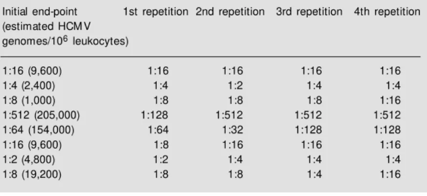
in the initial sample could be achieved with the simple procedures described. For ex-ample, in 232 separate assays the end-point dilution of the positive control was also within a single two-fold dilution. As a further con-trol of reproducibility, the assay was re-peated four times with eight of the patients selected to cover the range of estimated copy numbers encountered. The results are shown in Table 2. In all cases the end-point was always plus or minus a single dilution. Thus the estimates are taken as being plus or mi-nus this value.
The estimated HCMV copy number ranged from 60 to more than 300,000 copies/ 106 leukocytes. Of a total of 232
quantifica-tions performed, 76% had low levels of
HCMV DNA (<10,000 copies/106
leuko-cytes), and only 12% had high levels (>100,000 copies/106 leukocytes). Table 3
shows the relation between the blood levels of HCMV DNA and clinical status. The geometric mean of the highest level of HCMV DNA was 152,000 copies/106 leukocytes in
symptomatic patients. In contrast, in asymp-tomatic patients this value was 12,050 cop-ies/106 leukocytes.
Table 4 shows the clinical relevance of different blood levels of HCMV DNA for the detection of symptomatic infections. Because there were significant gaps in the follow-up of one of the symptomatic patients (patient 1), this analysis was performed considering only the other three symptomatic patients.
Figure 2 - Proportion of speci-mens positive for HCM V DNA at different times after renal trans-plantation.
P
ro
p
o
rt
io
n
o
f
H
C
M
V
-p
o
s
it
iv
e
s
a
m
p
le
s
50
40
30
20
10
0
2 4 6 8 10 12 14
0
Weeks after transplantation
Table 2 - Reproducibility of end-point determination.
Initial end-point 1st repetition 2nd repetition 3rd repetition 4th repetition (estimated HCM V
genomes/106 leukocytes)
1:16 (9,600) 1:16 1:16 1:16 1:16
1:4 (2,400) 1:4 1:2 1:4 1:4
1:8 (1,000) 1:8 1:8 1:8 1:16
1:512 (205,000) 1:128 1:512 1:512 1:512
1:64 (154,000) 1:64 1:32 1:128 1:128
1:16 (9,600) 1:8 1:16 1:16 1:16
1:2 (4,800) 1:2 1:4 1:4 1:4
1:8 (19,200) 1:8 1:8 1:4 1:16
Table 3 - Relation betw een HCM V quantification and clinical manifestations.
aOnly the highest copy numbers w ere considered. bOmitted from the calculations in Table 3 due to a significant gap in follow -up of this patient (low er left quadrant of Figure 3).
Viral loada (HCM V copies/ Number of patients 106 leukocytes)
Symptomatic Asymptomatic Total HCM V infection HCM V infection
<10,000 0 30 30
10,001-50,000 1b 10 11
50,001-100,000 0 4 4
>100,001 3 4 7
Total 4 48 52
Table 4 - Sensitivity, specificity and predictive values of HCM V quantification for HCM V disease.
PPV: Positive predictive value; NPV: negative predictive value. These values w ere calculated w ithout considering the patient for w hom incomplete follow -up data w ere available (low er left quadrant, Figure 3).
HCM V DNA level Sensitivity (% ) Specificity (% ) PPV (% ) NPV (% ) (copies/106 leukocytes)
>10,000 100 37 18 100
>50,000 100 83 27 100
Six patients were treated for rejection episodes and two of them developed HCMV DNA levels of over 100,000 copies/106
leu-kocytes, but did not exhibit symptoms attrib-utable to CMV disease. Although these pa-tients had high HCMV levels in blood, the results suggest that the activation of the virus following anti-rejection therapy had no clini-cal significance in these patients.
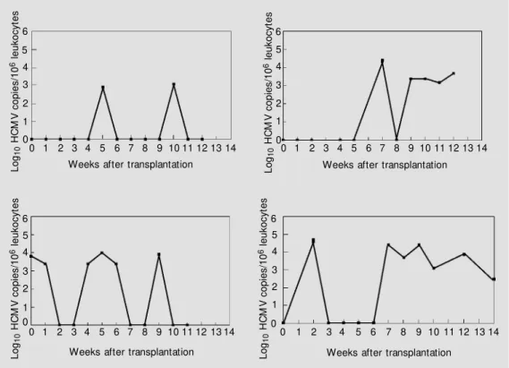
The time course of infection of the 4 symptomatic and of 4 asymptomatic patients is shown in Figures 3 and 4. The former group had high, protracted HCMV DNA levels, whereas the latter had intermittent or transient, low or moderate, levels of HCMV DNA in their blood. The point at which the symptomatic patients became IgM positive, confirming diagnosis of HCMV infection, is indicated in Figure 3.
D iscussio n
HCMV disease is a major cause of clini-cal complications in renal transplant patients, thus requiring careful monitoring for viral
infection (14-17). Recently, several studies have suggested that viral load in blood or urine is the main determinant of clinical manifestations (3,4,15,17) and quantitative or semi-quantitative assays are warranted to identify patients who require antiviral treat-ment. All of these studies were performed, however, in developed countries with rela-tively high frequencies of seronegative trans-plant recipients who are more prone to clini-cally significant HCMV disease following transplantation. Therefore we sought to es-tablish whether viral load, estimated by a simple and low-cost assay, would also be applicable to identify individuals at risk in the setting of transplant services in a devel-oping country, where virtually all donors and recipients are HCMV seropositive.
Our results demonstrate that all patients exhibited HCMV DNA in blood during the post-transplantation period, thus showing that under our conditions qualitative PCR is not suitable for monitoring renal transplant pa-tients. A similar conclusion was reached in a study in another locality with a high HCMV
L o g1 0 H C M V c o p ie s /1 0
6 l
e u k o c y te s 6 L o g1 0 H C M V c o p ie s /1 0
6 l
e u k o c y te s 6 4 2 1 0 3 5 4 2 1 0 3 5 Fever Antigenemia+ IgM + Ganciclovir
0 1 2 3 4 5 6 7 8 9 10 11 12 13 14 Weeks after transplantation
0 1 2 3 4 5 6 7 8 9 10 11 12 13 14 Weeks after transplantation
L o g1 0 H C M V c o p ie s /1 0
6 l
e u k o c y te s 6 4 2 1 0 3 5 Fever
IgM + and Leukopenia
0 1 2 3 4 5 6 7 8 9 10 11 12 13 14
Weeks after transplantation Log
1 0 H C M V c o p ie s /1 0
6 l
e u k o c y te s 6 4 2 1 0 3 5 Fever Leukopenia
0 1 2 3 4 5 6 7 8 9 10 11 12 13 14
Weeks after transplantation Figure 3 - Variation in HCM V viral
load over time in PBLs from symptomatic patients during the period of surveillance. Copy numbers are expressed as log10 genomes/106 leukocytes. Each graph indicates the main clinical signs and symptoms as w ell as the therapy used in these pa-tients.
IgM + Antigenemia+
Gastrointestinal ulcers w ith evidence of HCM V
Ganciclovir
seropositive rate (18). In various studies un-dertaken in developed countries, where the proportion of renal transplant recipients with evidence of blood HCMV DNA in the post-transplant period is around 50%, qualitative PCR has also been reported to be associated with a low predictive value for the develop-ment of HCMV disease (5,13). Peiris et al. (5) reported a 27% predictive value of HCMV qualitative PCR when analyzing 77 renal transplant patients. In a different study in which a total of 476 HCMV qualitative PCR assays were performed on samples from 134 solid abdominal organ recipients, the posi-tive predicposi-tive value was 55% (19). Because our predictive value for symptomatic HCMV disease would be even lower than in the previous reports, we focused our attention on a semi-quantitative HCMV PCR assay to find a better indicator for monitoring HCMV disease at a lower cost.
Despite the higher frequency of detec-tion of HCMV DNA in the patients studied here, only 7.7% developed disease. This is in contrast to other studies, in which the
inci-dence of HCMV disease, considering all donor/receptor (D/R) serological categories, is up to 25% (3,5,20). The lower incidence of the present population may reflect the fact that in this study the majority of recipients were seropositive, in contrast with other stud-ies in which the number of D+/R- patients was at least 20% (21). In the present study, two seronegative patients received organs from seropositive donors but did not de-velop symptomatic CMV infection. This observation is consistent with that made by others who have found that, although D+/R-patients are at a higher risk for developing HCMV disease, not all such patients do so. Indeed, a study conducted on British pa-tients showed that recipient seropositivity had an apparent protective effect against disease when analyzed by univariate logistic regression analysis. However, statistical sig-nificance was not maintained when viral load or viremia was controlled for in a mul-tivariate analysis (17).
Our results show substantial variations in HCMV load over time and among different
L
o
g1
0
H
C
M
V
c
o
p
ie
s
/1
0
6 l
e
u
k
o
c
y
te
s
6
L
o
g10
H
C
M
V
c
o
p
ie
s
/1
0
6 l
e
u
k
o
c
y
te
s
6
4
2
1
0 3 5
4
2
1
0 3 5
0 1 2 3 4 5 6 7 8 9 10 11 12 13 14 Weeks after transplantation
0 1 2 3 4 5 6 7 8 9 10 11 12 13 14
Weeks after transplantation
L
o
g1
0
H
C
M
V
c
o
p
ie
s
/1
0
6 l
e
u
k
o
c
y
te
s
6
4
2
1 0 3 5
0 1 2 3 4 5 6 7 8 9 10 11 12 13 14
Weeks after transplantation Log
1
0
H
C
M
V
c
o
p
ie
s
/1
0
6 l
e
u
k
o
c
y
te
s
6
4
2
1
0 3 5
0 1 2 3 4 5 6 7 8 9 10 11 12 13 14
Weeks after transplantation
patients. The mean values of maximum viral load detected in PBLs from asymptomatic versus symptomatic patients differed by more than 105 copies/106 leukocytes. The analysis
of the temporal course of HCMV DNA in symptomatic HCMV patients showed that in the majority of patients viral levels >100,000 copies/106 leukocytes were reached before
or at the onset of clinical symptoms. This indicates that early diagnosis can be achieved using a cut-off approach for semi-quantita-tive HCMV PCR. We found that an HCMV DNA level higher than 100,000 copies/106
leukocytes in a febrile patient is strongly suggestive of symptomatic HCMV infec-tion. The positive predictive value for CMV disease of a viral load of more than 100,000 HCMV copies/106 leukocytes for subsequent
disease was 43%, thus permitting the identi-fication of a subset of patients in which the probability of symptomatic disease is higher. This viral load test may lead to a more ration-al use of antivirration-al prophylaxis in this popula-tion. The sensitivity and specificity of the estimation of viral load reported here for HCMV disease using a cut-off of 100,000 copies per 106 leukocytes (100 and 92%,
respectively) is in agreement with recent data reported by others for solid organ trans-plant recipients using antigenemia (18) or quantitative PCR (22-24).
The importance of monitoring these pa-tients weekly must be stressed. Indeed, for optimal monitoring even more frequent test-ing may be warranted, particularly in cases where anti-rejection therapy is applied. In the patient shown in the upper left quadrant in Figure 3 who had received anti-rejection therapy, the number of viral genomes increased from an undetectable level to more than 100,000 copies within the period of one week. The semi-quantitative method used in this study is extremely cost-effective (our estimate for all the reagents required for a single assay is less than $10) and readily applicable in clinical laboratories that per-form PCR regularly. Results can be provided
in one day, allowing fast diagnosis of HCMV infection. The use of total PBLs for HCMV detection and quantification simplifies the preparation of DNA because they are easily recovered from whole blood.
The clinical application of this test is facili-tated by the substitution of the ethidium bro-mide-stained agarose gels with polyacrylamide gels stained with silver. Silver staining is ap-proximately one hundred times more sensitive than ethidium bromide staining and avoids the need for transillumination and photography, since these gels are conveniently dried and stored as a permanent record of the diagnosis. One of the main disadvantages of the use of silver staining has been that it is too laborious for routine use. This problem has been re-cently overcome by a method that shortens the staining to approximately 15 min with the use of very simple reagents (11). In addition, the application of this test was facilitated through the use of electrophoresis against molecular mass ladder to control for variations of tem-plate concentration rather than performing for example, ß-globin PCR on serial dilutions, thus significantly simplifying the approach for routine use.
Alternatives to the use of quantitative DNA methodologies are being explored for the quan-tification of HCMV infections. Among these, antigenemia is the best developed and charac-terized (25). Ongoing comparisons between DNA quantification and antigenemia in São Paulo suggest that the two methodologies pro-vide data of equal clinical value and that the choice of diagnostic approach will be made on technical grounds and according to the prefer-ence of individual diagnostic laboratories. The advantage of PCR-based assays is that they can be easily assimilated in laboratories where PCR is already being done for other purposes without significant investment in training.
Re fe re nce s
1. Ranjan D, Burke G, Esquenazi V, M ilgrom M , Koleitat N, Roth D, Gomez C, Olson L, Babischkin S, Gharagozloo H & M iller J (1991). Factors affecting the ten-year out-come of human renal allografts. The ef-fect of viral inef-fections. Transplantation, 51: 113-117.
2. Grundy JE, Lui SF, Super M , Berry NJ, Sw eny P, Fernando ON, M oorhead J & Griffiths PD (1988). Symptomatic infec-tion in seropositive kidney recipients: infection w ith donor virus rather than re-activation of recipient virus. Lancet, ii: 132-135.
3. Kühn JE, Wendland T, Schäfer P, M öhring K, Wieland U, Elgas M & Eggers HJ (1994). M onitoring of renal allograft re-cipients by quantitation of human cytome-galovirus genomes in peripheral blood leu-kocytes. Journal of M edical Virology, 44: 398-405.
4. Fox JC, Kidd IM , Griffiths PD, Sw eny P & Emery VC (1995). Longitudinal analysis of cytomegalovirus load in renal transplant recipients using a quantitative polymerase chain reaction: correlation w ith disease.
Journal of General Virology, 76: 309-319. 5. Peiris JSM , Taylor CE, M ain J, Graham K
& M adeley CR (1995). Diagnosis of cy-tomegalovirus (CM V) disease in renal al-lograft recipients: the role of semiquanti-tative polymerase chain reaction. Nephrol-ogy, Dialysis, Transplantation, 10: 1198-1205.
6. Drouet E, Colim on R, M ichelson S, Fourcade N, Niveleau A, Ducerf C, Boibieux A, Chevallier M & Denoyel G (1995). M onitoring levels of human cy-tomegalovirus DNA in blood after liver transplantation. Journal of Clinical M icro-biology, 33: 389-394.
7. Ljungman P & Plotkin S (1995). Workshop on CM V disease: definitions, clinical se-verity scores and new syndromes. Scan-dinavian Journal of Infectious Diseases, 99 (Suppl): 87-89.
8. Saiki RK, Gelfand DH, Stoffel S, Scharf SJ, Higuchi R, Horn GT, M ullis KB & Erlich HA (1988). Primer-directed enzymatic amplifi-cation of DNA w ith a thermostable DNA polymerase. Science, 239: 487-491. 9. Caballero OL, M enezes CL, Costa M C,
Fernandes SC, Anacleto TM , Oliveira RM , Viotti EA, Távora ER, Vilaça SS, Sabbaga
E, Paula FJ, Villa LL & Simpson AJG (1997). A single-step PCR protocol for di-agnosis and monitoring of human cytome-galovirus infection in renal transplant re-cipients. Journal of Clinical M icrobiology, 35: 3192-3197.
10. Chou Q, Russel M , Birch DE, Raymond J & Bloch W (1992). Prevention of pre-PCR mis-priming and primer dimerization im-proves low -copy-number amplifications.
Nucleic Acids Research, 20: 1717-1723. 11. Sanguinetti CJ, Dias Neto E & Simpson
AJG (1994). Rapid silver staining and re-covery of PCR products separated on polyacrylamide gels. Biotechniques, 17: 915-919.
12. Sykes PJ, Neoh SH, Brisco M J, Hughes E, Condon J & M orley AA (1992). Quantita-tion of targets for PCR by use of limiting dilution. Biotechniques, 13: 444-449. 13. Kulski JK (1994). Quantitation of human
cytomegalovirus DNA in leukocytes by end-point titration and duplex polymerase chain react ion. Journal of Virological M ethods, 49: 195-208.
14. Gerna G, Zipeto D, Parea M , Revello M G, Silini E, Percivalle E, Zavattoni M , Grossi P & M ilanesi G (1991). M onitoring of hu-m an cyt ohu-m egalovirus inf ect ions and ganciclovir treatment in heart transplant recipients by determination of viremia, antigenemia and DNAemia. Journal of In-fectious Diseases, 164: 488-498. 15. Gerdes JC, Spees EK, Fitting K, Hiraki M ,
Sheehan M , Duda D, Jarvi T, Roehl C & Robertson AD (1993). Prospective study utilizing a quantitative polymerase chain reaction for detection of cytomegalovirus DNA in the blood of renal transplant pa-tients. Transplantation Proceedings, 25: 1411-1413.
16. Falagas M E & Snydman DR (1995). Re-current cytomegalovirus disease in solid-organ transplant recipients. Transplanta-tion Proceedings, 27: 34-37.
17. Cope AV, Sw eny P, Sabin C, Rees L, Griffiths PD & Emery VC (1997). Quantity of cytomegalovirus viruria is a major risk factor for cytomegalovirus disease after renal transplantation. Journal of M edical Virology, 52: 200-205.
18. Lo CY, Ho KN, Yuen KY, Lui SL, Li FK, Chan TM , Lo WK & Cheng IK (1997). Di-agnosing cyt om egalovirus disease in
CM V seropositive renal allograft recipi-ents: A comparison betw een the detec-tion of CM V DNAemia by polymerase chain reaction and antigenemia by CM V pp65 assay. Clinical Transplants, 11: 286-293.
19. Abecassis M M , Koffron AJ, Kaplan B, Buckingham M , M uldoon JP, Cribbins AJ, Kaufman DB, Fryer JP, Stuart J & Stuart FP (1997). The role of PCR in the diagno-sis and management of CM V in solid or-gan recipients: w hat is the predictive value for the development of disease and should PCR be used to guide antiviral therapy? Transplantation, 63: 275-279. 20. Hokeberg I, Eriksson BM , Zw
eygberg-Wirgart B, Tufvesson G, Olding-Stenkvist E & Grillner L (1995). Diagnostic markers and risk factors of cytomegalovirus infec-tion and disease in renal allograft recipi-ents. Scandinavian Journal of Infectious Diseases, 27: 435-440.
21. Schnitzler M A, Woodw ard RS, Brennan DC, Spitznagel EL, Dunagan WC & Bailey TC (1997). The effects of cytomegalovi-rus serology on graft and recipient sur-vival in cadaveric renal transplantation: implications for organ allocation. Ameri-can Journal of Kidney Diseases,29: 428-434.
22. M endez J, Espy M , Smith TF, Wilson J, Wiesner R & Paya CV (1998). Clinical sig-nificance of viral load in the diagnosis of cytomegalovirus disease after liver trans-plantation. Transplantation, 64: 1477-1481.
23. Zaia JA, Gallez-Haw kins M G, Tegtmeier BR, ter Veer A, Li X, Niland JC & Forman SJ (1997). Late cytomegalovirus disease in marrow transplantation is predicted by virus load in plasma. Journal of Infectious Diseases, 176: 782-785.
24. Imbert M BM , Cantarovich D, Ferre AV, Richet B, Soulillou JP & Billaudel S (1997). Usefulness of DNA viral load quantifica-tion for cytomegalovirus disease monitor-ing in renal and pancreas/renal transplant recipients. Transplantation, 63: 1476-1481.