.
How does the nectar of stomata-free nectaries cross
the cuticle?
Elder Antônio Sousa Paiva1
Received: December 13, 2016 Accepted: May 24, 2017
ABSTRACT
In many glandular structures, departure from the cell is only one step in the process of exudate release to the plant surface. Here the set of events that lead nectar to the external environment is presented and discussed mainly for stomata-free nectaries. After being synthesized, the nectar or some of its component needs to be released to the environment where it performs its functions. Nectar precursors derived from cell metabolism need to cross several barriers, such as the cell membrane and cell wall, in order to become nectar. Th en the nectar must cross the cuticle or pass through stomata in order to be off ered to plant mutualists. Release through stomata is a simple mechanism, but the ways by which nectar crosses the cuticle is still controversial. Hydrophilic pathways in the cuticle and repetitive cycles of rupture or cuticle detachment are the main routes for nectar release in stomata-free nectaries. In addition to nectar, there are other exogenous secretions that must leave the protoplast and reach the plant surface to perform their function. Th e ways by which nectar is released discussed herein are likely relevant to understanding the release of other hydrophilic products of the secretory process of plants.
Keywords: animal-plant interactions, cuticular pores, nectar, nectar release, plant secretion
Introduction
For many secretory structures, once the secreted substances cross the cell wall they remain inside the gland or beneath a protective cuticle. Th erefore, the question arises: how do substances produced by secretory cells, like nectar, reach the exterior of the plant? In plant secretory systems, if secretory substances are discharged outside of
the cell, they are considered to be extracellular secretions (see Fahn 1979 for details). However, there are several ways by which secretory products leave the protoplast of cells and reach their fi nal destination.
Holocrine or merocrine modes of secretion are used to explain how secretory products are released from the protoplast of plant secretory cells, but they do not explain, e.g., how a nectar droplet is off ered to an ant over a nectary. What remains to be explained for most secretory systems
1 Departamento de Botânica, Instituto de Ciências Biológicas, Universidade Federal de Minas Gerais, 31270-901, P.O. Box 486, Belo Horizonte, MG,
Brazil, [email protected]
Elder Antônio Sousa Paiva
is how secretion passes the barrier imposed by the cell wall and the cuticle. Although a cell-cycle model was recently proposed to explain how secretory products cross the plant cell wall (Paiva 2016), some doubt regarding the way secretory substances reach the surface of the outer cell wall remain. This is particularly true when there is a cuticle that has to be traversed and, in part, results from the complexity of the cuticle, which is often erroneously interpreted.
The term “cuticle” is frequently employed to refer to a cuticle sensu lato, which was an undesirable generalization of something quite complex. The cuticle (sensu lato) cannot be interpreted as a homogeneous layer as can the cuticle proper (CP), which is basically composed of soluble polymeric lipids (see Jeffree 2006 for details). The cuticle sensu lato
is a multilayered structure that coats the outer cell walls of the epidermis, being composed of an outermost layer (cuticle proper), the cuticular layer (with embedded cellulose and other cell wall elements), and the innermost pectin layer. The hydrophobic character of the cuticle therefore increases from the innermost to the outermost layers and influences the way in which hydrophilic substances cross this barrier. In order to simplify terminology, the present work will employ “cuticle” to refer to cuticle sensu lato, as most authors do.
Therefore, herein some possible ways are proposed by which nectar could move from secretory cells towards the gland surface and perform its function, specifically in stomata-free nectaries (mostly extrafloral).
What about nectar exudation and
disposal outside of the plant?
Given the great morphological diversity of nectaries, the possible ways by which material to be secreted can be released from these glandular structures is equally diverse. Among extrafloral and floral nectaries (extranuptials and nuptials, respectively, sensu Delpino 1886) there is a gradation of forms from the simplest, consisting of glandular trichomes, to complex structures encompassing a diversity of plant tissues, including vascular tissues.
Regardless of the type of nectary, there are two cases to consider: nectaries with stomata, and stomata-free nectaries. The release of aqueous solutions, such as nectar, as well as other hydrophilic substances, through stomata is a simple and well-studied mechanism. In general, nectar simply passively flows through the stomata as a result of a concentration gradient and capillarity action. In general, nectaries that exuded nectar through stomata are able to release large volumes of nectar usually in a short time interval, compared to the stomata-free nectaries. In stomata-free nectaries, the production of larger volumes of nectar is relatively slow and nectar accumulation is time dependent (see Gaffal 2012).
Stomata-free nectaries
In stomata-free nectaries, after a hydrophilic substance (nectar) crosses the cell wall, how does it cross the usually hydrophobic cuticle? For some nectaries and other plant glands, the cuticle constitutes the last barrier to be crossed by secretions. According to Jeffree (2006), cuticular pores capable of functioning as pathways for the passage of fluid are practically confined to secretory cells in higher plants, and despite the possible presence of these pores, the release of secretion may still require the rupture of the cuticle. However, Tresmondi et al. (2017) presented evidences that secretion crosses the cuticle by micro-channels, without breaking the cuticle in some colleters.
Extranuptial nectaries, which are mostly stomata-free nectaries, commonly possess a barrier surrounding the secretory tissue, which impedes the apoplastic transport of secretion products. This barrier prevents the reflux of material into the inner tissues, and thus forces the unidirectional displacement of nectar towards the external environment (see Paiva 2009; Paiva 2011). This barrier is commonly a juxtaposed cell layer that exhibits changes to the cell wall that prevent the transport of solutes into the free space of the cell wall. Thus, the solutes are forced through the gland at a critical location, and drive the flow of secretion. According to Lüttge (1971), the cell wall incrustations in these barriers involve suberization, cutinization, and possibly lignification of the gland cell wall, which alters its chemical composition and structural features. This view is supported by the development of such an apoplastic barrier in a variety of plant structures (Fahn 1979 and examples therein), and the presence of similar barriers in animal gland systems.
The model proposed by Paiva (2016) to explain how some secretory products cross the cuticular barrier, is largely based on the restriction to secretion reflux inside glands. This reflux restriction is facilitated by a Casparian strip, an endoderm or an endoderm-like layer. So, the accumulation of secretion products inside glands appears to produce pressure that permits the flux of secretion across the cuticle (Fig. 1), as pointed out by Paiva (2016).
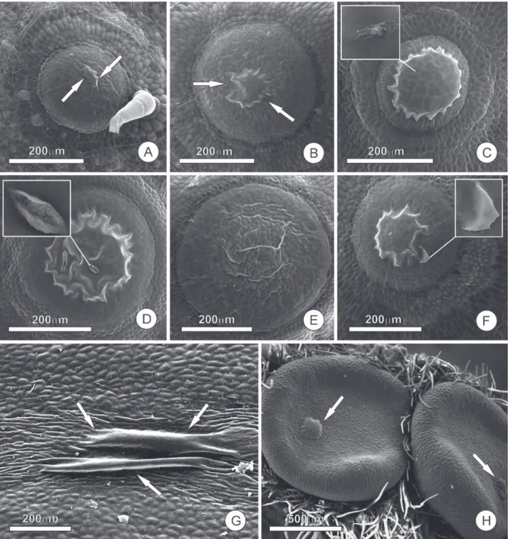
Figure 1. Accumulation of nectar and formation of the subcuticular space in extrafloral nectaries (NEFs); samples were prepared following standard SEM methods, fixed in 2.5 % glutaraldehyde in 0.1 M phosphate buffer, dehydrated in an ascending ethanol series and dried to the critical point in liquid CO2, metalized with gold and observed using a Quanta 200 scanning electron microscope.
A-F- NEFs of Luffa cilindrica (Cucurbitaceae) showing different stages of nectar release. In A-B, the arrows indicate the beginning of nectar accumulation and the subsequent formation of the subcuticular space. In C, the subcuticular space is full of nectar, nearly reaching the limits of the gland; the inset shows indications of cuticular ruptures. Cuticular ruptures and reduction in the subcuticular space due to the departure of nectar can be seen in D. In E, the cuticle, still distended, resumes its original position shortly after the extravasation of nectar. After the cuticle is recomposed and a new nectar accumulation cycle begins, a small cuticular fragment can be seen, in F, indicating the restoration of the cuticle from the previous cycle. In G and H, the secretory face of NEFs in Ouratea
Elder Antônio Sousa Paiva
subcuticular space, the volume of the nectar is defined by the extent of the cuticle that covers the gland and, naturally, the expandability of the cuticle. Nectar accumulation beneath the cuticle was reported by Pacini et al. (2003), Gama et al.
(2016) and Nepi (2007), the latter of which explained some modes of nectar release. This process of nectar release was described by Wunnachit et al. (1992) who stated: “cuticle became distended by secretion of nectar (…) no cuticular pores were observed and the nectar must tear the cuticle”. Reaching its expansion threshold, the cuticle ruptures and the nectar is released to the external environment (Fig. 1). Nectar accumulation in subcuticular space, and cuticle rupture for the release of nectar, were described by Roshchina & Roshchina (1993) for nectaries with thick cuticles and the absence of pores. It should be noted that the fact that the subcuticular space does not extend beyond the limits of the gland (Rocha et al. 2009; Possobom et al. 2015; Gama
et al. 2016) allows us to infer that pectinases, which are responsible for the weakening of the pectin layer and the consequent displacement of the cuticle, act on secretory cells and not neighboring cells. If this were not the case, the subcuticular space would tend to expand beyond the limits of the gland due to cuticular resistance. Although there is no experimental evidence, it seems quite likely that in most cases of merocrine secretion this cuticular rupture is cyclical, with the cuticle being restored after each nectar release event, thus beginning a new cycle. If this were not the case, and it occurred in any other way, the extrafloral nectary, whose secretory activity extends for long periods of time, would have persistent breaks in the cuticle, thereby allowing pathogen attacks or water loss from the gland tissue. However, this is not what is observed.
The cyclic model for cuticle restoration was suggested by Findlay & Mercer (1971), in trichome nectaries of
Abutilon (Malvaceae), wherein the nectar accumulates in a subcuticular space; the hydrostatic pressure within the hairs causes the opening of fine pores in the cuticle and the concomitant release of a nectar droplet with a sudden decrease in the hydrostatic pressure, whereupon the pores close and the periodic process starts again. Referring to Findlay & Mercer (1971), Jeffree (2006) interpreted these pores to be a kind of valve: “these pores seemed to have valve-like action, periodically releasing nectar accumulated between the cuticle and the cell wall”. This view is very interesting and we must consider that cuticles, due to their lipidic nature, are sufficiently plastic in order to restore their integrity after the release of any secretory products or after any other kind of mechanical damage. Some nectaries possess lipid droplets inside secretory cells even when lipid was not detected in the nectar (Paiva & Machado 2006; Gama et al. 2016). Thus, the role of lipids inside nectary cells during the cuticle restoration process needs to be investigated.
Some floral nectaries that lack stomata exhibit the formation of a kind of subcuticular space where nectar accumulates transiently before release (see
Weryszko-Chmielewska & Chwil 2016). Interestingly these subcuticular spaces are very small, being just vesicles on the cell surface (for details see Stpiczynska et al. 2003), and their formation is completely independent of the endodermal layer as discussed above and suggested for Luffa extrafloral nectaries (as shown in Fig. 1). These small cuticular swellings are thus distinct from large subcuticular spaces in both development and shape, but yet are similar in function and indicate the presence of an impermeable cuticle on the cell surface.
However, according to Paiva (2016), there are nectaries without stomata and from which nectar passes without the formation of a subcuticular space. In these cases, the presence of pores or hydrophilic pathways must be considered.
While there have been reports of the occurrence of cuticular canals in nectaries, many of them are questionable or not supported by convincing evidence. Nonetheless, the existence of such cuticular canals must be considered, although they seem to be restricted to the nectaries of just a few plant species. Indeed, Findlay & Mercer (1971) described such cuticular pores in the nectaries of Abutilon. Cuticular pores or canals are terms employed to describe hydrophilic pathways through the plant cuticle; however, these terms are vague because they do not always refer to a duct with a free lumen. In addition, most of them just cross the cuticular layer, and do not reach the cuticle proper. In most cases, canals are hydrophilic pathways composed of cell wall elements (usually cellulose and pectins), which extend into the cuticular layer to the proximity of the cuticle proper, but without reaching the outer surface of the cuticle. So, in fact these canals are note true ducts, but hydrophilic pathways into the cuticular layer, since ducts imply in a structure with an empty lumen. These hydrophilic pathways into the cuticular layer have been described for some nectaries, such as those of Platanthera chlorantha
(Orchidaceae) (Stpiczynska 2003), Galanthus nivalis
(Amaryllidaceae) (Weryszko-Chmielewska & Chwil 2016), nectary spurs of four representatives of Ranunculaceae (Antón & Kaminska 2015) and several other examples in the literature, mostly in stomata-free floral nectaries. Similar cuticular canals, which are polysaccharide material from cell wall protruded inside the cuticular layers, seems to act increasing the porosity of the cuticle and, consequently, constituting pathways for the secretion release in colleters (see Tresmondi et al. 2017).
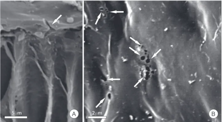
In the pericarpial nectaries of Spathodea campanulata
Figure 2. Cuticular canals in pericarpal nectaries of Spathodea campanulata (Bignoniaceae), seen using a scanning electron microscope; samples were prepared as described in figure 1. In A, a transverse section showing detail of the secretory epithelium; the arrow indicates a channel through the cuticle. In B, frontal view of the secretory face showing the cuticle with the opening of the cuticular canals (arrows).
For both nectaries with and without stomata, the accumulation of sugars inside the gland establishes the “water potential gradient” required for exuding nectar. According to Lüttge & Schnepf (1976), an alternative driving force may be the modification of sugars by gland-cell metabolism, or by the presence of invertases on the gland surface or in the released nectar itself. Cell wall invertases are important for phloem sugar unloading (Roitsch 1999) and are required for nectar production in Arabidopsis (Ruhlmann
et al. 2010).
Final remarks
Nectar release occurs differently in nectaries with stomata compared to stomata-free nectaries. Simply crossing the plasma membrane and cell wall is insufficient for the provision of nectar to consumers as the hydrophobic barrier of the cuticle must be surpassed. Stomata, cuticular pores, cuticle rupture or detachment (in repetitive cycles or not) are the main routes for nectar release, and are easily recognized by structural features, thus avoiding unnecessary speculation.
Acknowledgements
The author would like to acknowledge the Center of Microscopy at the Universidade Federal de Minas Gerais for
providing equipment and technical support for experiments involving electron microscopy. Special thanks go to Dr. D.M.T. Oliveira for her helpful comments. E. Paiva receives a research grant from CNPq (306790-2015-7).
References
Antón S, Kamińska M. 2015. Comparative floral spur anatomy and nectar secretion in four representatives of Ranunculaceae. Protoplasma 252:1587-1601.
Chakravarty HL. 1948. Extrafloral glands of Cucurbitaceae. Nature 4119: 576-577.
Delpino F. 1886. Funzione mirmecofila nel regno vegetale. Memorie della Reale Accademia delle Scienza di Bologna7: 215-323.
Fahn A. 1979. Secretory tissues in plants. New York, Academic Press. Findlay N, Mercer FV. 1971. Nectar production in Abutilon - I. Movement
of nectar through the cuticle. Australian Journal of Biological Sciences 24: 647-656.
Fineran BA, Gilbertson JM. 1980. Application of lanthanum and uranyl salts as tracers to demonstrate apoplastic pathways for transport in glands of the carnivorous plant Utricularia monanthos. European Journal of Cell Biology 23: 66-72.
Gaffal KP. 2012. How common is the ability of extrafloral nectaries to produce nectar droplets, to secrete nectar during the night and to store starch? Plant Biology 14: 691-695.
Gama TSS, Aguiar-Dias ACA, Demarco D. 2016. Transfer cells in trichomatous nectary in Adenocalymma magnificum (Bignoniaceae). Anais da Academia Brasileira de Ciências 88: 527-537.
Grout BWW, Williams A. 1980. Extrafloral nectaries of Dioscorea rotundata
Poir.: their structure and secretions. Annals of Botany 46: 255-258. Jeffree CE. 2006. The fine structure of the plant cuticle. In: Riederer M,
Elder Antônio Sousa Paiva
Lüttge U, Schnepf E. 1976. Elimination processes by glands. Organic substances. In: Lüttge U, Pitman MG. (eds.) Transport in plants II, Encyclopedia of Plant Physiology, New Series. Vol. 2B. New York, Springer. p. 244-277.
Lüttge U. 1971. Structure and function of plant glands. Annual Review of Plant Physiology 22: 23-44.
Nepi M. 2007. Nectary structure and ultrastructure. In: Nicolson SW, Nepi M, Pacini E. (eds.) Nectaries and Nectar. Dordrecht, Springer. p. 129-167.
Owen TP, Lennon KA. 1999. Structure and development of the pitchers from the carnivorous plant Nepenthes alata (Nepenthaceae). American Journal of Botany 86: 1382-1390.
Pacini E, Nepi M, Vesprini JL. 2003. Nectar biodiversity: a short review. Plant Systematics and Evolution 238: 7-21.
Paiva EAS. 2009. Ultrastructure and post-floral secretion of the pericarpial nectaries of Erythrina speciosa (Fabaceae). Annals of Botany 104: 937-944.
Paiva EAS. 2011. Petaline nectaries in Swietenia macrophylla (Meliaceae): distribution and structural aspects. Flora 206: 484-490.
Paiva EAS. 2016. How do secretory products cross the plant cell wall to be released? A new hypothesis involving cyclic mechanical actions of the protoplast. Annals of Botany 117: 533-540.
Paiva EAS, Machado SR. 2006. Ontogênese, anatomia e ultra-estrutura dos nectários extraflorais de Hymenaea stigonocarpa (Fabaceae-Caesalpinioideae). Acta Botanica Brasilica 20: 471-482.
Possobom CCF, Guimarães E, Machado SR. 2015. Structure and secretion mechanisms of floral glands in Diplopterys pubipetala (Malpighiaceae), a neotropical species. Flora 211: 26-39.
Rocha DI, Silva LC, Valente VMM, Francino DMT, Meira RMSA. 2009. Morphoanatomy and development of leaf secretory structures in
Passiflora amethystina Mikan (Passifloraceae). Australian Journal of
Botany 57: 619-626.
Roitsch T. 1999. Source-sink regulation by sugar and stress. Current Opinion in Plant Biology2: 198-206.
Roshchina VV, Roshchina VD. 1993. The excretory function of higher plants. Berlin, Springer Verlag.
Ruhlmann JM, Kram BW, Carter CJ. 2010. CELL WALL INVERTASE 4 is required for nectar production in Arabidopsis. Journal of Experimental Botany 61: 395-404.
Stpiczynska M. 2003. Nectar resorption in the spur of Platanthera chlorantha
Custer (Rchb.) Orchidaceae - structural and microautoradiographic study. Plant Systematics and Evolution 238: 119-126.
Stpiczynska M, Davies KL, Gregg A. 2003. Nectary structure and nectar secretion in Maxillaria coccinea (Jacq.) L.O. Williams ex Hodge (Orchidaceae). Annals of Botany 93: 87-95.
Tresmondi F, Canaveze Y, Guimarães E, Machado SR. 2017. Colleters in Rubiaceae from forest and savanna: the link between secretion and environment. The Science of Nature 104:17. DOI 10.1007/s00114-017-1444-x.
Weryszko-Chmielewska E, Chwil M. 2016. Flowering biology and structure of floral nectaries in Galanthus nivalis L. Acta Societatis Botanicorum Poloniae 85: 1-20.