Peripubertal orchidectomy transitorily
affects age-associated thymic involution
in rats
1Department of Physiology, Faculty of Pharmacy, University of Belgrade,
Belgrade, Serbia
2Immunology Research Center “Branislav Jankoviƒ”, Institute of Virology, Vaccines and Sera “Torlak”, Belgrade, Serbia
V. PeÓiƒ1, K. Radojeviƒ2, D. Kosec2, B. PleƒaÓ-Solaroviƒ1, M. PeriÓiƒ2, I. Pilipoviƒ2 and G. Leposaviƒ1,2
Abstract
The role of gonadal hormones in induction and, particularly, mainte-nance/progression of rat thymic involution, which normally starts around puberty, was reassessed by examining the effects of peripuber-tal orchidectomy on thymic weight and morphometric parameters at different times up to the age of 10 months. Up to 6 months post-castration both thymic weight and cellularity in orchidectomized (Cx) rats were greater than in age-matched control rats, sham Cx (Sx). The increase in thymic cellularity reflected an increase in thymocyte proliferation rate (the proportion of proliferating cells was 18.6 ± 0.7% in 2-month-old Cx (N = 5) vs 13.4 ± 0.3% (N = 5) in age-matched Sx rats) followed by reduced sensitivity to apoptotic signals (apopto-tic thymocytes were 9.8 ± 0.9% in 2-month-old Cx (N = 5) vs 15.5 ± 0.3% (N = 5) age-matched Sx rats). However, 9 months post-orchidec-tomy, neither thymic weight and cellularity nor any of the morpho-metric parameters analyzed differed between Cx and control rats. The reduction of thymic cellularity in Cx rats to control values may be related to increased sensitivity of their thymocytes to apoptotic signals in culture (72.6 ± 1.2% in 10-month-old vs 9.8 ± 0.9% in 2-month-old Cx rats) followed by reduced responsiveness to proliferative stimuli (14.1 ± 0.2% in 10-month-old vs 18.6 ± 0.7% in 2-month-old Cx rats). Thus, the study indicates that the effects of peripubertal orchidectomy on thymic weight and cellularity, as well as on the main morphometric indices, are long-lasting but not permanent, i.e., that removal of the testes can only postpone but not prevent age-related organ atrophy and consequently functional deterioration of the immune system. Correspondence
G. Leposaviƒ Faculty of Pharmacy 450 Vojvode Stepe 11221 Belgrade Serbia
Fax: +381-11-397-2840 E-mail:
Research supported by project grant No. 145049 from the Ministry of Science and Environmental Protection of the Republic of Serbia.
Received March 9, 2007 Accepted July 24, 2007
Key words
•Rat thymus •Aging •Orchidectomy •Stereological analysis •Thymocyte proliferation •Thymocyte apoptosis
Introduction
Thymic involution is one of the most clear-cut effects of aging on the immune system of both laboratory animals and hu-mans. In rodents, there is a sharp increase in
(1-3). Thus, as the thymus involutes, the output of newly generated T cells declines, with a reduction in the level of naive T cells in the periphery. These changes have been related to the age-associated decline in func-tion of the immune system, leading to an increased frequency of specific infections, malignant diseases and autoimmune disor-ders in old individuals (4). The fact that thymic involution is associated with immu-nosenescence and its various associated dis-eases has prompted many studies aimed at understanding the causes and mechanisms of thymic degeneration which may ultimately lead to the possibility of manipulation. This might not only prolong life, but also improve its quality, due to the absence of severe associated pathologies.
Since thymic involution in rodents be-comes obvious at the same time as the in-crease in production of gonadal steroids, a causal link between increasing sex steroid level and age-related thymic atrophy has been suggested (1,2). This notion is sup-ported by data indicating that androgens in males modulate both thymic weight and T-cell maturation by acting either directly on the thymocytes (5) or indirectly on thymic epithelial cells (TEC) (6) supporting T-cell differentiation/maturation. Namely, substan-tial alterations in the composition of thy-mocyte subsets at different stages of matura-tion (delineated by expression of the major differentiation antigens CD4, CD8, T-cell receptor), which are localized in clearly defined thymic compartments, have been observed in animals subjected to gonadec-tomy (7,8). A putative role for sex steroids in the induction of thymic involution is also supported by numerous experiments show-ing reversal of orchidectomy-induced thy-mic hypertrophy by androgen replacement (7). However, the long-term commonly held view that gonadal hormones have a pivotal role in the induction and maintenance of thymic involution has recently been disputed. Namely, thymic involution in humans is
pro-posed to be a sex steroid-independent event (9), while lack of gonadal hormones was shown not to produce a delay in age-associ-ated thymic involution in hypogonadal HPG/ Bm-hpg/hpg mice (10). The obvious dis-crepancy between these and numerous ear-lier studies may be reconciled by the fact that the role of sex steroids in thymic involu-tion was estimated by exploring the effects of gonadal hormone deprivation/adminis-tration for not more than a few months. Therefore, the present study was undertaken to reassess the putative role of gonadal hor-mones in the induction and, particularly, in the maintenance/progression of age-associ-ated thymic atrophy in rats. To this end, we followed the effects of peripubertal orchidec-tomy on the rat thymus for up to 9 months after surgery. Since it has been shown that the functional capacity of the aged residual thymus correlates with anatomical measure-ments of so-called true thymic tissue (4), we subjected this thymic component (i.e., cortical and medullary lymphoid tissue) from both orchidectomized (Cx) and control rats to a tentative morphometric analysis. Additionally, since changes in thymic cellu-larity mainly reflect alterations in the main homeostatic processes, i.e., apoptosis and proliferation, thymocytes from Cx and control rats were examined for their sensi-tivity to both apoptotic and proliferative sig-naling.
Material and Methods
Animals
One, 3, 6, or 9 months after orchidec-tomy, the rats were killed under ether anes-thesia by exsanguination. Thirty-day-old in-tact rats, as well as 2-, 4-, 7-, and 10-month-old Sx and non-operated rats were included in the study as controls. All groups consisted of 5 animals. Each thymus was aseptically isolated, trimmed of all excess body fat and gently blotted on gauze to remove excess blood. The two lobes were divided and weighed individually. The right lobe was used for preparation of thymocyte single cell suspensions and the left lobe was fixed in Bouin’s solution and further processed for morphometric analysis.
Computer-assisted morphometric measurements
Thymic specimens fixed in Bouin’s solu-tion and embedded in paraffin were serially cut into 5-µm thick sections. Every 40th section (approximately 20-30 sections per organ) was stained with hematoxylin and eosin. Morphometric measurements were made by a point counting method as previ-ously described (11) using an Olympus BX50 microscope and image analysis software (Micro Image, Version 4.0, Olympus Opti-cal Co. GmbH, Hamburg, Germany). The test areas were randomly chosen and each image, acquired using a digital camera, was saved, overlaid with the corresponding grid and analyzed.
Absolute volumes of the main thymic compartments, i.e., cortex, medulla and sub-capsular/interlobular connective/adipose tis-sue, were estimated from the volume of the processed and embedded organ and volume density (Vv) of the corresponding compart-ment. The relative amount of thymic tissue shrinking during processing and embedding (approximately 34%) in all groups was de-termined stereologically, as described ear-lier (11). Thus, all stereological data refer to fixed thymic tissue. Each thymic compart-ment Vv was determined at 40X
magnifica-tion using an orthogonal test grid with 130 points, and by dividing the number of test points hitting the analyzed structure by the total number of test points falling on the organ. The overall number of test areas was 100 per animal.
The total number of thymocytes in the thymic compartment was calculated from the numerical density (Nv) of thymocytes, as the number of cells per volume unit, and the absolute volume of that compartment. When calculating the overall number of cor-tical thymocytes it was taken into consider-ation that: i) two cortical subcompartments, i.e., outer and deep cortex (12) can be delin-eated morphologically and functionally, ii) three quarters of cortical thymocytes are situated in the deep cortex (13). The Nv of thymocytes was estimated at immersion magnification using a grid that corresponds to the multipurpose M42 test-system. The
test grid was placed randomly, but posi-tioned parallel to and just touching the cap-sule for the outer cortex and the cortico-medullary junction for the deep cortex anal-ysis, respectively. To estimate the Nv of medullary thymocytes, the grid was placed randomly throughout the medulla. For each thymic compartment 60 test areas per ani-mal were measured.
Preparation of thymocyte suspensions
Analysis of cycling cells
Propidium iodide binding to DNA was utilized to identify cells in active phases of the cell cycle. As previously described (14), 100 µL RPMI 1640 with 5 µg/mL concanava-lin A (ConA, Sigma-Aldrich Chemie, Tauf-kirchen, Germany) was added to 100 µL of a cell suspension containing 2.5 x 10⁵
thy-mocytes dispersed into plastic 96-well plates (Nunc A/S, Roskilde, Denmark) to attain a final ConA concentration of 2.5 µg/mL. All cultures were run in triplicate. Cells were harvested after 48 h of culture. A total of 0.5 x 10⁶ cells was fixed by the addition of 400
µL ice-cold absolute ethanol, on ice, for 30 min. The cells were then centrifuged at 300
g for 10 min and the supernatant was de-canted. After the addition of 1 mg/mL RNAse 1A (Sigma-Aldrich Chemie) in 250 µL phos-phate-buffered saline, pH 7.4, to the pellet and vortexing, the samples were incubated in a water bath at 37ºC for 20 min. Propi-dium iodide (Sigma-Aldrich Chemie) at 20 µg/mL in 250 µL was then added and the samples were gently mixed and incubated for at least 10 min at room temperature. Next, the cells were passed through a fine nylon mesh and analyzed with a FACScan flow cytometer (Becton Dickinson, Moun-tain View, CA, USA). The gate was set for singlet populations so that doublets and higher order cell aggregates would be ex-cluded from DNA analysis.
Detection of apoptotic thymocytes
Since apoptoticthymocytes are normally rapidly eliminated by phagocytes in vivo, the relative number of apoptotic cells was quantified after 18 h of cultivation, as previ-ously suggested (15). A 100-µL aliquot of the cell suspension (at a concentration of 2.5 x 10⁶/mL in RPMI 1640 complete medium)
was added to each well of a 96-well flat-bottom plate (Nunc A/S). The cells were incubated for 18 h at 37ºC in a 5% CO2
humidified atmosphere. Apoptotic thy-mocytes were detected using merocyanine 540 (MC540). Similarly to annexin-V, this lipophilic dye stains an altered packing or-der of phospholipids in the outer leaflet of the apoptotic cell plasma membrane. Thus, the percentage of apoptotic cells labeled with MC540 is shown to be equivalent to that obtained by annexin-V staining (16).
The procedure described by Mower and collaborators (17) was used for MC540 stain-ing. All samples were analyzed on the same day with the FACScan flow cytometer (Bec-ton-Dickinson) using CellQuest Software (Becton-Dickinson).
Statistical analysis
Data are reported as means ± SEM. Thy-mic weight and stereological parameters from Sx and non-operated rats of the same age were compared by the Mann-Whitney U-test. Since none of the values of the param-eters analyzed differed significantly between Sx and non-operated rats of the same age, the data for these two groups were pooled and presented as one group (Sx) in all graphs. The differences among groups of different ages were tested by non-parametric Krus-kal-Wallis one-way analysis of variance fol-lowed by the Mann-Whitney U-test when-ever differences were found. The SPSS 10.0 software for Windows was used for statisti-cal analysis.
Results
Histology
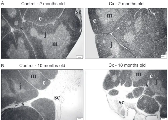
struc-tural changes were observed. The thickness of the thymic capsule and septa increased and a substantial quantity of adipose tissue accumulated under the capsule and in the interlobular spaces. The borderline between the cortex and medulla appeared less dis-tinct.
No prominent differences in thymic struc-ture were observed between Cx and age-matched controls.
Computer-assisted morphometric analysis
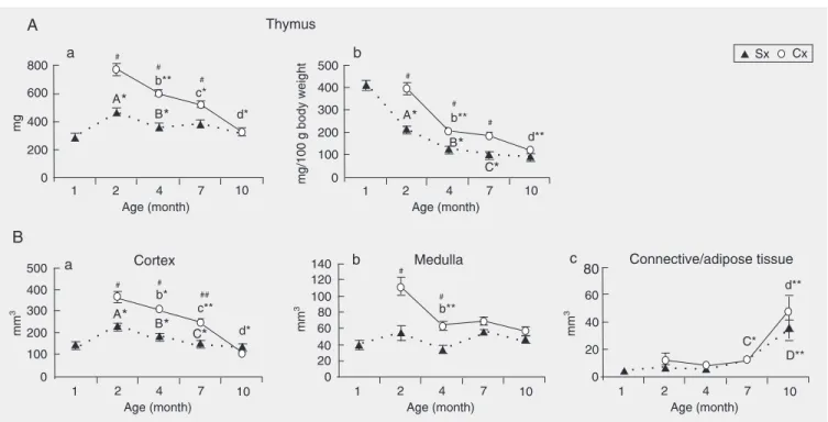
Age-associated changes in control rats. The results showed that the relative thymic weight (ratio to 100 g body weight) of con-trol rats progressively decreased from the age of 30 days to 7 months, and then re-mained at that level until the age of 10 months (Figure 2, Panel A, b). Furthermore, they revealed that: i) the absolute thymic weight of the same animals was maximal around 2 months of age; ii) between the ages of 2 and 4 months regressive changes took place, so that at 4 months of age thymic
weight was significantly reduced by an aver-age of 27% (Figure 2, Panel A, a). This decrease reflected a significant reduction in the size of the thymic cortex (Figure 2, Panel B, a). Absolute thymic weight did not de-crease further with age (Figure 2, Panel A, a). However, the volume of thymic cortex continued to shrink until the age of 7 months, and remained at that level for the next 3 months. In spite of the decrease in cortical volume between the ages of 4 and 7 months, the absolute thymic weight remained unal-tered due to an increase in the volume of thymic connective/adipose tissue (Figure 2, Panel B, c). Although further enlargement in the volume of this tissue was recorded be-tween the ages of 7 and 10 months, overall thymic weight remained unaffected.
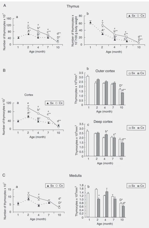
The relative thymic cellularity (ratio to 100 g body weight) showed a progressive decrease from 30 days up to 10 months of age (Figure 3, Panel A, b). The absolute thymic cellularity exhibited a similar pattern of changes to that shown by absolute organ weight except for an additional significant
Figure 1. Photomicrographs of thymus sections derived from 2-month-old (A) and 10-2-month-old (B) controls and rats orchidecto-mized at the age of 30 days (Cx). c = cortex; m = medulla; sc = subcapsular adipose tissue; j = cortico-medullary junction; s = septa; H-E staining, 40X. Bar = 100 µm.
Control - 2 months old Cx - 2 months old
A
Figure 2. Age-related changes in A, absolute thymic weight (a) and relative thymic weight (b), as well as B, volume of different thymic compartments: (a) cortex, (b) medulla, and (c) connective/adipose tissue, in rats orchidectomized at the age of 30 days (Cx) and control animals (Sx). Data are reported as means ± SEM for N = 5. A2-month-old Sx vs 30-day-old Sx; B4-month-old Sx vs 2-month-old Sx; C7-month-old Sx vs 4-month-old Sx; D10-month-old Sx vs 7-month-old Sx; #Cx vs age-matched controls; b4-month-old Cx vs 2-month-old Cx; c7-month-old Cx vs 4-month-old Cx; d10-month-old Cx vs 7-month-old Cx. *P < 0.05 and **P < 0.01; #P < 0.05 and ##P < 0.01 (Mann-Whitney U-test).
decrease between the ages of 7 and 10 months (the average thymic cellularity diminished by 66% from 2 to 10 months of age; Figure 3, Panel A, a). The age-associated changes in thymic cellularity reflected alterations in cortical thymocyte numbers at all time points except for the age of 10 months, when the reduction in organ cellularity was related to a significant decrease in the number of med-ullary thymocytes due to a decline in their numerical density (Figure 3, Panels B, C).
Effects of orchidectomy. Orchidectomy at the age of 30 days produced a significant increase in relative and absolute thymic weight (Figure 2, Panel A, a,b), as well as in relative and absolute thymocyte number at all time points except for the age of 10 months (Figure 3, Panel A, a,b). At that age neither the relative and absolute thymic weight nor any of the morphometric param-eters significantly differed between Cx and
Figure 3. A, Age-related changes in the (a) total and (b) relative number of thymocytes, as well as in the number of thymocytes (B, a and C, a) and in thymocyte numerical density in the (B, b,c) thymic cortex and (C, b) medulla in rats orchidectomized at the age of 30 days (Cx) and control animals (Sx). Data are reported as means ± SEM for N = 5. A 2-month-old Sx vs 30-day-old Sx; B4-month-old Sx vs 2-month-old Sx; C7-month-old Sx vs 4-month-old Sx; D10-month-old Sx
by 57%; Figure 2, Panel A, a,b). This de-crease was particularly pronounced between the ages of 4 and 10 months (the average weight was reduced by 45%). The reduction of absolute thymic weight mainly reflected the decrease in cortical volume (Figure 2, Panel B, a). A significant decrease in medul-lary volume (Figure 2, Panel B, b) was found to contribute to the reduction of thymic weight only in 4-month-old rats. In 10-month-old rats a significant enlargement of connec-tive-adipose tissue volume was also recorded (Figure 2, Panel B, c).
In Cx rats both absolute and relative thy-mic cellularity also exhibited a progressive decrease between the ages of 2 and 10 months (the average number was reduced by 82%; Figure 3, Panel A, a,b). The changes in thymic cellularity primarily reflected the re-duction in number of cortical thymocytes (Figure 3, Panel B, a) due to the progressive loss of these cells in the deep cortex (at all time points) and in the outer cortex (at 10 months of age), which was evident from the reduced Nv of thymocytes in the deep and outer cortical compartments, respectively (Figure 3, Panel B, b,c). Between the ages of 7 and 10 months a significant loss in medul-lary thymocytes leading to a decrease in Nv of thymocytes was also recorded (Figure 3, Panel C, a,b).
Thymocyte proliferation
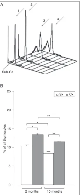
To clarify the mechanisms underlying age-dependent alterations in thymic cellu-larity in ConA+ thymocyte cultures from 2-and 10-month-old rats (i.e., at time points when the most striking changes in thymic weight and cellularity were recorded) the percentage of cells in the S/G2M active phases of the cell cycle was examined.
The results showed that, irrespective of age, in thymocyte cultures from Cx rats there was a greater percentage of cells in the S/ G2M phases of the cell cycle than in the corresponding cultures from age-matched
1 2
3 4
Sub-G1
A
B
Figure 4. Increased percentage of cycling cells (cells in the S/ G2M phases of the cell cycle) in concanavalin A-stimulated thy-mocyte cultures from adult rats orchidectomized at the age of 30 days (Cx) as determined by pro-pidium iodide binding to DNA. A, Representative overlaid histo-grams (3-D view) of propidium iodide staining of cells from thy-mocyte cultures of 1) old control rats (Sx), 2) 2-month-old Cx rats, 3) 10-month-2-month-old Sx rats, and 4) 10-month-old Cx rats. The arrow points at the sub-G1 peak that corresponds to apoptotic cells. B, Relative pro-portion of thymocytes in the S/ G2M phases of the cell cycle in concanavalin A-stimulated thy-mocyte cultures from control rats (Sx) and rats orchidectomized at the age of 30 days (Cx). Data are reported as means ± SEM for N = 5. *P < 0.05 and **P < 0.01 (Mann-Whitney U-test).
volume of the whole compartment, the over-all number of lymphoid cells in this com-partment remained unaltered (Figure 3, Panel C, a).
control rats (Figure 4, Panel B). Further-more, irrespective of gonadal presence, the percentage of cells in the S/G2M phases of the cell cycle was greater in thymocyte cul-tures from 2-month-old rats than in corre-sponding cultures from 10-month-old rats.
It should also be pointed out that the percentage of apoptotic cells forming a flow cytometric peak to the left of the G1 peak (Figure 4, Panel A), which is often referred to as the sub-G1 peak (18), was significantly (P < 0.01) greater in thymocyte cultures from 10-month-old rats (68.16 ± 1.4% in Sx rats and 75.28 ± 0.8% in Cx rats) than in the corresponding cultures from 2-month-old rats (25.71 ± 1.7% in Sx rats and 22.15 ± 2.34% in Cx rats).
Thymocyte apoptosis
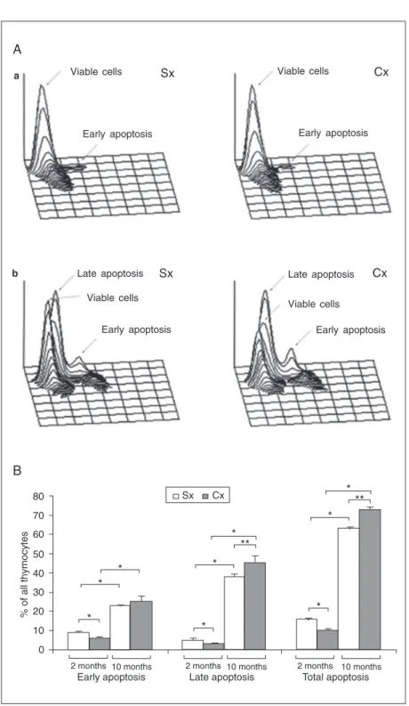
Since not only alterations in thymocyte proliferation, but also in thymocyte apopto-sis may affect thymic cellularity, thymocyte sensitivity to apoptotic signaling was esti-mated at the same time points at which thy-mocyte proliferative capacity was examined. In thymocyte cultures from 2-month-old Cx rats the percentage of apoptotic cells was significantly lower than in the age-matched controls. However, on the contrary, in thy-mocyte cultures from 10-month-old rats the percentage of apoptotic cells was increased in Cx compared to Sx rats (Figure 5, Panel B). Nevertheless, the percentage of apopto-tic cells was significantly lower in thymocyte cultures from both Cx and control 2-month-old rats than in the corresponding cultures from 10-month-old rats (Figure 5, Panel B). According to the intensity of MC540 fluorescence, on the one hand, and forward scatter, on the other (19), two subsets of apoptotic cells can be distinguished: 1) cells in early apoptosis, and 2) cells in advanced/ late apoptosis (Figure 5, Panel A). Cells in early apoptosis exhibit a high level of MC540 staining, while those in advanced/late apop-tosis show lower levels of MC540 staining
Figure 5. Effects of orchidectomy at the age of 30 days on the frequency of apoptotic cells in 18-h thymocyte cultures. The apoptotic cells were identified by MC540 binding. A, Three-dimensional plots of MC540-labeled thymocytes in cultures from (a) 2-month-old and (b) 10-month-old orchidectomized (Cx) and control rats (Sx). According to the intensity of MC540 thymocyte staining and forward scatter, two subsets of cells at distinct phases of apoptosis (early apoptosis and advanced/late apoptosis) were delineated. B, The histo-gram represents the overall percentage of apoptotic cells and the percentages of cells in early and advanced/late apoptosis in thymocyte cultures from Cx and Sx rats. Data are reported as means ± SEM for N = 5. *P < 0.05 and **P < 0.01 (Mann-Whitney U-test).
Viable cells Sx Viable cells Cx
Early apoptosis Early apoptosis
Sx Cx
Late apoptosis
Viable cells
Early apoptosis
Late apoptosis
Viable cells
Early apoptosis
A
a
b
and lower forward scatter. In thymocyte cul-tures from 2-month-old Cx rats the reduc-tion in the percentage of apoptotic cells re-flected decreases in the relative numbers of cells in both the early and late phases of apoptosis (Figure 5, Panel B). On the other hand, the higher percentage of apoptotic cells in 10-month-old Cx rats compared to age-matched control rats was mainly due to a rise in the proportion of cells in late apop-tosis (Figure 5, Panel B).
Discussion
This study describes the age-related changes in weight, cellularity and morpho-metric indices of the thymus in peripubertally Cx and control AO rats. We have confirmed that gonadal hormone removal in the peripu-bertal period leads to a significant increase in thymic weight and cellularity in young adult rats (8), and have shown that this organ reaches its maximum weight and cellularity one month after surgery. Finally, to the best of our knowledge, for the first time it has been demonstrated that in rats the effects of peripubertal orchidectomy on the thymus are long-lasting but not permanent, i.e., that removal of the testes can only postpone but not prevent the age-related organ atrophy.
The present findings confirm that, after an initial decline in early adulthood, thymic weight in rats (3), as in humans (9) and some other mammals (20), but not in mice (21-23), remains unaltered during a relatively long period of time (from 4 to 10 months of age). Although thymic weight in 10-month-old rats did not differ from values for 4- and 7-month-old animals, the total number of thymocytes in both thymic compartments, and hence the overall organ cellularity, were significantly reduced reflecting, most likely, an age-associated increase in thymocyte sen-sitivity to apoptotic stimuli followed by de-creased responsiveness to proliferative sig-naling. This assumption emerged from our findings obtained with thymocyte cultures,
which are fully consistent with previously reported data on cell sensitivity to apoptotic and proliferative signaling in thymocyte cul-tures from aged Wistar rats (14). The age-associated expansion of connective-adipose tissue described here, which compensates for the loss of lymphoid tissue, has also been observed in aged male Wistar rats (24). Therefore, these results support our previ-ous findings that the overall thymic weight of aging rats does not correlate with the amount of functional lymphoid thymic tis-sue (24).
nei-ther thymic weight nor any estimated mor-phometric parameter significantly differed from those in age-matched controls. This is in keeping with the very recent conclusion of Min and colleagues (10) that the orchidec-tomy-induced effect on thymic weight in mice is of limited duration. In other words, our results indicate that removal of the testes in rats, as in mice (10), postpones age-asso-ciated thymic involution, but does not pre-vent it completely. Furthermore, our results also indicate that a new balance between thymocyte apoptosis and proliferation was established in these animals. Differently from thymocyte cultures from 2-month-old rats, the percentage of apoptotic cells was signifi-cantly greater in thymocyte cultures from Cx rats compared with age-matched con-trols. Moreover, although the proportion of cells in the S/G2M phases of the cell cycle was significantly greater in ConA+ thy-mocyte cultures from 10-month-old Cx rats than in those from age-matched Sx rats, it was significantly less than in thymocyte cul-tures from 2-month-old Cx rats. A compen-satory intrathymic increase in androgen pro-duction sufficient to cause accelerated thy-mocyte apoptosis, but still not sufficient to prevent proliferation of these cells may be assumed to explain these findings. In favor of this hypothesis are data suggesting that both lymphocytes (31) and TEC may pro-duce steroid hormones (32). Furthermore, analysis of thymocyte differentiation/matu-ration under the same experimental condi-tions showed that alteracondi-tions in the relative proportion of thymocytes (delineated by ex-pression of CD4, CD8 and T-cell receptor) become quantitatively more pronounced with duration of gonadal deprivation (8). Thus, the present findings, taken together with those previously published (8), indicate that, although 9 months after orchidectomy a new balance between the main thymic homeo-static processes (i.e., apoptosis and prolif-eration) is established, so that thymic cellu-larity does not significantly differ between
Cx and control rats, the thymocyte differen-tiation kinetics remains substantially altered, producing an accumulation of cells at one maturational step and depletion at the subse-quent downstream steps. Moreover, the age-associated decline in thymic weight and cel-lularity in Cx rats was even more pronounced than that in non-operated animals and, in contrast to control rats, both thymic weight and cellularity exhibited a progressive re-duction between the ages of 2 and 10 months. This suggests that: i) age-associated thymic atrophy, which is initially sensitive to circu-lating androgen levels, becomes independ-ent of them with progression of the process and ii) the kinetics of age-associated thymic involution depends on the presence of circu-lating gonadal hormones.
that the adrenals of rats and mice do not synthesize androgens (34), so that in these animals orchidectomy is not followed by increased serum levels of either dehydroepi-androsterone or testosterone (35) and this hypothesis should be rejected. Third, it may be assumed that thymic involution is medi-ated by distinct mechanisms acting during different periods of ontogenesis or that some other, non-androgen-mediated mechanism (e.g., growth hormone-dependent) (36), may take over the androgen role in long-term androgen-deprived rats. At present there are no data available to strongly support this hypothesis. Finally, it may also be hypoth-esized that gonadal hormones are important in the initial phases of thymic regression, while later accumulating age-related func-tional or structural defects (37) are responsi-ble for the maintenance/progression of thy-mic involution. In agreement with this hypo-thesis are data showing that: i) normal, age-related androgen depletion (38) exerts no
beneficial effect on the aged thymus (39), and ii) castration of old rats does not restore thymic weight to the level found in young animals (40).
In conclusion, this study has clearly shown that gonadal ablation cannot prevent thymic involution, suggesting that even if circulating gonadal hormones are predomi-nantly responsible for the initiation of age-associated thymic involution, maintenance of their circulating levels is not necessary to secure maintenance/progression of the age-associated regressive thymic changes. To further clarify the role of androgens in the initiation, and particularly in the mainte-nance/progression of age-related thymic at-rophy, experiments addressing not only thy-mic effects evoked by long-lasting with-drawal of circulating androgens, but also those induced by blockade of intrathymi-cally synthesized/released androgen action, should be undertaken.
References
1. Grossman CJ. Interactions between the gonadal steroids and the immune system. Science 1985; 227: 257-261.
2. Bodey B, Bodey B Jr, Siegal SE, Kaiser HE. Involution of the mammalian thymus, one of the leading regulators of aging. In Vivo
1997; 11: 421-440.
3. Quaglino D, Capri M, Bergamini G, Euclidi L, Zecca L, Ronchetti IP. Age-dependent remodeling of rat thymus. Morphological and cyto-fluorimetric analysis from birth up to one year of age. Eur J Cell Biol
1998; 76: 156-166.
4. Pawelec G, Effros RB, Caruso C, Remarque E, Barnett Y, Solana R. T cells and aging (update February 1999). Front Biosci 1999; 4: D216-D269.
5. Viselli SM, Olsen NJ, Shults K, Steizer G, Kovacs WJ. Immuno-chemical and flow cytometric analysis of androgen receptor expres-sion in thymocytes. Mol Cell Endocrinol 1995; 109: 19-26. 6. Olsen NJ, Olson G, Viselli SM, Gu X, Kovacs WJ. Androgen
recep-tors in thymic epithelium modulate thymus size and thymocyte de-velopment. Endocrinology 2001; 142: 1278-1283.
7. Kendall MD, Fitzpatrick FT, Greenstein BD, Khoylou F, Safieh B, Hamblin A. Reversal of ageing changes in the thymus of rats by chemical or surgical castration. Cell Tissue Res 1990; 261: 555-564.
8. Leposaviƒ G, Pejciƒ-Karapetroviƒ B, Kosec D. Alterations in thymo-poiesis in intact and peripubertally orchidectomized adult rats of different age. Mech Ageing Dev 2002; 123: 401-411.
9. Steinmann GG, Klaus B, Muller-Hermelink HK. The involution of the ageing human thymic epithelium is independent of puberty. A mor-phometric study. Scand J Immunol 1985; 22: 563-575.
10. Min H, Montecino-Rodriguez E, Dorshkind K. Reassessing the role of growth hormone and sex steroids in thymic involution. Clin Immunol 2006; 118: 117-123.
11. PleƒaÓ-Solaroviƒ B, Laliƒ Lj, Leposaviƒ G. Age-dependent mor-phometrical changes in the thymus of male propranolol-treated rats.
Ann Anat 2004; 186: 141-147.
12. Lind EF, Prockop SE, Porritt HE, Petrie HT. Mapping precursor movement through the postnatal thymus reveals specific microenvi-ronments supporting defined stages of early lymphoid development.
J Exp Med 2001; 194: 127-134.
13. Kendall MD, Al-Shawaf AA. Innervation of the rat thymus gland.
Brain Behav Immun 1991; 5: 9-28.
14. Leposaviƒ G, PeÓiƒ V, Kosec D, Radojeviƒ K, Arsenoviƒ-Ranin N, Pilipoviƒ I, et al. Age-associated changes in CD90 expression on thymocytes and in TCR-dependent stages of thymocyte maturation in male rats. Exp Gerontol 2006; 41: 574-589.
15. Kamath AB, Nagarkatti PS, Nagarkatti M. Characterization of phe-notypic alterations induced by 2,3,7,8-tetrachlorodibenzo-p-dioxin on thymocytes in vivo and its effect on apoptosis. Toxicol Appl Pharmacol 1998; 150: 117-124.
Immunol Methods 2002; 261: 129-139.
17. Mower DA Jr, Peckham DW, Illera VA, Fishbaugh JK, Stunz LL, Ashman RF. Decreased membrane phospholipid packing and de-creased cell size precede DNA cleavage in mature mouse B cell apoptosis. J Immunol 1994; 152: 4832-4842.
18. Ormerod MG. Investigating the relationship between the cell cycle and apoptosis using flow cytometry. J Immunol Methods 2002; 265: 73-80.
19. Cohen JJ. Programmed cell death in the immune system. Adv Immunol 1991; 50: 55-85.
20. Hale LP, Clark AG, Li J, Greer PK, Byers Kraus V. Age-related thymic atrophy in the guinea pig. Dev Comp Immunol 2001; 25: 509-518.
21. Nabarra B, Andrianarison I. Ultrastructural study of thymic microen-vironment involution in aging mice. Exp Gerontol 1996; 31: 489-506. 22. Lau LL, Spain LM. Altered aging-related thymic involution in T cell receptor transgenic, MHC-deficient, and CD4-deficient mice. Mech Ageing Dev 2000; 114: 101-121.
23. Li L, Hsu HC, Grizzle WE, Stockard CR, Ho KJ, Lott P, et al. Cellular mechanism of thymic involution. Scand J Immunol 2003; 57: 410-422.
24. PleƒaÓ-Solaroviƒ B, PeÓiƒ V, Radojeviƒ K, Leposaviƒ G. Morpho-metrical characteristics of age-associated changes in the thymus of old male Wistar rats. Anat Histol Embryol 2006; 35: 380-386. 25. Ishimi Y, Yoshida M, Wakimoto S, Wu J, Chiba H, Wang X, et al.
Genistein, a soybean isoflavone, affects bone marrow lymphopoie-sis and prevents bone loss in castrated male mice. Bone 2002; 31: 180-185.
26. Gill J, Malin M, Sutherland J, Gray D, Hollander G, Boyd R. Thymic generation and regeneration. Immunol Rev 2003; 195: 28-50. 27. Windmill KF, Lee VW. Influences of surgical castration on the
thy-mus of male rats. J Reprod Immunol 1999; 44: 29-39.
28. Olsen NJ, Viselli SM, Fan J, Kovacs WJ. Androgens accelerate thymocyte apoptosis. Endocrinology 1998; 139: 748-752. 29. Olsen NJ, Viselli SM, Shults K, Stelzer G, Kovacs WJ. Induction of
immature thymocyte proliferation after castration of normal male mice. Endocrinology 1994; 134: 107-113.
30. Guevara Patino JA, Marino MW, Ivanov VN, Nikolich-Zugich J. Sex steroids induce apoptosis of CD8+CD4+ double-positive thymocytes via TNF-alpha. Eur J Immunol 2000; 30: 2586-2592.
31. Zhou Z, Shackleton CH, Pahwa S, White PC, Speiser PW. Promi-nent sex steroid metabolism in human lymphocytes. Mol Cell Endo-crinol 1998; 138: 61-69.
32. Khosla S, Ovalle WK. Morphology and distribution of cystic cavities in the normal murine thymus. Cell Tissue Res 1986; 246: 531-542. 33. Sakabe K, Kawashima I, Urano R, Seiki K, Itoh T. Effects of sex
steroids on the proliferation of thymic epithelial cells in a culture model: a role of protein kinase C. Immunol Cell Biol 1994; 72: 193-199.
34. van Weerden WM, Bierings HG, van Steenbrugge GJ, de Jong FH, Schroder FH. Adrenal glands of mouse and rat do not synthesize androgens. Life Sci 1992; 50: 857-861.
35. Belanger B, Belanger A, Labrie F, Dupont A, Cusan L, Monfette G. Comparison of residual C-19 steroids in plasma and prostatic tissue of human, rat and guinea pig after castration: unique importance of extratesticular androgens in men. J Steroid Biochem 1989; 32: 695-698.
36. Kelley KW, Brief S, Westly HJ, Novakofski J, Bechtel PJ, Simon J, et al. GH3 pituitary adenoma cells can reverse thymic aging in rats.
Proc Natl Acad Sci USA 1986; 83: 5663-5667.
37. Montecino-Rodriguez E, Clark R, Dorshkind K. Effects of insulin-like growth factor administration and bone marrow transplantation on thymopoiesis in aged mice. Endocrinology 1998; 139: 4120-4126. 38. Kaler LW, Neaves WB. The androgen status of aging male rats.
Endocrinology 1981; 108: 712-719.
39. Taub DD, Longo DL. Insights into thymic aging and regeneration.
Immunol Rev 2005; 205: 72-93.
•
Deposit and Distribution of Cell Lines•
Cell Cryopreservation•
Development and Distribution of Human and Animal Primary Cell Cultures•
Development of New Monoclonal Antibody Secreting Hybridomas•
Monoclonal Antibodies, in Cell Culture Supernatants or Purified•
Identification of Microbiological Contaminants•
Decontamination of Cell Lines•
Cytotoxicity Tests•
Characterization of Cell Lines•
Training in “in vitro” Culture of Human and Animal Cells•
Consultation Assistance in Cell CultureContact us for specific services not listed above
Banco de Células do Rio de Janeiro - BCRJRio de Janeiro Cell Bank
Associação Técnico Científica Paul Ehrlich
Hospital Universitário Clementino Fraga Filho/4oandar/Sala 4 a 9
Programa Avançado de Biologia Celular Aplicada à Medicina - PABCAM
Universidade Federal do Rio de Janeiro - UFRJ, Cidade Universitária
Ilha do Fundão, CEP 21941-097 Rio de Janeiro, RJ
Tel: (21) 2562.24.67/2562.64.84/2562.64.83
[email protected]/http://www.bcrj.hucff.ufrj.br/