online | memorias.ioc.fiocruz.br
In vivo antileishmanial activity and chemical profile of
polar extract from
Selaginella sellowii
Dayane Priscilla de Souza Queiroz1, Carlos Alexandre Carollo2,
Mônica Cristina Toffoli Kadri3, Yasmin Silva Rizk1, Vanessa Carneiro Pereira de Araujo1,
Paulo Eduardo de Oliveira Monteiro1, Patrik Oening Rodrigues4, Elisa Teruya Oshiro1,
Maria de Fátima Cepa Matos5, Carla Cardozo Pinto de Arruda1/+
1Universidade Federal de Mato Grosso do Sul, Centro de Ciências Biológicas e da Saúde, Laboratório de Parasitologia Humana,
Campo Grande, MS, Brasil 2Universidade Federal de Mato Grosso do Sul, Centro de Ciências Biológicas e da Saúde,
Laboratório de Produtos Naturais e Espectrometria de Massas, Campo Grande, MS, Brasil 3Universidade Federal de Mato Grosso do Sul,
Centro de Ciências Biológicas e da Saúde, Laboratório de Biofisiofarmacologia, Campo Grande, MS, Brasil
4Universidade Federal de Mato Grosso do Sul, Centro de Ciências Biológicas e da Saúde, Laboratório de Tecnologia Farmacêutica,
Campo Grande, MS, Brasil 5Universidade Federal de Mato Grosso do Sul, Centro de Ciências Biológicas e da Saúde,
Laboratório de Biologia Molecular e Culturas Celulares, Campo Grande, MS, Brasil
The polar hydroethanolic extract from Selaginella sellowii (SSPHE) has been previously proven active on intra-cellular amastigotes (in vitro test) and now was tested on hamsters infected with Leishmania (Leishmania) amazo-nensis (in vivo test). SSPHE suppressed a 100% of the parasite load in the infection site and draining lymph nodes at an intralesional dose of 50 mg/kg/day × 5, which was similar to the results observed in hamsters treated with N-methylglucamine antimonate (Sb) (28 mg/Kg/day × 5). When orally administered, SSPHE (50 mg/kg/day × 20) suppressed 99.2% of the parasite load in infected footpads, while Sb suppressed 98.5%. SSPHE also enhanced the release of nitric oxide through the intralesional route in comparison to Sb. The chemical fingerprint of SSPHE by high-performance liquid chromatography with diode-array detection and tandem mass spectrometry showed the presence of biflavonoids and high molecular weight phenylpropanoid glycosides. These compounds may have a synergistic action in vivo. Histopathological study revealed that the intralesional treatment with SSPHE induced an intense inflammatory infiltrate, composed mainly of mononuclear cells. The present findings reinforce the potential of this natural product as a source of future drug candidates for American cutaneous leishmaniasis.
Key words: antileishmanial activity - plant extracts - natural products - experimental leishmaniasis
doi: 10.1590/0074-02760150307 Financial support: CNPq, FUNDECT + Corresponding author: carla.arruda@ufms.br Received 13 August 2015
Accepted 25 January 2016
American cutaneous leishmaniasis (ACL) is an infec-tious, noncontagious disease caused by different species of protozoa of the genus Leishmania Ross, 1903, that af-fects the skin, cartilage, and mucous membranes of the upper respiratory tract (Reithinger et al. 2007). Drugs used in the treatment of leishmaniasis have a number of drawbacks, such as high degrees of toxicity, the develop-ment of resistance on the part of the parasite, and high costs (Santos et al. 2008). Pentavalent antimonials are the first choice for treatment while other drugs, such as pent-amidine, amphotericin B, and paromomycin are used as a second option in resistant cases, despite the considerable degree of toxicity to the host (Mitropoulos et al. 2010).
A number of plant-derived extracts have been tested in experimental leishmaniasis, looking for the better ef-fects and less toxicity showed by these natural products (Fournet et al. 1996, Pontin et al. 2008, Ezatpour et al.
2015). Different secondary metabolites with consider-able structural variety have demonstrated antileishman-ial activity while offering a low degree of toxicity and allowing other forms of administration, such as deriva-tives of hydroquinones, naphthoquinones, terpenoids, flavonoids, alkaloids, and lignans (Fournet & Muñoz 2002). Recently, the hydroethanolic extract from Selagi-nella sellowii was proven active on Leishmania (Leish-mania) amazonensis intracellular amastigotes (Rizk et al. 2014). This noncytotoxic extract contained amento-flavone and robustaamento-flavone, two compounds of the main bioactive class in Selaginella genus, the biflavonoids (Lin et al. 1994, Silva et al. 1995, Sun et al. 1997, Aguilar et al. 2008, 2013, Lee et al. 2008).
The aim of the present study was to investigate the in vivo antileishmanial activity of the hydroethanolic ex-tract from S. sellowii in hamsters, a susceptible model for experimental cutaneous leishmaniasis, where it was administered by intralesional and oral route.
MATERIALS AND METHODS
do Sul (MS), Brazil in good health and free of infections or parasites common to rodents, maintained in individu-ally ventilated cages equipped with mini-isolators, fed a balanced feed (Nuvilab CR-1; Nuvital, Brazil) with free access to water. This study received approval from the lo-cal Animal Experimentation Ethilo-cal Committee (UFMS) under protocol 402/2012.
Plant material - Plant specimens of S. sellowii Hier-on. 1990 (Selaginellales: Selaginellaceae) were collected in MS, in June 2009. Voucher material was deposited in the CGMS Herbarium/UFMS under registration 27218 (Genetic Heritage Management Council/Brazilian Mi-nistry of the Environment license 010273/2013-1), after identification by Dr Arnildo Pott (Botany Laboratory, CCBS/UFMS). Crude extract was obtained from the whole dried pulverised plant. Plant material (66 g) was extracted in a pressurised liquid extractor (ASE-150; Dionex, USA), first with dichloromethane to remove apolar compounds, followed by a mixture of ethyl ac-etate:methanol (8:2) and finally ethanol:water (7:3), ob-taining the hydroethanolic extract - polar hydroethanol-ic extract from S. sellowii (SSPHE) with yield of 8.9% (w/w) (Rizk et al. 2014). SSPHE was endotoxin free.
Fingerprint of SSPHE by high-performance liquid chromatography with diode-array detection and tandem mass spectrometry (HPLC-DAD-MS/MS) - The SSPHE was solubilised in methanol:water 1:1 (2 mg/mL) and a 2 µL sample was injected in an Ultra Fast Liquid Chromato-graph Shimadzu LC-20AD coupled with a DAD and ESI-qTOF microTOF-Q III (BrukerDaltonics, USA) detectors coupled in-line. The DAD was monitoring between 240-800 and mass spectrometer operates in negative mode (120-1200 Da and collision energy 45-65 V). The stationary and mobile phases were a C-18 column (2.6 μ, 150 x 2.2 mm) (Kinetex, USA) protected by a pre-column with the same material, a gradient elution program using water (phase A) and acetonitrile (phase B), both with 1% of acetic acid: 0-2 min, 3% of B; 2-25 min, 3-25% of B; 25-40 min, 25-80% of B, followed by washing and reconditioning of the column (8 min). Flow rate: 0.3 mL/min. The compounds amento-flavone and robustaamento-flavone were identified by comparison with standards (Rizk et al. 2014). Other compounds were putatively identified, based on their molecular mass, frag-mentation, and ultraviolet (UV) spectrum.
Parasites - A standard strain of L. (L.) amazonensis (IFLA/BR/1967/PH8) was used for the establishment of in-fection. Promastigote forms were cultured at 25ºC in Sch-neider’s Insect Medium (Sigma, USA) supplemented with 20% foetal calf serum (FCS) (Cultilab, Brazil) and 140 µg/ mL gentamicin (Sigma). The parasites were maintained in vivo through serial passages in hamsters (M. auratus).
Infection and treatment of infected animals - Ninety animals were infected subcutaneously in the left hind footpad with 1 x 10⁶ L. amazonensis promastigotes. Treatment began 28 days post-infection when the infec-tion was well established. The animals were divided into six groups according to the route of administration and type of treatment. The groups treated through the intral-esional route received five injections of SSPHE [50 mg/
kg in 0.05 mL phosphate-buffered saline (PBS)/Tween 80 10%], PBS/Tween 80 10% or N-methylglucamine an-timonate (Sb) (Glucantime®; Sanofi-Aventis, Brazil) (28 mg/kg), respectively, in the infection site with a four-day interval between administrations. The groups submitted to oral administration received 0.2 mL of SSPHE (50 mg/kg/day in PBS/Tween 80 10%), Sb (28 mg/kg/ day), or PBS/Tween 10% by gavage, daily, for 20 days.
Evaluation of effects - The kinetics of the cutaneous lesion was evaluated weekly after infection until one week after the end of treatment. Footpad thickness was measured using a caliper with an accuracy of 0.01 mm (Worker, Brazil) and was expressed as the difference between the infected footpad and the mean of five noninfected footpads.
Parasite load was evaluated at the inoculation site and popliteal draining lymph nodes one week after the end of treatment. The organs were removed, weighed, and homogenised in 1 mL of Schneider’s Insect Medium (Sigma) supplemented with 20% FCS (Sigma) and 140 µg/mL gentamicin (Sigma). The limiting dilution assay was performed in duplicate, as previously described (Ti-tus et al. 1985). The parasite load was calculated using the geometric mean reciprocal of positive titres obtained for the homogenate of each organ divided by the respec-tive weight and the number of parasites per nanogram of tissue was then calculated. The parasite suppression index (SI) was calculated using the following formula:
SI = mean number of parasites in (or weight of) treated hamsters x 100 - 100 mean number of parasites in (or weight of) untreated hamsters
Nitric oxide (NO) evaluation - Cells obtained from the peritoneum of control and treated animals were collected, quantified, and resuspended in RPMI-1640 medium (Sigma) supplemented with 10% FCS (Gibco, USA) and 140 µg/mL gentamicin (Sigma) at a concentra-tion of 1 x 10⁵ mL-1. Cells were incubated for 48 h at 37ºC in a humid atmosphere containing 5% CO2. Afterwards, 100 µL of the supernatants were collected and incubated with an equal volume of Griess reagent (1% sulfanil-amide/0.1% naphthalene diamine dihydrochloride/2.5% H3PO4) for 10 min at room temperature for the quantifi-cation of the accumulation of nitrite (Ding et al. 1988). Absorbance was determined at 540 nm. The conversion to µM of NO2- was obtained by comparing the samples to a standard curve obtained with known concentrations (1-10 µM) of sodium nitrite diluted in RPMI medium.
Histopathological study - Infected and treated foot-pads were removed and fixed in 10% buffered formalin for subsequent embedment in paraffin. Sections (5 µm) were performed on a microtome (Zeiss Hyrax M25) and stained with haematoxylin-eosin. Photomicrographs were taken on an image capturing microscope (Leica DM5500B); the nature of the inflammatory infiltrate and the presence of parasites were analysed.
per group and the data were analysed using ANOVA, followed by Tukey post-test. Differences were consid-ered significant at p < 0.05 (represented by an asterisk).
RESULTS
Fingerprint of SSPHE - HPLC-DAD-MS/MS analy-sis of SSPHE showed the presence of two classes of com-pounds: biflavonoids and caffeoyl-hexoside derivatives of high molecular weight (Fig. 1A). The main biflavonoids, amentoflavone, and robustaflavone were identified in a previous work (Rizk et al. 2014); now two new biflavo-noids are observed (Table I). The peak at 34.3 min with an m/zof537.0821 [M-H]- is compatible with the formula C30H18O10 (537.0827, error 1.2 ppm). The fragmentation of m/z 537 generates the ions at m/z 284 (C15H8O6) and m/z
269 (C15H9O5). This peak was putatively identified as hi-nokiflavone based on their fragments and UV spectrum and the peak at 35.9 min m/z 551.0977 (compatible with the formula C31H20O10 -551.0984, error 1.3 ppm) showed a similar UV spectrum and fragments of 34.3 min and was putatively identified as OMe-hinokiflavone.
Polar compounds between 11-17 min were also ob-served in the chromatogram. These compounds showed an UV spectrum characteristic of the caffeoyl/feruloyl group (Grassi-Zampieron et al. 2010) and a molecular weight range of 990-1638 Da (Table I). The fragmenta-tion patterns of these peaks are similar to the sequential losses of caffeoyl acids and hexose moieties (Fig. 1B, C). The compounds were putatively determined as di, tri, or tetracaffeoyl acids with tetra, penta, or hexahexosides,
based on the molecular formula and fragmentation; however, the groups’ position could not be determined. Molecular weight, formula, and fragmentation of these compounds are shown in the Table I.
Effect of SSPHE throughout progression of cutane-ous lesion - L. amazonensis promastigotes induced a progressive increase in thickness of the infected footpad in most hamsters. Intralesional treatment with SSPHE resulted in progressively greater thickness towards the end of treatment in comparison to the group that re-ceived only PBS/Tween by the same administration route (Fig. 2). However, thickness of the footpads treated with SSPHE was significantly reduced one week after the end of the treatment in comparison to untreated foot-pads. Sb administered by the same route also induced a gradual increase in footpad thickness, with a signifi-cantly reduction one week after the end of treatment, at which point no significant difference was found in foot-pads treated with Sb and SSPHE.
Treatment with SSPHE administered orally resulted in a significant lesser footpad thickness in comparison to that of untreated animals, especially one week after the end of treatment (Fig. 3). The group that received Sb through the same administration route exhibited a pro-gressive increase in footpad thickness. Moreover, no re-duction in footpad thickness was found in the Sb-treated and untreated groups one week after the end of treatment (Fig. 3). No significant difference in footpad thickness was found between the animals that received Sb by the oral route and untreated animals.
Effect of SSPHE treatment on parasite load - Treat-ment with SSPHE by the intralesional route led to a sig-nificant reduction in parasite burden at the infection site in comparison to the untreated group. Indeed, no pro-mastigotes were found in the serial dilution of the organs analysed, indicating an SI of 100% (Table II). The same result was observed in animals treated with Sb by the
in-T A B L E I R et en ti o n t im es ( R t) , m ax im al a b so rp ti o n w av el en g th ( U V /V IS ), f o rm u la , a n d m o le cu la r w ei g h t ( m /z ) o f c o m p o u n d s o f t h e p o la r h y d ro et h an o li c e x tr ac t f ro m S el a g in el la s el lo w ii (S S P H E ) Rt U V/ V IS M o le cu la r f o rm u la [M-H ] -(m /z) M S/ M S (m /z) C o m p ou n d 12 .8 29 7 /3 29 C42 H54 O27 9 89 .2 7 89 8 2 7 (C 33 H4 7 O24 ), 6 6 5 ( C23 H41 O21 ), 5 0 3 ( C18 H31 O16 ), 34 1 ( C12 H21 O11 ), 1 61 ( C9 H5 O3 ) p u ta ti v e d ic af fe o y l-O -t et ra -h ex os id e 13 .0 29 7 /3 29 C42 H54 O27 9 8 9. 2 7 8 5 8 2 7 (C 33 H4 7 O24 ), 6 6 5 ( C23 H41 O21 ), 5 0 3 ( C18 H31 O16 ), 34 1 ( C12 H21 O11 ), 1 61 ( C9 H5 O3 ) p u ta ti v e d ic af fe o y l-O -t et ra -h ex os id e 13 .3 2 9 7/ 3 2 4 C72 H86 O43 818 .2 2 4 3 a 11 51 ( C51 H59 O3 0 ), 9 8 9 ( C42 H53 O27 ), 8 2 7 ( C33 H4 7 O24 ), 1 61 ( C9 H5 O3 ) p u ta ti v e t et ra -ca ff e o y l-O -h ex a-h ex o si d e 13 .5 2 9 7/ 3 2 4 C57 H70 O35 6 5 6 .18 0 9 a 9 8 9 (C 42 H53 O27 ), 8 2 7 ( C33 H4 7 O24 ), 6 6 5 ( C23 H41 O21 ), 1 61 ( C9 H5 O3 ) p u ta ti v e t ri -c af fe o y l-O -p e n ta -h ex o si d e 14 .9 2 9 7/ 3 2 4 C72 H86 O43 818 .2 2 3 3 a 11 51 ( C51 H59 O3 0 ), 9 8 9 ( C42 H53 O27 ), 8 2 7 ( C33 H4 7 O24 ), 1 61 ( C9 H5 O3 ) p u ta ti v e t et ra -ca ff e o y l-O -h ex a-h ex o si d e 15 .4 2 9 7/ 3 2 4 C66 H76 O3 8 7 3 7. 1 9 6 4 a 11 51 ( C51 H59 O3 0 ), 9 8 9 ( C42 H53 O27 ), 8 2 7 ( C33 H4 7 O24 ), 6 6 5 ( C23 H41 O21 ), 1 61 ( C9 H5 O3 ) p u ta ti v e t et ra -ca ff e o y l-O -p e n ta -h ex o si d e 31 .8 2 6 9 /33 4 C30 H18 O10 5 3 7. 0 8 2 8 3 75 ( C21 H11 O7 ), 3 31 ( C20 H11 O5 ) a me n to fl av o ne 32 .4 2 6 9 /33 4 C30 H18 O10 5 3 7. 0 81 9 3 31 ( C20 H11 O5 ), 3 0 9 ( C17 H9 O6 ) ro b u st af la vo n e 3 4. 3 2 6 9 /33 4 C30 H18 O10 5 3 7. 0 8 21 2 8 4 (C 15 H8 O6 ), 2 6 9 ( C15 H9 O5 ) pu ta ti v e h in ok if la v o n e 35 .9 2 6 9 /33 4 C31 H20 O10 5 51 .0 9 7 7 2 8 3 (C 15 H7 O6 ), 2 5 5 ( C1 4 H7 O5 ) p u ta ti v e OM e -h in o k if lav o n e a : [ M -H ]
-2; M
S/ M S : t a n d e m m a ss s p e ct ro m et ry .
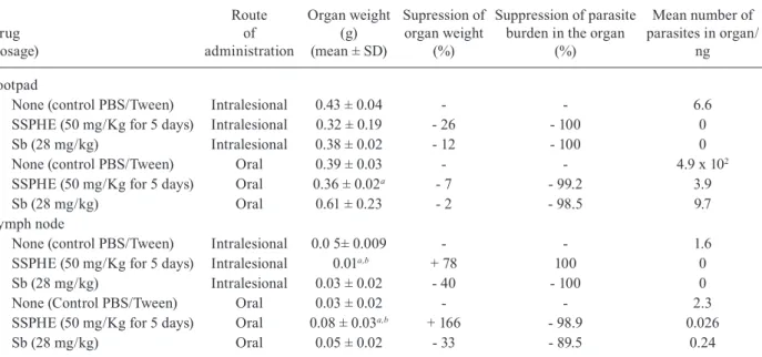
tralesional route. Oral treatment with SSPHE and Sb also induced a significant reduction in parasite burden at the infection site in comparison to the group that received PBS/Tween (99.2 and 98.5%, respectively). Both treat-ments through both administration routes induced a re-duction in the weight of the infected footpads in compari-son to the untreated group, especially in animals treated with SSPHE through the intralesional route (Table II).
In the popliteal draining lymph nodes, complete sup-pression of the parasite load occurred one week after treatment with SSPHE and Sb through the intralesional route. However, treatment with SSPHE induced an in-crease in the weight of these organs, while Sb treatment induced a 40% reduction in weight. Through the oral route, SSPHE also induced an increase in the weight, with a 98.9% reduction in the parasite load, whereas Sb treatment led to a reduction in lymph node weight, with an 89.5% reduction in the parasite load (Table II).
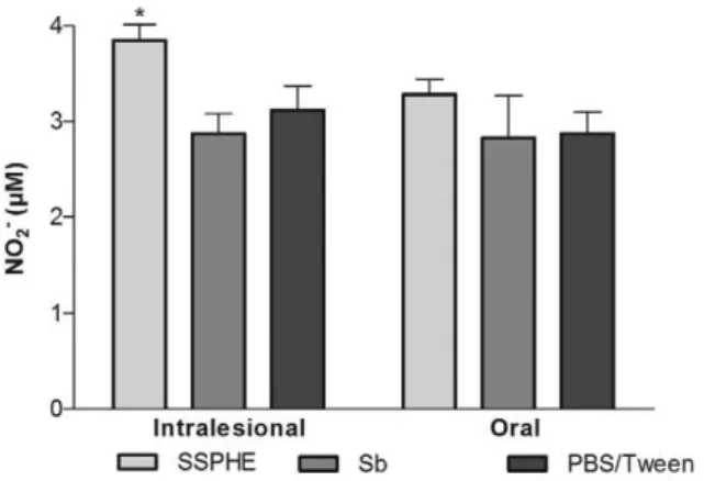
Effect of SSPHE on NO production - Treatment with SSPHE through the intralesional route induced a signifi-cant increase in NO production by peritoneal cells derived from infected animals in comparison to the group treated with Sb. Treatment with SSPHE through the oral route also induced an increase in NO production in comparison to the groups that received Sb and PBS/Tween, but this increase did not achieve statistical significance (Fig. 4).
Histopathological study - Footpads treated with SSPHE through the intralesional route revealed few cells with amastigotes. In contrast, numerous parasitised mac-rophages were observed in the control group (Fig. 5A, B). Infection associated with intralesional treatment resulted in intense inflammatory infiltrate composed of mononuclear cells and a few granulocytes. A few parasites were found in the footpads of animals that received SSPHE through the oral route (Fig. 5C). The inflammatory infiltrate in this case was composed of mononuclear cells. Animals treated with Sb by intralesional route showed nonparasitised tis-sue (Fig. 5E); by the oral route, however, several heavily infected macrophages were observed (Fig. 5F).
Fig. 3: kinetics of cutaneous lesion induced by Leishmania amazonen-sis after treatment with polar hydroethanolic extract from Selaginella sellowii (SSPHE) administered by the oral route (50 mg/kg/day dur-ing 20 days). Controls received N-methylglucamine antimonate (Sb) or phosphate-buffered saline (PBS)/Tween by the same route. Ham-sters were infected in the left hind footpad with L. amazonensis pro-mastigotes and treatment started four weeks after infection, ending seven weeks after infection. The data represent the mean ± standard deviation of 15 animals per group. *: p < 0.05 for SSPHE-treated vs. control animals (PBS/Tween); #: p < 0.05 for SSPHE-treated vs.
Sb-treated group. Student’s t test.
TABLE II
Effect of polar hydroethanolic extract from Selaginella sellowii (SSPHE) (50 mg/kg) administered by oral and intralesional routes in hamsters infected with Leishmania amazonensis one week after the end of treatment
Drug (dosage)
Route of administration
Organ weight (g) (mean ± SD)
Supression of organ weight
(%)
Suppression of parasite burden in the organ
(%)
Mean number of parasites in organ/
ng
Footpad
None (control PBS/Tween) Intralesional 0.43 ± 0.04 - - 6.6
SSPHE (50 mg/Kg for 5 days) Intralesional 0.32 ± 0.19 - 26 - 100 0
Sb (28 mg/kg) Intralesional 0.38 ± 0.02 - 12 - 100 0
None (control PBS/Tween) Oral 0.39 ± 0.03 - - 4.9 x 10²
SSPHE (50 mg/Kg for 5 days) Oral 0.36 ± 0.02a - 7 - 99.2 3.9
Sb (28 mg/kg) Oral 0.61 ± 0.23 - 2 - 98.5 9.7
Lymph node
None (control PBS/Tween) Intralesional 0.0 5± 0.009 - - 1.6
SSPHE (50 mg/Kg for 5 days) Intralesional 0.01a,b + 78 100 0
Sb (28 mg/kg) Intralesional 0.03 ± 0.02 - 40 - 100 0
None (Control PBS/Tween) Oral 0.03 ± 0.02 - - 2.3
SSPHE (50 mg/Kg for 5 days) Oral 0.08 ± 0.03a,b + 166 - 98.9 0.026
Sb (28 mg/kg) Oral 0.05 ± 0.02 - 33 - 89.5 0.24
a: p < 0.05 for treated vs. positive control [phosphate-buffered saline (PBS)/Tween]; b: p < 0.05 for treated when compared to
Fig. 4: effect of oral and intralesional treatment with polar hydroetha-nolic extract from Selaginella sellowii (SSPHE) (50 mg/kg) on nitric oxide (NO)production by peritoneal cells isolated from Leishmania amazonensis-infected hamsters. N-methylglucamine antimonate (Sb) and phosphate-buffered saline (PBS)/Tween were used as controls. The data represent the mean and ± standard deviation of four animals per group. Asterisk means p < 0.05 for SSPHE intralesional treatment vs. Sb intralesional treatment. Student’s t test.
DISCUSSION
Biflavonoids are a frequent class in the genus Selagi-nella and have been considered a chemical marker for this genus (Silva et al. 1995, Aguilar et al. 2013, Schulz et al. 2013). Recently, Rizk et al. (2014) have isolated the biflavonoids amentoflavone and robustaflavone from S. selowii. In the present work, four biflavonoids were identified in SSPHE. Amentoflavone (31.8 min) and ro-bustaflavone (32.4 min) are C-link flavone dimmers and the other minor biflavonoids are O-linked flavones (34.3 min hinokiflavone and 35.9 min OMe-hinokiflavone). Romani et al. (2002) have demonstrated that O-linked and methylated biflavonoids are more likely to be re-tained in a C-18 column than C-linked ones, what is in agreement with the observed retention times.
Other compounds detected in the extract were the caffeoyl-hexoside derivatives. The lower retention time of this class suggests the presence of polar groups in the molecules. Correlation of retention time with physico-chemical properties was demonstrated in several models (Tellez et al. 2009, Eugster et al. 2014). Caffeic acid linked to sugar groups has been described in the literature (Ham-erski et al. 2005) however the number of sugars is limited to three units. In the present work, compounds with four, five, or six units of hexose linked to four, five, or six units of caffeic acid were found in SSPHE. Partial structure de-termination was based on the fragments obtained from high resolution MS/MS spectrum; all compounds exhib-ited sequential losses of the hexose/caffeic acid moiety. Above described data together with the molecular formu-la allowed the putative identification of these compounds as caffeoyl-hexoside derivatives. This is the first relate of these compounds in the literature.
The in vitro antileishmanial activity of SSPHE on in-tracellular amastigotes was satisfactory and proved not to be cytotoxic to the mammalian cells tested (Rizk et al. 2014). Thus, the extract was used for in vivo testing.
It is important to note that SSPHE administered orally at a very high dose (2 g/Kg) did not cause acute toxicity in the animals (unpublished observations).
The treatment schedule in the present study was similar to that described by Fournet et al. (1996) and Patrício et al. (2008). It is important to note that no para-sitic forms were detected in the infection site or drain-ing lymph nodes usdrain-ing the limitdrain-ing dilution method in animals treated with SSPHE through the intralesional administration route, suggesting that the extract reduces the parasite load by 100%. The overall reduction in para-site load has been described by Arruda et al. (2009), who demonstrated the in vitro and in vivo antileishmanial activity of limonene, which is a cyclohexanoid monoter-pene found in the oil of citric plants.
We demonstrated that the intralesional injection of SSPHE reduced the parasite load in the infected footpads and draining lymph nodes, but also induced a signifi-cant, progressive increase in footpad thickness through-out treatment, whereas oral treatment with SSPHE led to a significant reduction in both parasite load and foot-pad thickness. The progressive increase in the footfoot-pad lesions may have resulted from a pro-inflammatory ef-fect induced by the intralesional injection of SSPHE, to-gether with the inflammatory response to the infection itself. This phenomenon has been described by Patrício et al. (2008), who also found a reduction in parasite bur-den despite the increase in footpad thickness, after the intralesional administration of a crude hydroalcoholic extract from Chenopodium ambrosioides (rich in flavo-noid and terpeflavo-noid compounds) in mice infected with L. (L.) amazonensis. In the present work, histopathological study corroborated this hypothesis, revealing that the intralesional treatment with SSPHE induced an intense inflammatory infiltrate composed mainly of mono-nuclear cells. It should be stressed, however, that foot-pads treated with an intralesional injection of SSPHE exhibited a significant reduction in thickness one week after the end of treatment in comparison to nontreated footpads, returning to values similar to those measured prior to infection. In this same timeframe, no signifi-cant differences were found between the Sb and SSPHE groups submitted to the intralesional route. However, no significant difference was found in footpads treated with orally administered Sb in comparison to untreated foot-pads, while a significant reduction in footpad thickness was found among those treated with orally administered SSPHE in comparison to controls.
Fig. 5: histopathological study of the site of infection in hamsters infected in the left hind footpad with Leishmania amazonensis promastigotes and treated with polar hydroethanolic extract from Selaginella sellowii (SSPHE) (50 mg/kg) by intralesional and oral routes (A, C). Vacuolated macro-phages with rare amastigotes are observed (arrows). The tissue fragments were obtained seven days after the end of treatments. Control nontreated group received phosphate-buffered saline/Tween by the same routes (B, D). There is a mononuclear infiltrate in the dermis, composed mainly of parasitised, vacuolated macrophages (arrows). Animals treated with N-methylglucamine by intralesional route showed nonparasitised tissue (E); by the oral route, several heavily infected macrophages (arrow) are observed (F). The figures are representative of five animals analysed in each group. Haematoxylin-eosin staining (A-F) 400X (A, E) and 1,000X (B-D, F) magnification.
time, increased interleukin (IL)-2, IL-12 and interferon-gamma cytokines were found, unlike decreased IL-4 and IL-10, evincing the T-helper 1 profile classically as-sociated with protection in leishmaniasis. This class of compounds could be also active against Leishmania, as established by Abdel-Mageed et al. (2012), who found phenylpropanoid glycosides active against Leishmania (Leishmania) donovani promastigotes.
The presence of biflavonoids and caffeoyl-hexoside de-rivatives in SSPHE suggests an immunomodulatory action from these compounds associated with the control of infec-tion. The increase in NO production by peritoneal cells iso-lated from animals treated with SSPHE by the intralesional route corroborates the immunomodulatory activity toward resistance to the parasite. The stimulation of NO production in murine macrophages infected with L. (L.) amazonensis has already been described elsewhere as an inhibitory ef-fect of a treatment of plant origin (Pereira et al. 2005). The increased NO production induced by SSPHE in vivo
cor-roborates the findings of Rizk et al. (2014), who showed the increase in NO production by peritoneal macrophages infected and treated by SSPHE in vitro.
Gupta et al. (1992) evaluated different experimental models for leishmaniasis and found that practically no treatment schedule provides adequate information for understanding the overall effectiveness of a potential antileishmanial drug, once it depends on the interaction between the parasite and the immune system. Indeed, it is well documented that the cure of animals from infec-tion occurs due to the combined effect of drug acinfec-tion and immunological status (Sacks et al. 1987).
cutane-ous leishmaniasis. Further studies with purified fractions have been carried out to establish which compound is re-sponsible for the immunomodulatory properties.
ACKNOWLEDGEMENTS
To Dr Arnildo Pott, for the identification of species, and Dr Vanessa Matos, from University Hospital, UFMS, for kindly providing the Glucantime® medication.
REFERENCES
Abdel-Mageed WM, Backheet EY, Khalifa AA, Ibraheim ZZ, Ross SA 2012. Antiparasitic antioxidant phenylpropanoids and iridoid glycosides from Tecoma mollis. Fitoterapia 83: 500-507. Aguilar MI, Mejia IA, Menchaca C, Vazquez I, Navarrete A, Chavez
MI, Reyes-García A, Rios-Gómez R 2013. Determination of bi-flavonoids in four Mexican species of Selaginella by HPLC. J AOAC Int96: 712-716.
Aguilar MI, Romero MG, Chávez MI, King-Díaz B, Lotina-Hennsen B 2008. Biflavonoids isolated from Selaginella lepidophylla in-hibit photosynthesis in spinach chloroplasts. J Agric Food Chem 56: 6994-7000.
Arruda DC, Miguel DC, Yasunaka JKUY, Katzin AM, Uliana SRB 2009. Inhibitory activity of limonene against Leishmania para-sites in vitro and in vivo. Biomed Pharmacother 63: 643-649. Ding AH, Nathan CF, Stuer DJ 1988. Release of reactive nitrogen
intermediates and reactive oxygen intermediates from mouse peritoneal macrophages: comparison of activating cytokines and evidence for independent production. J Immunol 141: 2407-2412. Eugster PJ, Boccard J, Debrus B, Breant L, Wolfender JL, Martel S,
Carrupt PA 2014. Retention time prediction for dereplication of natural products (CxHyOz) in LC-MS metabolite profiling. Phy-tochemistry 108: 196-207.
Ezatpour B, Dezaki ES, Mahmoudvand H, Azadpour M, Ezzatkhah F 2015. In vitroand in vivoantileishmanial effects of Pistacia khinjuk
against Leishmania tropica and Leishmania major. eCAM 2015: 6 pp. Fournet A, Ferreira ME, Arias AR, Ortiz ST, Fuentes S, Nakayama H,
Schinini A, Hocquemiller R 1996. In vivo efficacy of oral and intral-esional administration of 2-substituted quinolines in experimental treatment of new world cutaneous leishmaniasis caused by Leish-mania amazonensis. Antimicrob Agents Chemother 40: 2447-2451. Fournet A, Muñoz V 2002. Natural products as trypanocidal,
an-tileishmanial, and antimalarial drugs. Curr Top Med Chem 2: 1215-1237.
Grassi-Zampieron R, Franca LV, Carollo CA, Vieira MD, Oliveros-Bastidas A, Siqueira JM 2010. Comparative profiles of Achyro-cline alata (Kunth) DC. and A. satureioides (Lam.) DC., Asterace-ae, applying HPLC-DAD-MS. Rev Bras Farmacogn 20: 575-579. Gupta S, Zehra K, Nigam V, Katiyar JC 1992. Antileshmanial drug
testing: appraisal on existing techniques. Indian J Parasitol 16: 1-7. Hamerski L, Bomm MD, Silva DHS, Young MCM, Furlan M, Eberlin MN, Castro-Gamboa I, Cavalheiro AJ, Bolzani VS 2005. Phen-ylpropanoid glucosides from leaves of Coussarea hydrangeifolia
(Rubiaceae). Phytochemistry 66: 1927-1932.
Kunert O, Swamy RC, Kaiser M, Presser A, Buzzi S, Rao AVNA, Schu-hly W 2008. Antiplasmodial and leishmanicidal activity of biflavo-noids from Indian Selaginella bryopteris. Phytochem Lett 1: 171-174. Lee CW, Kim HS, Choi HJ, Kim JW, Kim HK, Moon HT, Lee SY,
Oh WK, Woo ER 2008. Biflavonoids isolated from Selaginella tamariscina regulate the expression of matrix metalloproteinase in human skin fibroblasts. Bioorg Med Chem16: 732-738. Lin RC, Skaltsounis A-L, Seguin E, Tillequin F, Koch M 1994. Phenolic
constituents of Selaginella doederleinii. Planta Med 60: 168-170.
Mitropoulos P, Konidas P, Durkin-Konidas M 2010. New World cuta-neous leishmaniasis: updated review of current and future diag-nosis and treatment. J Am Acad Dermatol 63: 309-322.
Patrício FJ, Costa GC, Pereira PVS, Aragão-Filho WC, Sousa SM, Frazão JB, Pereira WS, Maciel MCG, Silva LA, Amaral FMM, Rêbelo JMM, Guerra RNM, Ribeiro MNS, Nascimento FRF 2008. Eficacy of the intralesional treatment with Chenopodium ambrosioides in the murine infection by Leishmania amazonen-sis. J Ethnopharmacol 115: 313-319.
Pereira WKV, Lonardoni MVC, Grespan R, Caparroz-Assef SM, Cuman RKN, Bersani-Amado CA 2005. Immunomodulatory effect of Canova medication on experimental Leishmania ama-zonensis infection. J Infect 51: 157-164.
Pontin K, Silva Filho AS, Santos FS, Silva M, Cunha W, Nanayakkara N, Bastos S, Albuquerque S 2008. In vitro and in vivo antileish-manial activities of a Brazilian green propolis extract. Parasitol Res103: 487-492.
Reithinger R, Dujardin JD, Louzir H, Pirmez C, Alexander B, Brook-er S 2007. Cutaneous leishmaniasis. Lancet Infect Dis 7: 581-596. Rizk YS, Fischer A, Cunha MC, Rodrigues PO, Marques MCS, Matos
MFC, Kadri MCT, Carollo CA, de Arruda CCP 2014. In vitro activity of the hydroethanolic extract and biflavonoids isolated from Selaginella sellowii on Leishmania (Leishmania) amazo-nensis. Mem Inst Oswaldo Cruz 109: 1050-1056.
Romani A, Galardi C, Pinelli P, Mulinacci N, Heimler D 2002. HPLC quantification of flavonoids and biflavonoids in Cupressaceae leaves. Chromatographia 56: 469-474.
Sacks DL, Lal SL, Shrivastava SN, Blackwell J, Neva FA 1987. An analysis of T-cell responsiveness in Indian kala-azar. J Immunol 138: 908-913.
Santos DO, Coutinho CER, Madeira MF, Bottino CG, Vieira RT, Nascimento SB, Bernardino A, Bourguignon SC, Corte-Real S, Pinho RT, Rodrigues CR, Castro HC 2008. Leishmaniasis treat-ment - a challenge that remains: a review. Parasitol Res 103: 1-10. Schulz C, Homberg J, Stutzel T 2013. Taxonomic revision of
Selagi-nella subg. EricetorumSyst Bot 38: 5-14.
Sharma P, Rastogi S, Bhatnagar S, Shrivastava JK, Dube A, Guru PY, Kulshershtha DK, Dhawan BN 2003. Antileishmanial action of a plant Tephrosia pupurea Linn, extracts and its fractions against experimental visceral leishmaniasis. Drug Dev Res 60: 285-293. Silva GL, Chai HY, Gupta MP, Farnsworth NR, Cordell GA, Pezzuto
JM, Beecher CW, Kinghorn AD 1995. Cytotoxic biflavonoids from Selaginell willdenowii. Phytochemistry 40: 129-134. Sun C-M, Syu W-Jr, Huang Y-T, Chen C-C, Ou J-C 1997. Selective
cytotoxicity of ginkgetin from Selaginella moellendorffii. J Nat Prod 60: 382-384.
Tellez A, Roses M, Bosch E 2009. Modeling the retention of neutral compounds in gradient elution RP-HPLC by means of polarity parameter models. Anal Chem 81: 9135-9145.
Titus RG, Marchand M, Boon T, Louis JA 1985. A limiting dilution assay for quantifying Leishmania major in tissues of infected mice. Parasite Immunol 7: 545-555.
Weniger B, Vonthron-Se’ne’cheau C, Kaiser M, Brun R, Anton R 2006. Comparative antiplasmodial, leishmanicidal, and antitrypanoso-mal activities of several biflavonoids. Phytomedicine 13: 176-180. Zeng S, Wang DC, Cao YG, An N, Zeng FQ, Han CT, Song Y, Deng X