Antioxidant effect of 4-nerolidylcatechol
and
a
-tocopherol in erythrocyte ghost
membranes and phospholipid bilayers
K.S. Fernandes
1, A.H.M. Silva
1, S.A. Mendanha
1, K.R. Rezende
2and A. Alonso
11Instituto de Fı´sica, Universidade Federal de Goia´s, Goiaˆnia, GO, Brasil 2Laborato´rio de Biofarma´cia e Farmacocine´tica de Substaˆncias Bioativas, Faculdade de Farma´cia,
Universidade Federal de Goia´s, Goiaˆnia, GO, Brasil
Abstract
4-Nerolidylcatechol (4-NC) is found inPothomorphe umbellataroot extracts and is reported to have a topical protective effect against UVB radiation-induced skin damage, toxicity in melanoma cell lines, and antimalarial activity. We report a comparative study of the antioxidant activity of 4-NC anda-tocopherol against lipid peroxidation initiated by two free radical-generating
systems: 2,29-azobis(2-aminopropane) hydrochloride (AAPH) and FeSO4/H2O2, in red blood cell ghost membranes and in egg
phosphatidylcholine (PC) vesicles. Lipid peroxidation was monitored by membrane fluidity changes assessed by electron paramagnetic resonance spectroscopy of a spin-labeled lipid and by the formation of thiobarbituric acid-reactive substances. When lipoperoxidation was initiated by the hydroxyl radical in erythrocyte ghost membranes, both 4-NC anda-tocopherol acted
in a very efficient manner. However, lower activities were observed when lipoperoxidation was initiated by the peroxyl radical; and, in this case, the protective effect ofa-tocopherol was lower than that of 4-NC. In egg PC vesicles, malondialdehyde
formation indicated that 4-NC was effective against lipoperoxidation initiated by both AAPH and FeSO4/H2O2, whereasa
-tocopherol was less efficient in protecting against lipoperoxidation by AAPH, and behaved as a pro-oxidant for FeSO4/H2O2.
The DPPH (2,2-diphenyl-1-picrylhydrazyl) free-radical assay indicated that two free radicals were scavenged per 4-NC molecule, and one free radical was scavenged pera-tocopherol molecule. These data provide new insights into the antioxidant
capacity of 4-NC, which may have therapeutic applications for formulations designed to protect the skin from sunlight irradiation.
Key words: Electron paramagnetic resonance; 4-Nerolidylcatechol; Spin label; Lipid peroxidation; DPPH (2,2-diphenyl-1-picrylhydrazyl)
Introduction
The compound 4-nerolidylcatechol (4-NC) can be isolated from the root ofPothomorphe umbellata L. Miq. (1), a native plant of the Brazilian Atlantic Forest that is more commonly known as ‘‘pariparoba’’.P. umbellataroot extracts have demonstrated antioxidant activity against spontaneous brain lipid peroxidationin vitro, as evaluated by malondialdehyde (MDA) formation and chemilumines-cence emission. Part of this protection has been attributed to 4-NC (2). As an antioxidant,P. umbellataroot extracts and 4-NC have also demonstrated topical protective activities against UVB radiation-induced skin damage and prevented a-tocopherol depletion in the skin of hairless mice following UVB exposure (3,4).P. umbellata
root extracts, and, to a lesser extent, 4-NC can inhibit activity of the metalloproteinases MMP-2 and MMP-9 (5),
which are family members of a group of matrix-degrading enzymes, and whose synthesis is increased in response to UV irradiation. These metalloproteinases play impor-tant roles in morphogenesis, angiogenesis, arthritis, skin ulcer formation, tumor invasion, and metastasis (6,7). Furthermore, it has been shown that 4-NC is able to induce apoptosis in melanoma cell lines (8,9) and that 4-NC derivatives have significant antimalarial and cytotoxic potential (10,11).
Both vitamin E and 4-NC molecules are hydrophobic (Figure 1); thus, in its role as an antioxidant, 4-NC is likely to protect the cell membrane from lipid peroxidation. Lipid peroxidation is initiated by reactive oxygen species (ROS), such as hydroxyl radicals, (NOH), alkoxyl radicals (LON), and the peroxyl radical (LOON), which can abstract a
Correspondence: A. Alonso, Instituto de Fı´sica, Universidade Federal de Goia´s, 74001-970 Goiaˆnia, GO, Brasil. Fax: + +55-62-3521-1014. E-mail: [email protected]
hydrogen atom from the methylene group of an unsatu-rated fatty acid, forming a carbon-centered radical that combines with oxygen to form a peroxyl radical that is able to abstract another hydrogen atom (12,13). Peroxidation propagates until a chain-breaking antiox-idant such as vitamin E is able to stop the chain reaction or until two free radicals annihilate each other to terminate the chain reaction by forming cyclic peroxide (LOOL) (12). Extensive lipid peroxidation in the cell causes a loss of membrane fluidity, a decrease in membrane potential, and electrolyte leakage (12,14). Membrane stiffness associated with lipid peroxidation has been assessed by electron paramagnetic resonance (EPR) spectroscopy with spin labeling (14-17) and by the spectroscopy of fluorescent probes (18,19), which has proved to be very reliable in detecting the occurrence of lipid peroxidation.
We used EPR spectroscopy to evaluate the protective effects of 4-NC against lipid peroxidation initiated by the free radical-generating systems 2,29-azobis(2-aminopropane) hydrochloride (AAPH) or FeSO4/H2O2in isolated erythrocyte membranes. For comparison, the effect of a-tocopherol was evaluated in parallel. The antioxidant potentials of 4-NC anda-tocopherol were also tested by analyzing MDA formation in egg phosphatidylcholine (PC) vesicles and erythrocyte ghost membranes using the same initiators of lipid peroxidation.
Material and Methods
Chemicals
a-tocopherol, the 5-doxyl stearic acid (5-DSA) spin label, (++)-catechin (Figure 1), 2,2-diphenyl-1-picrylhydrazyl (DPPH), egg phosphatidylcholine, AAPH, iron sulfate, and hydrogen peroxide were purchased from Sigma (USA). All of the other chemicals were of the highest grade available and the buffers were prepared with a Milli-Q water system (Millipore, Germany).
Purification of 4-NC
P. umbellata roots were purchased from a herbarium
supply company (Flora Medicinal, Vale do Ribeira, SP, Brazil). The certified roots (1 kg) were dried, milled, and
exhaustively extracted by ultrasonication for 1 h with dichloromethane, yielding a crude extract (184.2 g) after solvent evaporation. The crude extract was initially chro-matographed [thin-layer chromatography (TLC); SiO2; dichloromethane:acetone, 99:1 (v/v)] in parallel with pre-vious authenticP. umbellataextracts showing a character-istic composition profile. Next, it was chromatographed on a flash-filtering silica gel 60 (0.063-0.2 mm, Merck, USA) column (906330 mm) and eluted with a hexane:ethyl acetate gradient elution. The apolar fractions that were obtained were recombined and submitted to a flash chromatography silica gel (0.063-0.2 mm, Merck) column (906330 mm) on a Bu¨chi Sepacore prep-MPLC System (Bu¨chi Labortechnik, Switzerland). The sample constituents were eluted with dichloromethane:cyclohexane:methanol (5:2:1, v/v/v) on a Sephadex LH-20 (GE Healthcare Bio-Sciences, Japan) column (496920 mm) at a flow rate of 40.0 mL/min and monitored at 220/280 nm (Bu¨chi UV detector model C-660). Fractions containing 4-NC were detected by TLC analysis and further purified on a Sephadex LH-20 GE column (496920 mm; 220 nm) to yield a pale yellow oil (1.2 g) that was chemically identified as 4-NC by [1H]- and [13C]-NMR. Sample purity was assessed with a peak purity analysis using high-performance liquid chromatography with a photodiode array detector. The chromatographic conditions were as follows: RP-C18 Phenomenex column (15064.6 mm; 4mm) using ACN:MeOH:H2O (54:20:26, v/v/v) as the mobile phase and a flow rate of 1.0 mL/min. The peak similarity at three peak spectra (upslope, downslope, and apex) gave values greater than or equal to 0.99.
DPPH assay
The free radical-scavenging activity of 4-NC was determined by its ability to reduce the free-radical DPPH following the method of Brand-Williams et al. (20). Small aliquots of ethanol solutions of 4-NC, a-tocopherol, or (++)-catechin were added to 75mM DPPH in methanol. These solutions were prepared daily. The total stoichio-metric value of n (the number of DPPH that each antioxidant molecule is able to reduce) for each antioxidant was calculated from the absorbance decay measurements at 515 nm after a 30-min incubation with the various solutions.
Preparation of liposomes
Egg PC was dissolved in a chloroform:methanol mixture (2:1, v/v). A lipid film was formed in a glass tube by evaporating the solvent with a nitrogen gas flux. The film was then kept under vacuum overnight to remove any residual organic solvent. To obtain uniform unilamellar vesicles, the lipid film was hydrated with an appropriate volume of Tris-HCl buffer (50 mM Tris, 0.1 mM diethylene triamine pentaacetic acid, 0.05 mM KCl, 0.35% HCl, pH 7.4) and then extruded through 0.4-mM pore size polycarbonate filters using a mini-extruder (Avanti Polar Lipids, Inc., USA). The extrusion process, consisting of Figure 1.Structure of (++)-catechin,a-tocopherol,
approximately 21 serial passes, was performed at room temperature (liquid crystalline phase).
Preparation of erythrocyte ghosts
Human blood, obtained from blood banks (unknown donors), was diluted in phosphate-buffered saline (PBS; 10 mM phosphate, 154 mM NaCl, pH 7.4) and centri-fuged at 200gfor 10 min at 46C. The plasma and white blood cells were carefully removed by aspiration after each wash (3 times). For the ghost preparation, the isolated red blood cells (RBCs) were incubated in lysis buffer (5 mM phosphate, pH 8.0) overnight. On the next day, the samples were centrifuged at 20,000gfor 10 min at 46C. To remove hemoglobin, the addition of a lysis buffer and subsequent centrifuging was repeated five times (without incubation). To recover the original shape of the erythrocyte membranes, the sample was centri-fuged in PBS twice. At the end of this procedure, concentrated erythrocyte membranes with a whitish color were obtained and stored at -206C until use.
Incorporation of antioxidants in membranes
Small aliquots of 4-NC ora-tocopherol in ethanol were placed in glass tubes, and the ethanol was evaporated using a nitrogen gas flux. Fifty microliters of egg PC liposomal membranes (initial concentration 16 mM) or erythrocyte ghost membranes (,4 mg protein/mL) was added to the antioxidant film. The membrane suspension was gently agitated and incubated for 1.5 h at 376C to allow complete incorporation of antioxidants into the membranes. Control measurements were performed before and after this period of incubation. In all of these experiments, concentrations of the antioxidants 4-NC and a-tocopherol ranged from 1 to 100mM.
Induction of lipid peroxidation
To induce oxidative stress, the egg PC liposomal membranes that were previously treated with different concentrations of antioxidant molecules (1 to 100mM) were added to a solution of 30 mM AAPH/Tris-HCl buffer or to a neutral solution of 0.1/1 mM FeSO4/H2O2 in 50 mM NaCl. The final concentration of egg PC liposomal membranes was 1.6 mM in both experiments. Similarly, erythrocyte ghost membranes with or without antioxidant (1 to 100mM) were added to a solution of AAPH in PBS or to a neutral solution of FeSO4+H2O2(erythrocyte ghost final concentration of 0.134 mg protein/mL). All of the samples in these four types of experiments were incubated for 3 h at 376C and were gently agitated every 30 min.
Assessment of antioxidant capacity of 4-NC and
a-tocopherol
The formation of thiobarbituric acid-reactive sub-stances (TBARS) was measured according to a previously described protocol (21). After the oxidation process, the
suspension of egg PC vesicles or erythrocyte ghost membranes was centrifuged at 20,000 g. Two milliliters of solution containing 15% trichloroacetic acid, 0.375% thiobarbituric acid, and 0.25 M HCl was added to the supernatant of each sample. Subsequently, the samples were heated at 956C in a water bath for approximately 30 min and rapidly cooled to stop the TBARS reaction. The suspension was centrifuged at 20,000 g and 256C for 10 min. The supernatant absorbance was read at 532 nm using a T80++ spectrophotometer (PG Instruments, England). MDA concentrations were calculated using a molar extinction coefficient of 1.566105?M-1?cm-1.
EPR spectroscopy
After the oxidative stress protocol, erythrocyte ghosts diluted in PBS were centrifuged at 20,000gand 46C for 10 min. A small aliquot (1mL) of spin label 5-DSA in ethanol solution (5 mg/mL) was added to the precipitated membranes (50mL). For EPR measurement, the ghost suspension was transferred to a capillary tube and flame-sealed. EPR measurements were performed using a Bruker ESP 300 spectrometer (Bruker, Germany) equipped with an ER 4102 ST resonator at room temperature (246-266C). The instrument settings were as follows: microwave power: 2 mW; modulation fre-quency: 100 kHz; modulation amplitude: 1.0 G; magnetic field scan: 100 G; sweep time: 168 s, and detector time constant: 41 ms. The EPR spectrum simulations were performed using the nonlinear least-squares fitting (NLLS) program developed by Budil et al. (22). In the spectral calculations, the NLLS program includes the magnetic
g- andA-tensors and the rotational diffusion tensor (R), which are expressed in a system of Cartesian axes fixed in the spin-labeled molecule. To reduce the number of parameters in the fittings and to simplify the simulation, the average rotational diffusion rate,Rbar, was calculated by the fitting program using the relationRbar=(Rper2Rpar)1/3, where Rper is the perpendicular and Rpar is the parallel component of the rotational diffusion (22). Rbar was converted to the parameter rotational correlation time, tc, following the relationship tc=1/6 Rbar. In this study, the spectra were simulated with a model of a single spectral component. Similar to previous studies (23,24), the magnetic parameters were determined based on a global analysis of the overall spectra obtained in this study, and all of the EPR spectra were simulated using the same predetermined parameters. The input parameters of tensorsAandgwereAxx: 7.0 G;Ayy: 7.2 G;Azz: 31.0 G;
gxx: 2.0083;gyy: 2.0060;gzz: 2.0027.
Results
DPPH radical-scavenging activity
and a-tocopherol, reaching a stable value of equilibrium after 5 min, compared with (++)-catechin, which reached equilibrium after approximately 10 min. 4-NC, which has two available hydroxyl groups, had a stoichiometric value of n=1.9±0.4 (mean±SD), which represents the reduc-tion of almost two DPPH molecules.a-tocopherol, which has only one available hydroxyl group, reduced more than one oxidant molecule (n=1.2±0.2). A previously reported explanation for this finding is that thea-tocopherol radical may undergo dimerization, and the newly formed molecule might then be able to reduce a second DPPH molecule (20). For comparison, we also measured the extensively studied scavenger (++)-catechin, which, due to its five available hydroxyl groups, showed the highest DPPH scavenging activity (n=4.3±1.4).
Antioxidant activity of 4-NC assessed by the TBARS assay
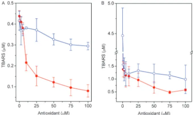
The protective effect of 4-NC and a-tocopherol on oxidative stress in egg PC vesicles and erythrocyte ghost membranes initiated by AAPH or FeSO4+H2O2 was evaluated using the TBARS assay. MDA production as a function of 4-NC concentration is shown in Figure 2 for egg PC vesicles and in Figure 3 for erythrocyte membranes. In egg PC vesicles, the amount of MDA formed with Fe(II)/H2O2was twice that formed with AAPH. For both oxidants, the level of MDA decreased gradually with the concentration of 4-NC. a-tocopherol showed a small protective effect against oxidation by AAPH, and, in the case of Fe(II)/H2O2,a-tocopherol was a pro-oxidant (Figure 2). In erythrocyte ghost membranes, the level of MDA formed with Fe(II)/H2O2 in the absence of an antioxidant was nearly triple that formed with AAPH (Figure 3). When lipoperoxidation was initiated by AAPH,
the level of MDA decreased slowly with increasing antioxidant concentrations (Figure 3A), and protection of a-tocopherol against lipoperoxidation was less efficient than the protection of 4-NC.
For membranes oxidized by Fe(II)/H2O2(Figure 3B), MDA formation decreased abruptly with increasing con-centration of both antioxidants. These results show that, in ghost membranes, 4-NC had a greater protective effect against attack by aqueous hydroxyl radicals, which are generated by the Fenton reaction, than against attack by peroxyl radicals, which arise from thermal decomposition of AAPH by its reaction with oxygen. At a concentration of 2.5mM 4-NC, the protection rate for hydroxyl radicals was approximately 95%, whereas the same 4-NC concentra-tion showed a maximum protecconcentra-tion of approximately 13% for peroxyl radicals.
Protective effect of 4-NC evaluated by EPR spectroscopy
EPR spectra of 5-DSA in RBC ghosts, as shown in Figure 4, show a reduction of membrane fluidity upon exposure to 50 mM AAPH for 3 h at 376C. The change in membrane fluidity was assessed by the spectral EPR parameter 2A// - the outer hyperfine splitting. This is a practice parameter that is measured directly in EPR spectra and has been widely used to monitor membrane fluidity, although, in principle, it is a static parameter associated with orientation distribution of spin labels in the membrane. Another EPR parameter obtained by spectral simulation, the rotational correlation time, has the advantage of taking into account the spectrum as a whole, and provided essentially the same results. For simplicity, only data obtained by 2A//are presented in this study. Membrane alteration was completely prevented by
Figure 2. Effect of 4-NC (filled circles) anda-tocopherol (open circles) on MDA production in egg PC vesicles oxidized with AAPH (A) or FeSO4+H2O2(B). Egg PC vesicles (1.6 mM) were incubated with several concentrations of 4-NC for 1.5 h at 376C. Lipid peroxidation was initiated by a 3-h incubation with 30 mM AAPH at 376C or a 1-h incubation with 0.1/1 mM FeSO4/H2O2at 376C. 4-NC: 4-nerolidylcatechol; MDA: malondialdehyde; AAPH: 2,29-azobis(2-aminopropane)hydrochloride;PC:phosphatidylcholine.
preincubation with the antioxidant 4-NC for 1.5 h at 376C (Figure 4C). The EPR spectra of 5-DSA in RBC ghosts were not altered in the presence of antioxidants 4-NC or a-tocopherol up to a concentration of 100mM. A small effect in membrane fluidity was observed for these compounds at concentrations approximately 10 times higher.
The 2A// value increased with increasing concentra-tions of AAPH in the RBC ghost suspension and reached a maximum level of approximately 2 G at a concentration of approximately 50 mM AAPH (Figure 5A). Interestingly, the results from the MDA test showed a behavior similar to that of the EPR 2A// parameter. Based on the plot shown in Figure 5A, 50 mM AAPH was chosen to induce lipoperoxidation in this system. As shown in Figure 5B, preincubation with increasing concentrations of the antioxidant 4-NC completely prevented the change in membrane fluidity due to lipid peroxidation, whereas the
corresponding preincubation with a-tocopherol provided only partial protection. A decrease of 2 G in the parameter 2A// relative to the oxidized sample was observed with approximately 50mM 4-NC, whereas the maximum decrease in this spectral parameter fora-tocopherol was only 1 G.
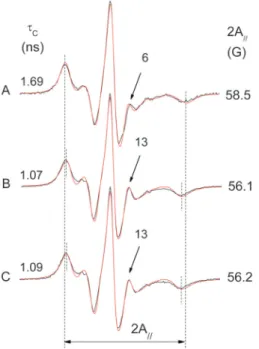
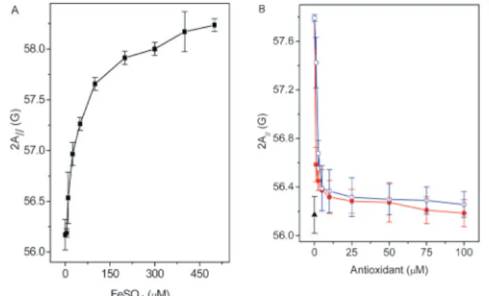
Figure 6A-C shows the EPR spectra of 5-DSA in RBC ghosts for three samples: oxidized with 200mM/2 mM FeSO4/H2O2 (spectrum A), preincubated with 100mM 4-NC for 1.5 h at 376C and then oxidized in the same manner (spectrum B), and a non-oxidized control sample (spectrum C). The EPR parameter 2A//of 5-DSA for the oxidized RBC membranes increased by almost 2 G (spectrum A), and 100mM 4-NC was able to prevent the corresponding reduction of membrane fluidity (spectrum C). The iron concentration dependence of 2A// for the 5-DSA probe in RBC ghost suspension containing 2 mM H2O2 is shown in Figure 7A. The maximum 2A// value observed (approximately 2 G) is typical for lipoperoxida-tion (14-16). A concentralipoperoxida-tion of 200mM/2 mM FeSO4/ H2O2was chosen to examine the antioxidant effects of 4-NC and a-tocopherol as a function of their concentra-tion (Figure 7B). In this case, very small concentraconcentra-tions of both antioxidants were sufficient to almost completely prevent the change in membrane fluidity caused by lipid peroxidation.
Discussion
Several studies have assessed the degree of lipid peroxidation using EPR spectroscopy of spin-labeled Figure 4.Experimental (black line) and best-fit (red line) EPR
spectra of 5-DSA in erythrocyte ghost membranes:A, oxidized with 50 mM AAPH;B, control sample (not oxidized);C, a sample pre-treated with 100mM 4-NC and then oxidized (50 mM AAPH). The EPR parameter 2A//represents the separation in magnetic-field units between the first and last resonance lines of the spectrum. The vertical lines indicate the 2A//for the oxidized sample (the total magnetic field scan range of the spectrum was 100 G). The values of both the 2A//and the rotational correlation time (tC) parameters (shown in the figure for each spectrum) were greater for the oxidized sample, indicating a reduction in molecular dynamics or increased molecular order. Another indication of this loss of probe mobility in the oxidized sample is the smaller relative intensity of the resonance line, which is indicated by an arrow (and intensity value) in each spectrum. The estimated experimental errors for 2A//andtC are 0.5 G and 0.2 ns, respectively. EPR: electron paramagnetic resonance; AAPH: 2,29-azobis(2-aminopropane) hydrochloride; 4-NC: 4-nerolidylcatechol.
lipids incorporated into membranes. For instance, the chilling stress in coffee seedlings caused by exposure of the plant to 106C for 6 days in darkness led to membrane stiffness of the plant root, growth inhibition, changes in metabolic rates, and MDA formation. These alterations were interpreted to be a result of lipid peroxidation (14,16). The increase in the 2A// parameter of 5-DSA measured directly in intact root-tip segments was approxi-mately 2 G for coffee seedlings exposed to chilling stress. A similar increase in the 2A// value, generated by iron-induced lipoperoxidation in the mitochondrial membrane, was prevented by 25mM dipyridamole, a coronary vasodilator (15). These data are consistent with the maximum increase in the 2A//parameter of approximately 2 G observed here for ghost membranes oxidized with AAPH or Fe(II)/H2O2. Membrane rigidity in rat liver microsomes, as assessed by fluorescence spectroscopy, has also been associated with lipid peroxidation that is induced by combining FeCl3-ADP-NADPH (19) or Fe2++ -ADP-ascorbic acid (18). EPR spectroscopy of the spin label 5-DSA was used to monitor cell membrane fluidity in the human hepatocellular carcinoma cell line HepG2 in a
study that showed that tea catechins can protect cells from lead-induced lipid peroxidation, decreased mem-brane fluidity, and cell viability (17). In the memmem-brane model of sonicated soybean phospholipid vesicles, the effect of lipid peroxidation with iron/ascorbate on mem-brane fluidity was dependent on the nitroxide position along the fatty-acid chain. Maximum rigidity was observed for the positional isomers 10- and 12-DSA (25).
The EPR spin-labeling technique is a reliable comple-mentary method to assess the degree of lipid peroxidation in cell membranes. Lipid peroxidation causes a reduction in membrane fluidity, which is well characterized by EPR spectra. Cell membrane fluidity can be modified by the addition of hydrophobic molecules in the millimolar concentration range that are distributed throughout the bilayer. On a time scale of fast rotational motion, these molecules generally destabilize the lipid tail packing and weaken the hydrogen-bonded network of the polar inter-face. The cases of molecules with opposite effects in the membrane are less common. For example, cholesterol, when added (millimolar range) into the membrane, facil-itates lipid packing, generating molecular order or decreased fluidity, which is detectable by the spin label method. In our study systems, the oxidant and the antioxidant compounds were added to the membrane in micromolar concentrations, with the exception of AAPH (100 mM), which is a water-soluble molecule that does not alter membrane fluidity. The present study also suggests that EPR spectroscopy can be useful to assess lipid peroxidation in more complex systems, such as cells, as Figure 6.Experimental (black line) and best-fit (red line) EPR
spectra of 5-DSA in erythrocyte ghost membranes:A, oxidized with 0.2/2 mM FeSO4/H2O2; B, pre-treated with 100mM 4-NC and then oxidized;C, control sample (non-oxidized). The vertical lines help to visualize the EPR parameter 2A//, the value of which is greater for the oxidized sample, indicating a more rigid membrane. The rotational correlation time (tC) increased and the relative intensity of the resonance lines, indicated by arrows, decreased with increasing membrane rigidity. EPR: electron paramagnetic resonance; 5-DSA: 5-doxyl stearic acid; 4-NC: 4-nerolidylcatechol.
has been performed previously in the root tip segments of coffee seedlings (14,16).
Our results show that in erythrocyte ghost mem-branes, very small concentrations ofa-tocopherol or 4-NC were sufficient to prevent the lipid peroxidation caused by hydroxyl radicals, but when the lipid peroxidation was induced by peroxyl radicals, the antioxidants, especially a-tocopherol, were much less effective. It has been demonstrated that the hydrophilic radicals generated from AAPH attack the protein component of the RBC mem-brane, and thata-tocopherol can inhibit lipid peroxidation. However, it does not prevent protein oxidation (26,27). Protein degradation could hinder protection against lipid peroxidation for these lipid-soluble antioxidants. However, our results fora-tocopherol in egg PC vesicles and ghost membranes were quite different.a-tocopherol showed a pro-oxidant effect in egg PC vesicles when subjected to oxidation by the Fe(II)/H2O2system (Figure 2B), whereas, for 4-NC, the results for the two membranes were consistent. Yamamoto and Niki (28) have shown that a-tocopherol incorporated into soybean vesicles reduces the ferric ion to the more reactive ferrous ion and that a-tocopherol may act either as an antioxidant or as a pro-oxidant depending on the experimental conditions. Consistent with our findings, the authors observed that a-tocopherol incorporated into intact erythrocyte mem-branes did not reduce ferric ion in the aqueous phase. Importantly, the erythrocyte membrane is more rigid than the egg PC bilayer, and thus a-tocopherol should find greater stability in the erythrocyte membrane, having less ability to fluctuate on the membrane to reach the ferric ions in the aqueous phase.
Interestingly, antioxidants also behave as pro-oxidants under certain conditions, as explained above. Recently, it was demonstrated that 4-NC is a cytotoxic compound with the capacity to induce apoptosis in metastatic melanoma cell lines (8). The main mechanisms of action suggested for this apoptosis-inducing activity were the formation and accumulation of ROS, leading to DNA damage, induction of the tumor suppressor p53, an increase in the pro-apoptotic protein Noxa, and caspase-dependent apoptosis (9). However, we believe that further studies are necessary to demonstrate that the formation of ROS by 4-NC is the cause, rather than a consequence, of induced apoptosis in melanoma cells. P. umbellata
extracts have also been shown to possess cytotoxic activity, with a high potency for growth inhibition in human cancer cell lines and antitumor activity in Ehrlich ascites carcinoma in male Swiss mice (29).
The physiological function of a-tocopherol as an antioxidant has recently been the topic of polemic reviews (30-32). The ability ofa-tocopherol to act as an antioxidant in vivo has been questioned. In fact, a-tocopherol has been shown to function as a cell-signaling agent (30). Other authors argue that as a chain-breaking antioxidant, vitamin E acts only as the main protector
against in vivo lipid peroxidation (31,32). An important feature ofa-tocopherol is its ability to act as an antioxidant at very low concentrations in biological membranes. Atkinson et al. (32) reviewed the amount ofa-tocopherol reported for several membranes, as expressed in the molar percentage relative to the amount of phospholipids in each membrane. Measurements of a-tocopherol in the mem-branes of rat liver mitochondria, human platelets, rat lung, rat brain cortex, and rat liver microsomes ranged from 0.1 to 1.0 mol%. Considering that the phospholipid concentra-tion in RBC membranes is 0.76 mg/mg protein (33), our data showed that both 4-NC anda-tocopherol have a large protective effect at 0.1 mol% against 200mM/2 mM FeSO4/H2O2(Figure 7B), whereas an appreciable protec-tion from AAPH was observed at 0.4 mol% 4-NC, and a moderate level of protection from AAPH was observed at 2.5 mol%a-tocopherol (Figure 5B). This finding indicates that ourin vitroresults are consistent with thein vivodata reported fora-tocopherol activity in terms of its concentra-tion in the membrane (32).
The irradiation of human skin with solar-simulated ultraviolet light depletes a-tocopherol from the stratum corneum, the outermost skin layer (34). At doses below the 0.75 minimal erythema dose, the amount of a-tocopherol was depleted by almost 50% in human stratum corneum and by 85% in murine stratum corneum (34). These authors (34) suggested that the high susceptibility ofa-tocopherol to ultraviolet radiation might be partially due to a lack of co-antioxidants in the stratum corneum (35). P. umbellata root extract gel, containing 0.1% 4-NC prevented a-tocopherol depletion in the skin of hairless mice following ultraviolet irradiation (3,4). In response to UVB radiation in the skin of hairless mice, MMP-2 and -9 were inhibited in the presence of 4-NC (5). Recently, the single application of a topical a-tocopherol-enriched rinse-off product led to significantly increased levels of vitamin E in the stratum corneum, and contributed to the protection of human skin against lipid peroxidationin vivo(36). These findings suggest that 4-NC could be used in topical formulations for treatment of human melanomas or as a topical agent to protect the skin from sunlight irradiation. Due to its hydrophobic nature, 4-NC can be easily incorporated into the intercellular membranes of the stratum corneum and can be associated with fluidity and permeation enhancers such as the monoterpenes 1,8-cineole, limonene, and a-terpineol (23,24), which are low in toxicity and could serve as a delivery system for this compound to the epidermis.
a protective effect against hydroxyl and peroxyl radicals, whereas a-tocopherol had a minor protective effect against peroxyl radicals and had a pro-oxidant effect when lipoperoxidation was induced by the free radicals generated by iron/hydrogen peroxide. As a free-radical scavenger, 4-NC displayed a greater ability than a -tocopherol to reduce DPPH molecules: 4-NC was able to reduce two free radicals, whereas only one free radical was reduced bya-tocopherol.
Acknowledgments
The authors are grateful to INGOH, the Goiano Institute of Oncology and Hematology and Hemolabor, and the Clinical Analysis Laboratories for supplying the blood. Research supported by CNPq, CAPES and FAPEG. K.S. Fernandes, S.A. Mendanha and A.H.M. Silva are recipients of fellowships from CAPES. A. Alonso gratefully acknowledges the CNPq for a research grant.
References
1. Kijjoa A, Giesbrecht AM, Akissue MK, Gottlieb OR, Gottlieb HE. 4-Nerolidylcatechol from Pothomorphe umbellata. Planta Med1980; 39: 85-87, doi: 10.1055/s-2008-1074908. 2. Barros SBM, Teixeira DS, Aznar AE, Moreira JA Jr, Ishiiand I, Freitas PCD. Antioxidant activity of ethanolic extracts of Pothomorphe umbellata.Cienc Cult1996; 48: 114-116. 3. Ropke CD, Meirelles RR, da Silva V, Sawada TC,
Barros SB. Pothomorphe umbellata extract prevents alpha-tocopherol depletion after UV-irradiation.Photochem Photobiol 2003; 78: 436-439, doi: 10.1562/0031-8655(2003)078,0436:PUEPTD.2.0.CO;2.
4. Ropke CD, Sawada TC, da Silva V, Michalany NS, de Moraes Barros SB. Photoprotective effect ofPothomorphe umbellata root extract against ultraviolet radiation induced chronic skin damage in the hairless mouse.Clin Exp Dermatol2005; 30: 272-276, doi: 10.1111/j.1365-2230.2005.01749.x.
5. Ropke CD, da Silva V, Kera CZ, Miranda DV, de Almeida RL, Sawada TC, et al.In vitroandin vivoinhibition of skin matrix metalloproteinases byPothomorphe umbellataroot extract. Photochem Photobiol 2006; 82: 439-442, doi: 10.1562/2005-06-29-RA-596.
6. Birkedal-Hansen H. Proteolytic remodeling of extracellular matrix.Curr Opin Cell Biol1995; 7: 728-735, doi: 10.1016/ 0955-0674(95)80116-2.
7. Inomata S, Matsunaga Y, Amano S, Takada K, Kobayashi K, Tsunenaga M, et al. Possible involvement of gelatinases in basement membrane damage and wrinkle formation in chronically ultraviolet B-exposed hairless mouse.J Invest Dermatol 2003; 120: 128-134, doi: 10.1046/j.1523-1747.2003.12021.x.
8. Brohem CA, Sawada TC, Massaro RR, Almeida RL, Rivelli DP, Ropke CD, et al. Apoptosis induction by 4-nerolidylcatechol in melanoma cell lines. Toxicol In Vitro 2009; 23: 111-119, doi: 10.1016/j.tiv.2008.11.004. 9. Brohem CA, Massaro RR, Tiago M, Marinho CE, Jasiulionis
MG, de Almeida RL, et al. Proteasome inhibition and ROS generation by 4-nerolidylcatechol induces melanoma cell death.Pigment Cell Melanoma Res2012; 25: 354-369, doi: 10.1111/j.1755-148X.2012.00992.x.
10. Pinto AC, Silva LF, Cavalcanti BC, Melo MR, Chaves FC, Lotufo LV, et al. New antimalarial and cytotoxic 4-nerolidylcatechol derivatives. Eur J Med Chem 2009; 44: 2731-2735, doi: 10.1016/j.ejmech.2008.10.025.
11. Rocha E Silva LF, Silva Pinto AC, Pohlit AM, Quignard EL, Vieira PP, Tadei WP, et al.In vivoandin vitroantimalarial activity of 4-nerolidylcatechol. Phytother Res 2011; 25: 1181-1188, doi: 10.1002/ptr.3424.
12. Gutteridge JM. Lipid peroxidation and antioxidants as
biomarkers of tissue damage.Clin Chem1995; 41: 1819-1828.
13. Moon JK, Shibamoto T. Antioxidant assays for plant and food components.J Agric Food Chem2009; 57: 1655-1666, doi: 10.1021/jf803537k.
14. Alonso A, Queiroz CS, Magalhaes AC. Chilling stress leads to increased cell membrane rigidity in roots of coffee (Coffea arabicaL.) seedlings. Biochim Biophys Acta1997; 1323: 75-84, doi: 10.1016/S0005-2736(96)00177-0.
15. Nepomuceno MF, Alonso A, Pereira-da-Silva L, Tabak M. Inhibitory effect of dipyridamole and its derivatives on lipid peroxidation in mitochondria.Free Radic Biol Med1997; 23: 1046-1054, doi: 10.1016/S0891-5849(97)00135-4. 16. Queiroz CGS, Alonso A, Mares-Guia M, Magalha˜es AC.
Chilling-induced changes in membrane fluidity and antioxidant enzyme activities inCoffea arabicaL. roots.Biol Plantarum 1998; 41: 403-413, doi: 10.1023/A:1001802528068. 17. Chen L, Yang X, Jiao H, Zhao B. Tea catechins protect
against lead-induced cytotoxicity, lipid peroxidation, and membrane fluidity in HepG2 cells.Toxicol Sci 2002; 69: 149-156, doi: 10.1093/toxsci/69.1.149.
18. Choe M, Jackson C, Yu BP. Lipid peroxidation contributes to age-related membrane rigidity. Free Radic Biol Med 1995; 18: 977-984, doi: 10.1016/0891-5849(94)00217-8. 19. Garcia JJ, Reiter RJ, Karbownik M, Calvo JR, Ortiz GG, Tan
DX, et al. N-acetylserotonin suppresses hepatic microsomal membrane rigidity associated with lipid peroxidation.Eur J Pharmacol 2001; 428: 169-175, doi: 10.1016/S0014-2999(01)01342-5.
20. Brand-Williams W, Cuvelier ME, Berset C. Use of a free radical method to evaluate antioxidant activity. Lebensm-Wiss u-Technol1995; 28: 25-30.
21. Gilbert HS, Stump DD, Roth EF Jr. A method to correct for errors caused by generation of interfering compounds during erythrocyte lipid peroxidation.Anal Biochem 1984; 137: 282-286, doi: 10.1016/0003-2697(84)90086-1. 22. Budil DE, Lee S, Saxena S, Freed JH.
Nonlinear-least-squares analysis of slow-motion EPR spectra in one and two dimensions using a modified Levenberg-Marquardt algorithm. J Magn Reson A 1996; 120: 155-189, doi: 10.1006/jmra.1996.0113.
23. dos Anjos JL, de Sousa ND, Alonso A. Effects of ethanol/l-menthol on the dynamics and partitioning of spin-labeled lipids in the stratum corneum.Eur J Pharm Biopharm2007; 67: 406-412, doi: 10.1016/j.ejpb.2007.02.004.
103-112, doi: 10.1016/j.ijpharm.2007.08.024.
25. Bruch RC, Thayer WS. Differential effect of lipid peroxida-tion on membrane fluidity as determined by electron spin resonance probes.Biochim Biophys Acta1983; 733: 216-222, doi: 10.1016/0005-2736(83)90525-4.
26. Miki M, Tamai H, Mino M, Yamamoto Y, Niki E. Free-radical chain oxidation of rat red blood cells by molecular oxygen and its inhibition by alpha-tocopherol. Arch Biochem Biophys 1987; 258: 373-380, doi: 10.1016/0003-9861(87)90358-4. 27. Zou CG, Agar NS, Jones GL. Oxidative insult to human red
blood cells induced by free radical initiator AAPH and its inhibition by a commercial antioxidant mixture. Life Sci 2001; 69: 75-86, doi: 10.1016/S0024-3205(01)01112-2. 28. Yamamoto K, Niki E. Interaction of alpha-tocopherol with
iron: antioxidant and prooxidant effects of alpha-tocopherol in the oxidation of lipids in aqueous dispersions in the presence of iron.Biochim Biophys Acta1988; 958: 19-23, doi: 10.1016/0005-2760(88)90241-X.
29. Sacoman JL, Monteiro KM, Possenti A, Figueira GM, Foglio MA, Carvalho JE. Cytotoxicity and antitumoral activity of dichloromethane extract and its fractions fromPothomorphe umbellata. Braz J Med Biol Res 2008; 41: 411-415, doi: 10.1590/S0100-879X2008000500010.
30. Azzi A. Molecular mechanism of alpha-tocopherol action. Free Radic Biol Med 2007; 43: 16-21, doi: 10.1016/j.
freeradbiomed.2007.03.013.
31. Traber MG, Atkinson J. Vitamin E, antioxidant and nothing more.Free Radic Biol Med 2007; 43: 4-15, doi: 10.1016/ j.freeradbiomed.2007.03.024.
32. Atkinson J, Epand RF, Epand RM. Tocopherols and tocotrienols in membranes: a critical review. Free Radic Biol Med 2008; 44: 739-764, doi: 10.1016/j.freerad-biomed.2007.11.010.
33. Younsi M, Quilliot D, Al-Makdissy N, Delbachian I, Drouin P, Donner M, et al. Erythrocyte membrane phospholipid composition is related to hyperinsulinemia in obese non-diabetic women: effects of weight loss.Metabolism2002; 51: 1261-1268, doi: 10.1053/meta.2002.35184.
34. Thiele JJ, Traber MG, Packer L. Depletion of human stratum corneum vitamin E: an early and sensitivein vivomarker of UV induced photo-oxidation.J Invest Dermatol1998; 110: 756-761, doi: 10.1046/j.1523-1747.1998.00169.x.
35. Packer L. Vitamin E is nature’s master antioxidant.Sci Med 1994; 1: 54-63.