Radiology Case. 2015 Apr; 9(4):14-22
Jo
u
rna
l o
f
Ra
d
io
lo
g
y
Ca
se
Re
p
o
rt
s
w
w
w
.Ra
di
ol
ogyC
as
es
.c
om
14
Placental site trophoblastic tumor:
a case report and review of the literature
Rita Lucas
1*, Teresa Margarida Cunha
2, Filipa Batista Santos
31. Department of Radiology, Hospital de Santo António dos Capuchos, CHLC, Lisboa, Portugal
2. Department of Radiology, Instituto Português de Oncologia de Lisboa Francisco Gentil, Lisboa, Portugal
3. Department of Pathology, Instituto Português de Oncologia de Lisboa Francisco Gentil, Lisboa, Portugal
* Correspondence:Rita Lucas, Alameda Santo António dos Capuchos, 1169-050 Lisboa, Portugal ( ritalucas1@gmail.com)
Radiology Case. 2015 Apr; 9(4):14-22 :: DOI: 10.3941/jrcr.v9i4.2146
ABSTRACT
We present a case of a gravida 1 para 1 woman, who presented with an 11-month history of amenorrhea after cesarean delivery. The patient was taking birth control pills at the time of presentation. She was observed with a slight elevation of serum β-hCG level, an enlarged heterogeneous uterus and hematometra. A biopsy was performed, and the patient was diagnosed with placental site trophoblastic tumor; the patient then underwent surgery. Placental site trophoblastic tumor is the rarest form of gestational trophoblastic disease, derived from intermediate trophoblast cells. It does not have a pathognomonic appearance; therefore, correlation with medical history, as well as results of laboratory tests and pathological analysis is mandatory. It is a relatively chemoresistant tumor, posing considerable therapeutic challenges; patients with localized disease are managed with surgery and those with metastatic disease require additional chemotherapy. Herein, we review the main features of this entity and top differential diagnosis, as the rarity of this tumor is associated with imaging and pathological pitfalls, reinforcing the need for further experience in this field.
CASE REPORT
A 34-year-old gravida 1 para 1 woman, presented with amenorrhea after her recent delivery via caesarean section 11 months previously. Amenorrhea was initially attributed to progestin-only contraceptives (desogestrel, 75µg, daily) that the patient had been consuming during 6 months of breastfeeding. Despite ceasing hormonal therapy and changing to combined estrogen-progestin contraceptive pills (drospirenone [3mg] and ethinyloestradiol [0.03mg] daily for 21 days followed by a 7 day break), which the patient had been consuming prior to pregnancy and was comfortable with, her amenorrhea persisted.
Gynecologic examination was unremarkable and physical examination revealed only right lower quadrant abdominal pain.
Serum β-hCG (human chorionic gonadotropin) level was slightly elevated at 68.4mIU/mL, and all other routine laboratory test results were within normal limits.
Imaging findings
Radiology Case. 2015 Apr; 9(4):14-22
Jo
u
rna
l o
f
Ra
d
io
lo
g
y
Ca
se
Re
p
o
rt
s
w
w
w
.Ra
di
ol
ogyC
as
es
.c
om
15
posterior axes. The endometrial cavity was filled with fluid and debris (hematometra) and no endometrial thickening was identified. No myometrial nodular lesions were observed, although the myometrial echotexture was diffusely heterogeneous. Both ovaries were normal.
Pelvic magnetic resonance (MR) imaging revealed a normal diffuse low-intensity myometrial signal on T1-weighted images, and moderate hyperintensity of the uterine myometrium, with loss of zonal uterine anatomy, on T2-weighted images. (Fig. 2A-2C). On dynamic T1-T2-weighted post-contrast images, the uterine body moderately enhanced, although to a lesser degree than the cervix (Fig. 2D) and it showed a high signal on diffusion-weighted images with a low signal on the ADC map (Fig. 3).
Management
The initial endometrial suction curettage specimen was not adequate for histological or immunohistochemical characterization. Therefore, endometrial sampling via resectoscopy was performed, which was complicated by uterine wall perforation.
A laparotomy was immediately performed to thoroughly evaluate the abdominal and pelvic cavities, and a palpable uterine wall lesion was detected and biopsied. The histological examination revealed intermediate trophoblastic cells with scant hyalinization, moderate atypia, and sparse hemorrhage.
The immunohistochemical evaluation was positive for α -inhibin, cytokeratin CAM 5.2, placental lactogenic hormone, and p63; it was only focally positive for β-hCG, with a Ki-67 index of 9%, supporting the diagnosis of placental site trophoblastic tumor (PSTT).
The patient underwent hysterectomy with ovarian conservation and bilateral pelvic lymphadenectomy.
The final pathology report confirmed the diagnosis of PSTT, revealing an enlarged uterus measuring 9.5cm along the long axis. Most of the uterine body was substituted by a yellowish tumor that infiltrated almost the entire myometrial thickness, without invading the cervix (Fig. 4). The serosa was intact, and no lymph node metastases were observed. The endometrial cavity was irregularly delineated and filled with blood clots. Microscopic evaluation revealed intermediate trophoblastic cells with moderate nuclear pleomorphism invading the myometrium (Fig. 5).
Follow-up
One month after surgery, the β-hCG level was <1mIU/mL.
At the time of the study, the patient had been in remission for 9 months without adjuvant chemotherapy, and she was undergoing monthly serum β-hCG follow-up evaluations.
Etiology and demographics
Gestational trophoblastic disease represents a wide spectrum of proliferative disorders characterized by proliferation of trophoblasts, including premalignant and malignant lesions [1].
PSTT is a slow-growing malignant monomorphic tumor that arises from placental intermediate trophoblasts, which are responsible for embryo implantation, and have features of both cytotrophoblasts and syncytiotrophoblasts [2,3].
Kurman and Scully first described this tumor it in 1976, under the term trophoblastic pseudotumor in accordance with the apparent benign nature of this lesion [4]. With further evidence of its malignant behavior, the nomenclature was changed to PSTT in 1981, and it has since then been adopted by the World Health Organization [5].
PSTT is the rarest subtype of gestational trophoblastic disease, with an incidence of approximately 1-5 per 100,000 pregnancies, accounting for approximately 0.2% of all cases, as suggested by population-based studies [1,6,7].
Because of the rarity of this type of tumor, the current medical knowledge is mainly based on the results of a few published trophoblastic disease center series [1,6-12] and sparse reports of small series or singular cases [13-16].
PSTT mostly affects women of reproductive age (the average age at presentation is 32-35 years) [1,11,12], and it is usually diagnosed following a normal or ectopic pregnancy, miscarriage, or gestational trophoblastic disease [6,7,12]. However, its occurrence in post-menopausal women has also been described [6].
Risk factors for PSTT are still not well understood.
Clinical presentation
The most common presenting symptom is irregular vaginal bleeding, followed by abdominal pain and amenorrhea [1]. Less frequent presentations include galactorrhea, virilization, nephrotic syndrome, polycythemia and symptoms related to the metastatic involvement of distant organs including the brain and lungs [6].
Laboratory and pathological diagnosis
In most patients with PSTT, serum levels of β-hCG are not highly elevated, which differs from other forms of gestational trophoblastic disease, as this tumor originates from intermediate trophoblastic cells. On the other hand, the expression of human placental lactogen (hPL) is usually high on histological sections and in the serum, and upregulation of b1-glycoprotein and CA-125 is also common [17].
Pathological evaluation is necessary for a definite diagnosis of PSTT, although it is not always sufficient; therefore, expertise in this field is critical to avoid incorrect diagnosis and therapeutic intervention [12].
Radiology Case. 2015 Apr; 9(4):14-22
Jo
u
rna
l o
f
Ra
d
io
lo
g
y
Ca
se
Re
p
o
rt
s
w
w
w
.Ra
di
ol
ogyC
as
es
.c
om
16In the present case, as the patient was taking hormonal birth control pills, the amenorrhea could have represented the absence of withdrawal bleeding related to hormonal side effects, and this could have contributed to the delay in the diagnosis.
Imaging findings
A small number of reports have described the imaging findings characteristic of PSTT [15,16,18-22].
On ultrasonography images, a solid mass within the endometrial cavity or in the myometrium may be identified, with more or less prominent blood vessels on color Doppler imaging. In certain cases, as in the present patient, no obvious abnormality is observed besides an enlarged uterus with a heterogeneous echotexture with echogenic foci and cystic areas of hemorrhage, reflecting the lack of sensitivity and specificity of this technique in this setting [20-23].
The role of computed tomography (CT) is limited to the detection of metastatic disease [21].
On MR imaging, PSTT has a nonspecific appearance. A frequent observation is the distortion of the junctional zone caused by a myometrial or endometrial mass, which is typically isointense to normal myometrium on T1-weighted images and isointense to slightly hyperintense to the myometrium on T2-weighted images; a diffusely heterogeneous pattern of the uterus with loss of zonal anatomy is also observed [15,18].
Two different vascular patterns of PSTT have been described, as follows: the hypervascular type, which is associated with numerous signal voids and marked dilatation of gonadal vessels, and enhances avidly after contrast administration; and the hypovascular type, which is typically smaller in size, and is hyperintense to normal myometrium on both T1- and T2-weighted images, with moderate enhancement after gadolinium administration [17,19], as in the present case.
The tumor is usually confined to the uterine body, but can extend into adjacent organs such as the ovary, parametrium, rectum or bladder and can involve lymph nodes, before metastasizing to lung, liver, brain and other organs. Metastases can occur years after the diagnosis [6,17].
Differential diagnosis
The differential diagnosis is made with other malignant forms of gestational trophoblastic disease, particularly invasive moles and choriocarcinoma, but also trophoblastic proliferations, which regress spontaneously [17].
Invasive moles are locally invasive nonmetastatic neoplasms that affect approximately 10% of women after undergoing treatment for complete (or partial) hydatidiform mole. The usual presentation includes persistently elevated β -hCG levels, and ultrasonography may reveal an enlarged uterus with a heterogeneous echogenic mass and multiple tiny
cystic foci corresponding to hydropic villi rather than hemorrhagic areas. Occasionally, myometrial invasion and even uterine wall perforation are observed [3]. The characteristic finding on MR imaging is a poorly defined mass with predominantly low signal intensity on T1-weighted images, high signal intensity on T2-weighted images and avid enhancement [24].
Choriocarcinomas usually develop from a complete hydatidiform mole, but can also occur after a normal pregnancy or abortion and can metastasize. They are characterized by elevated β-hCG levels and an enlarged uterus. Tumor margins are characteristically nodular and well defined. MR imaging shows a wide spectrum of appearances, reflecting the degree of hemorrhage and necrosis. After contrast administration, the enhancement of the solid periphery of the tumor is lower than that observed in an invasive mole [3,12,18].
Trophoblastic proliferations regressing spontaneously are nodular or plaque-like trophoblastic benign lesions that are typically circumscribed and smaller than PSTTs. However, to the best of our knowledge, little is known about the imaging features of these entities and the differential diagnosis is mainly based on pathological analysis showing mitotic inactivity [25].
Treatment and prognosis
In contrast to other trophoblastic tumors, PSTT is relatively insensitive to chemotherapy, and surgery is the primary therapeutic approach in patients presenting with disease limited to the uterus [1].
Patients with uterine-confined disease may be managed by hysterectomy, whereas those with metastatic disease at the time of diagnosis cannot be cured by surgery alone, and they require combination chemotherapy [7].
In premenopausal women, the standard practice is to preserve the ovaries unless there is evidence of ovarian disease or the patient has a family history of ovarian cancer. Some studies support focal resection of localized uterine disease to preserve fertility [14], however there is a risk of treatment failure because of the dissemination of microscopic disease through the rest of the uterus [26, 27].
Although most patients with PSTT can be cured, a recurrence rate of approximately 30% has been reported [7].
There is a lack of consensus in the literature regarding the key prognostic factors for PSTT. The largest retrospective study recently published concluded that the only significant independent predictor of survival was the time interval between the preceding pregnancy and diagnosis. In that study, the results of an extensive multivariate analysis indicated a cut-off point of 48 months or more as a predictor of poor prognosis [7].
Radiology Case. 2015 Apr; 9(4):14-22
Jo
u
rna
l o
f
Ra
d
io
lo
g
y
Ca
se
Re
p
o
rt
s
w
w
w
.Ra
di
ol
ogyC
as
es
.c
om
17
suggested an association between poor prognosis and factors such as a high-risk score, high β-hCG levels (>10,000 UI/L), age ≥40 years, or high mitotic index [6,7].
Although PSTT produces only small amounts of β-hCG and no correlation between serum β-hCG levels and tumor burden or malignant behavior has been observed, indicating its poor predictive value, it is the best available serum marker for monitoring response to treatment and follow-up [6,13].
Placental site trophoblastic tumor is a rare, chemoinsensitive, gestational trophoblastic tumor characterized by the lack of significantly elevated β-hCG levels and commonly associated with amenorrhea or irregular bleeding. The diagnosis is not always straightforward, and MR imaging represents a useful tool when the results of other modalities are indeterminate, although a high level of clinical suspicion is still required.
1. Hyman DM, Bakios L, Gualtiere G et al. Placental site trophoblastic tumour: Analysis of presentation, treatment, and outcome. Gynecol Oncol. 2013; 129:58-62. PMID: 23274560
2. Kurman RJ. The morphology, biology, and pathology of intermediate trophoblast: a look back to the present. Hum Pathol. 1991; 22: 847-55. PMID: 1655617
3. Wagner B, Dickey G. Gestational Trophoblastic Correlation. Radiographics. 1996 Jan; 16(1):131-48. PMID: 10946695
4. Kurman RJ, Scully RE, Norris HJ. Trophoblastic pseudotumour of the uterus: an exaggerated form of "syncytial endometritis" simulating a malignant tumour. Cancer. 1976; 38:1214-19 PMID: 182351
5. Scully RE, Young RH. Trophoblastic pseudotumour: a reappraisal. Am J Surg Pathol. 1981; 5:75-6.
6. Hassadia A, Gillespie A, Tidy J et al. Placental site trophoblastic tumour: clinical features and management. Gynecol Oncol. 2005; 99:603-7. PMID: 16085293
7. Schmid P, Nagai Y, Agarwal R et al. Prognostic markers and long-term outcome of placental-site trophoblastic tumours: a retrospective observational study. Lancet. 2009; 374:48-55. PMID: 19552948
8. Feltmate CM, Genest DR, Wise L, Bernstein MR, Goldstein DP, Berkovitz RS. Placental site trophoblastic tumour: a 17-year experience at the New England Trophoblastic Disease Center. Gynecol Oncol. 2001; 82:415-9. PMID: 11520134
9. Papadopoulos AJ, Foskett M, Seckl MJ et al. Twenty-five years clinical experience with placental site trophoblastic tumours. J Reprod Med. 2002; 47:460-4. PMID: 12092014
10. Baergen RN, Rutgers JL, Young RH, Osann K, Scully RE. Placental site trophoblastic tumour: a study of 55 cases and review of the literature emphasizing factors of prognostic significance. Gynecol Oncol. 2006; 100:511-20. PMID: 16246400
11. van Trommel NE, Lok CA, Bulten H, Thomas CM, Massuger LF. Long-term outcome of placental site trophoblastic tumour in The Netherlands. Gynecol Oncol. 2006 Mar; 100(3):511-20. PMID: 16246400
12. Moutte A, Doret M, Hajri, T et al. Placental site and epithelioid trophoblastic tumours: Diagnostic pitfalls. Gynecol Oncol. 2013 Mar; 128(3):568-72. PMID: 23159816
13. Piura B, Rabinovich A, Meirovitz M, Shaco-Levy R. Placental site trophoblastic tumour: report of four cases and review of literature. Int J Gynecol Cancer. 2007 Jan-Feb; 17(1):258-62. PMID: 17291263
14. Machtinger R, Gotlieb WH, Korach J et al. Placental site trophoblastic tumour: outcome of five cases including fertility preserving management. Gynecol Oncol. 2005; 96:56-61. PMID: 15589580
15. Brandt KR, Coakley KJ. MR appearance of placental site trophoblastic tumour: a report of three cases. AJR Am Roentgenol. 1998 Feb; 170(2):485-7. PMID: 9456970
16. Marques, V; Cunha, TM; Coutinho, S; Oliveira, P. A Placental site trophoblastic tumour {Online}, Eurorad - European Radiology Online Database - "Certified Radiological Teaching Cases" 2010, May 18; 8427:1-7. URL: http://www.eurorad.org/case.php?id=8427 DOI: 10.1594/EURORAD/CASE.8427
17. Kim SJ, Placental site trophoblastic tumour, Best Pract Res Clin Obstet Gynaecol. 2003 Dec; 17(6):969-84. PMID: 14614893
18. Jung SE, Byun JY, Lee JM et al. MR imaging of maternal diseases in pregnancy. AJR. 2001; 177:1293-300 PMID: 11717069
19. Sumi Y, Ozaki Y, Shindoh N et al. Placental site trophoblastic tumour: imaging findings. Radiation Medicine. 1999; 17:427-30. PMID: 10646979
20. Sakhel K, Khalil A, Kaspar H, Azar G, Mansour A, Nassar A. Placental site trophoblastic tumour in a patient with secondary infertility and radiological findings consistent with a leiomyoma: a case report. Int J Gynecol Cancer. 2004 Jul-Aug; 14(4):694-6. PMID: 15304170
21. Allen SD, Lim AK, Seckl MJ, Blunt DM, Mitchell AW, Radiology of gestational trophoblastic neoplasia. Clin Radiol. 2006 Apr; 61(4):301-13. PMID: 16546459
Radiology Case. 2015 Apr; 9(4):14-22
Jo
u
rna
l o
f
Ra
d
io
lo
g
y
Ca
se
Re
p
o
rt
s
w
w
w
.Ra
di
ol
ogyC
as
es
.c
om
18
Figure 1: Ultrasonography images of the uterus of a 34-year-old female patient with placental site trophoblastic tumor. Transabdominal sonography with a 5-MHz curvilinear probe, midline sagittal transabdominal view (1A); and transvaginal sonography with a 10-MHz endocavitary probe, midline sagittal view (1B) depict an acutely retroverted uterus measuring 90×67×45mm in the longitudinal longitudinal, transverse and antero-posterior axes, with diffuse heterogeneous myometrial echotexture and hematometra distending the endometrial cavity. Note the irregular contour of the endometrial cavity in the fundus region (white arrow).
22. Zhou Y, Lu H,Yu C, Tian Q, Lu W. Sonographic characteristics of placental site trophoblastic tumour. Ultrasound Obstet Gynecol. 2013 Jun; 41(6):679-84. PMID: 22807194
23. Huang F, Zheng W, Liang Q, Yin T. Diagnosis and treatment of placental site trophoblastic tumour - case report. Int J Clin Exp Pathol. 2013; 6(7):1448-51. PMID: 23826431
24. Elsayes KM. Trout AT, Friedkin AM, Imaging of the Placenta: A Multimodality Pictorial Review. Radiographics. 2009 Sep-Oct; 29(5):1371-91. PMID: 19755601
25. Young RH, Kurman RJ, Scully RE. Placental site nodules and plaques: a clinicopathologic analysis of 20 cases. Am J Surg Pathol. 1990 Nov; 14(11):1001-9. PMID: 2240354
26. Pfeffer PE, Sebire N, Lindsay I, McIndoe A, Lim A, Seckl MJ. Fertility-sparing partial hysterectomy for placental-site trophoblastic tumour. Lancet Oncol. 2007 Aug; 8(8):744-6. PMID: 17679085
27. Taylor JS, Viera L, Caputo TA et al. Unsuccessful planned conservative resection of placental site trophoblastic tumour. Obstet Gynecol. 2013 Feb; 121 (2 Pt 2 Suppl 1):465-8. PMID: 23344410
Radiology Case. 2015 Apr; 9(4):14-22
Jo
u
rna
l o
f
Ra
d
io
lo
g
y
Ca
se
Re
p
o
rt
s
w
w
w
.Ra
di
ol
ogyC
as
es
.c
om
19
Figure 2: Pelvic magnetic resonance images of a 34-year-old female patient with placental site trophoblastic tumor.
Axial T1-weighted image (2A) reveals hyperintense filling of the endometrial cavity related to the presence of blood (white arrow). Axial oblique (perpendicular to the long axis of endometrial cavity) (2B) and sagittal (2C) T2-weighted images reveal heterogeneous hypersignal of the myometrium of the uterine corpus (asterisk), with loss of the zonal anatomy (white arrowhead). On a sagittal T1-weighted dynamic contrast-enhanced MR image obtained 2 minutes after contrast medium injection (2D), the tumor enhances to a lesser degree (white circle) than the normal cervix.
Radiology Case. 2015 Apr; 9(4):14-22
Jo
u
rna
l o
f
Ra
d
io
lo
g
y
Ca
se
Re
p
o
rt
s
w
w
w
.Ra
di
ol
ogyC
as
es
.c
om
20
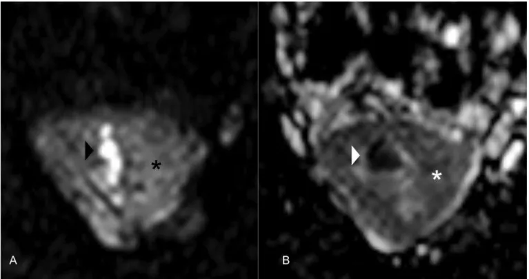
Figure 3: Diffusion-weighted magnetic resonance (MR) images of the uterus of a 34-year-old female patient with placental site trophoblastic tumor.
In the axial diffusion-weighted MR image (b=1000sec/mm2) (3A), the tumor shows high signal intensity with deep myometrial invasion (black asterisk).
In the axial apparent diffusion coefficient (ADC) map (3B), the area of high signal intensity in the diffusion-weighted MR image shows low signal intensity, a finding that is consistent with restricted diffusion (white asterisk).
Note the high intensity of the endometrial cavity in the diffusion-weighted MR image (black arrowhead) and the corresponding low intensity on the ADC map (white arrowhead) related to the presence of blood products causing marked restriction.
[Philips Intera Pulsar 1.5T: Axial diffusion-weighted images (TR=2500; TE=70,9); flip angles of 90º, performed with b values of
1000sec/mm2].
Figure 4: Specimen obtained from the uterus of a 34-year-old female patient with placental site trophoblastic tumor.
Radiology Case. 2015 Apr; 9(4):14-22
Jo
u
rna
l o
f
Ra
d
io
lo
g
y
Ca
se
Re
p
o
rt
s
w
w
w
.Ra
di
ol
ogyC
as
es
.c
om
21
Etiology Unknown
Incidence 1–5 per 100,000 pregnancies
Gender ratio Women
Age predilection 3rd decade
Risk factors Unknown
Treatment Hysterectomy for localized disease;
Hysterectomy plus chemotherapy for advanced disease
Prognosis Remains uncertain
May depend on time since preceding pregnancy and diagnosis
Findings on imaging
Ultrasound
Enlarged heterogeneous uterus, with echogenic areas and cystic foci; solid endometrial/myometrial lesion with more or less prominent blood vessels on color Doppler imaging.
MRI
Myometrial/endometrial mass:
–hypervascular type: T1-WI isointense to normal myometrium; T2-WI slightly hyperintense with distortion of the junctional zone, numerous signal voids and marked dilatation of gonadal vessels; marked enhancement;
–hypovascular type: usually smaller in size, hyperintense to normal myometrium on both T1 and T2-WI; moderate enhancement.
Table 1: Summary table of placental site trophoblastic tumor characteristics.
Figure 5: Sample of the uterus of a 34-year-old female patient with placental site trophoblastic tumor.
Radiology Case. 2015 Apr; 9(4):14-22
Jo
u
rna
l o
f
Ra
d
io
lo
g
y
Ca
se
Re
p
o
rt
s
w
w
w
.Ra
di
ol
ogyC
as
es
.c
om
22
US MR
Placental site trophoblastic tumor
Enlarged heterogeneous uterus, with echogenic areas and cystic foci.
Solid endometrial/ myometrial lesion with more or less prominent blood vessels on color Doppler imaging.
Hypervascular tumor type:
– T1-WI isointense.
– T2-WI slightly hyperintense.
– Avidly enhancing endometrial/ myometrial lesion with numerous signal voids and marked dilatation of gonadal vessels.
Hypovascular tumour type:
– T1-WI and T2-WI hyperintense to normal myometrium.
– Endometrial/myometrial lesion, smaller in size.
– Moderate enhancement and absence of signal voids or prominent vascularity.
Capable of metastasizing (particularly to pelvic lymph nodes).
Invasive mole Heterogeneous, echogenic,
hypervascular endometrial lesion.
Poorly defined intrauterine lesion that deeply invades the myometrium with evidence of disruption of the junctional zone.
T1-WI isointense with focal hyperintense areas within the myometrium (hemorrhage).
T2-WI mixed signal intensity, with tiny cystic inner structures related to the molar component.
Enhances intensely.
May be associated with dilated vessels within the tumor, myometrium and parametrium.
Locally invasive, nonmetastasizing.
Choriocarcinoma Heterogeneous lesion enlarging the
uterus and resembling invasive mole associated with areas of necrosis and hemorrhage but may also manifest as a discrete central infiltrative lesion. Usually markedly hypervascular on
color Doppler imaging.
Intrauterine lesion with nodular well-defined margins. T1WI isointense with focal hyperintense areas within
the myometrium (hemorrhage).
T2-WI heterogeneous high signal (necrosis). Myometrial invasion is visible as high-signal-intensity foci within the myometrium, which demonstrate enhancement on postcontrast images.
Peripheral intense enhancement with a necrotic center. Capable of metastasizing, frequently manifesting with
lung and pelvic metastases.
Table 2: Differential diagnosis table for placental site trophoblastic tumor.
β-hCG = Human chorionic gonadotropin hPL = Human placental lactogen MR = Magnetic resonance
PSTT = Placental site trophoblastic tumor US = Ultrasonography
Placental site trophoblastic tumor; Gestational trophoblastic disease; Uterus; Ultrasound; Magnetic resonance
Thanks to Dr. Rafael Cabrera for sharing the pathology results.
Online access
This publication is online available at:
www.radiologycases.com/index.php/radiologycases/article/view/2146
Peer discussion
Discuss this manuscript in our protected discussion forum at:
www.radiolopolis.com/forums/JRCR
Interactivity
This publication is available as an interactive article with scroll, window/level, magnify and more features.
Available online at www.RadiologyCases.com
Published by EduRad
www.EduRad.org ABBREVIATIONS
KEYWORDS