fine wrinkling and discrete perifollicular papular protu-sions. Moreover, our patient could be differentiated from papular acne scars, white fibrous papulosis of the neck and morphea guttate. The histological features of all of these diseases are distinct from those of PE.
There is no consensus whether PE is a unique entity or just a special demonstration of nevus anesticus, eruptive collagenoma and a mild form of Buschke-Ollendorff syn-drome [6]. We believe our patient adds another case sup-porting PE as a unique entity, according to Del Pozoet al.
[4]. No systemic associations have been described with PE to date. Furthermore, there is no reliable treatment for PE.
■
Acknowledgements. Financial support: none. Conflict of interest: none.
1
Department of Dermatology, first Affiliated Hospital of Nanjing University of TCM,
HanZhong Road, Jiang Su Province, Nanjing, China, 210029
2
Department of Dermatology, first Affiliated Hospital of Nanjing Medical University,
GuangZhou Road,
Jiang Su Province, Nanjing, China <tancheng@medmail.com.cn>
Cheng TAN1 Wen-Yuan ZHU2 Zhong Sheng MIN1
1. Bordas X, Ferra´ndiz C, Ribera M,et al.Papular elastorrhexis: a variety of nevus anelasticus?Arch Dermatol1987; 123: 433-4. 2. Ryder HF, Antaya RJ. Nevus anelasticus, papular elastorrhexis, and eruptive collagenoma: clinically similar entities with focal absence of elastic fibers in childhood.Pediatr Dermatol 2005; 22: 153-7.
3. Lee SH, Park SH, Song KY, et al.Papular elastorrhexis in child-hood improved by intralesional injections of triamcinolone.J Dermatol
2001; 28: 569-71.
4. Del Pozo J, Martı´nez W, Sacrista´n F,et al.Papular elastorrhexis, a distinctive entity?Am J Dermatopathol2008; 30: 188-90.
5. Buechner SA, Itin P. Papular elastorrhexis. report of five cases.
Der-matology2002; 205: 198-200.
6. Assmann A, Mandt N, Geilen CC. Buschke-Ollendorff syndrome--differential diagnosis of disseminated connective tissue lesions. Eur J Dermatol2001; 11: 576-9.
doi:10.1684/ejd.2009.0693
Cutaneous polyarteritis nodosa in a child
following hepatitis B vaccination
Polyarteritis nodosa (PAN) is a rare vasculitis in child-hood, characterized by necrotizing inflammatory changes in small and medium sized arteries [1]. The classification criteria for childhood vasculitis included cutaneous poly-arteritis nodosa (CPAN) and microscopic polyangiitis as two distinct additional categories to PAN [2]. CPAN can be distinguished from classical PAN by the absence of systemic involvement and a benign, but chronic and relapsing course [3, 4].
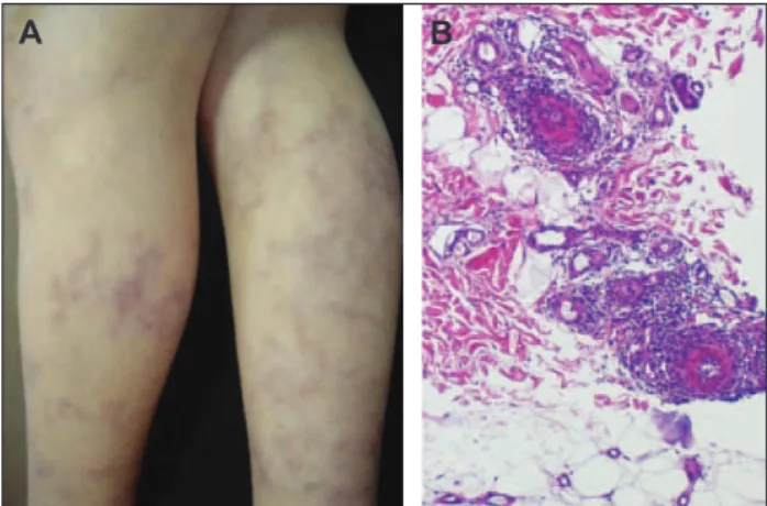
An 11-year-old boy presented with a 3 month history of extensive livedo reticularis mainly affecting the lower extremities (figure 1A) but also the abdomen and upper extremities. Dermatological findings were accompanied by asthenia, myalgias and anorexia. The patient reported
worsening of the lesions with the cold. There were no cutaneous nodules or additional lesions on physical exam-ination. This clinical picture had appeared one week after injection of the third dose of hepatitis B vaccine. Histol-ogy of the livedo reticularis on the thigh revealed an inflammatory arteritis of medium and small sized arteries of the lower dermis, consistent with the diagnosis of PAN
(figure 1B). Immunohistochemical exploration using an anti-antigen HBs antibody was negative. Laboratory investigations revealed anemia, elevated erythrocyte sedi-mentation rate and C-reactive protein. Renal and liver function tests were normal and anti-streptolysin O titre was negative. Immunological work-up revealed positive circulating immune complexes (78.61 U/L) and hyper-gammaglobulinemia (1760 mg/dL). The determination of antinuclear antibodies, antineutrophil cytoplasmic antibo-dies, antiphospholipid antiboantibo-dies, cryoglobulins and com-plement were normal. Serological results only showed positive anti-HBs antibodies. The test of HBV-DNA by PCR was negative. Radiological evaluation including chest X-ray, electrocardiogram, echocardiogram, abdomi-nal ultrasound, chest, abdomiabdomi-nal and pelvic CT, reabdomi-nal magnetic resonance angiography and pulmonary function tests were normal. The diagnosis of CPAN was made. The patient was treated with prednisolone (1 mg/kg/day) suc-cessfully reduced after 6 weeks of treatment, azathioprine and omeprazole, which relieved the symptoms but did not much benefit the cutaneous lesions. In a follow-up of 2 years, the boy was asymptomatic apart from a persisting livedo reticularis on the lower extremities, however, less inflammatory. The laboratory examinations conducted during this period were normal. He has not been taking any medication for one year.
CPAN is characterized by the presence of painful cutane-ous nodules associated with livedo reticularis and ulcera-tion [5]. Livedo reticularis appeared to be an important clinical feature in most series and, although rare, might be the only cutaneous manifestation, as in our case [4]. Extra-cutaneous symptoms like fever, anorexia, myalgia and arthralgia are frequent [5].
Most cases of childhood CPAN have been associated with streptoccocal infection [3, 6]. Associations with hepatitis B virus infection and hepatitis B vaccination have never been described in children. Twenty-seven cases of
vascu-A B
Figure 1. Livedo reticularis on the lower extremities (A). Histology revealed neutophilic-rich, medium-sized vasculitis in lower dermis (H&E, ×100)(B).
litis occurring after this vaccination have been published and among them only two cases of CPAN have been reported [4]. Both were young women with no relevant past medical history. One patient developed fever and infiltrated nodules on the legs two weeks after a five-year booster of hepatitis B vaccination and remission was obtained with oral corticosteroids. The other pre-sented with livedo reticularis on the legs that appeared one month after the first injection of hepatitis B vaccina-tion. She was treated with colchicine that had no effect on the livedo. The cause was never formally demonstrated because vascular deposits of HBs antigen were not found. Some authors believe that the physiopathology might be related to vascular deposits of excess circulating immune complexes of antigens, which, in certain condi-tions, could persist, form a deposit and activate the com-plement in the vessels [4]. To our knowledge this associa-tion has been described only twice in the literature and this is the first case reported in a child.
■
Acknowledgements. Financial support: none. Conflict of interest: none.
1
Dept of Dermatology and Venereology, Hospital de São Marcos, Apartado 2242,
4701-965 Braga, Portugal
2
Pediatric Department, Hospital de São Marcos, Braga, Portugal
3
Pathology Department, Hospital de São Marcos, Braga, Portugal
<filipamanuelventura@hotmail. com>
Filipa VENTURA1 Henedina ANTUNES2 Celeste BRITO1 Fernando PARDAL3 Teresa PEREIRA1 Ana Paula VIEIRA1
1. Ozen S, Anton J, Arisoy N,et al.Juvenile polyarteritis: results of a multicenter survey of 110 children.J Pediatr2004; 145: 517-22. 2. Dillon M, Ozen S. A new international classification of childhood vasculitis.Pediatr Nephrol2006; 21: 1219-22.
3. Fathalla BM, Miller L, Brady S, et al. Cutaneous polyarteritis nodosa in children.J Am Acad Dermatol2005; 53: 724-8. 4. Bourgeais AM, Dore MX, Croue A,et al.Cutaneous polyarteritis nodosa following hepatitis B vaccination. Ann Dermatol Venereol
2003; 130: 205-7.
5. Sunderko¨tter C, Sindrilaru A. Clinical classification of vasculitis.
Eur J Dermatol2006; 16: 114-24.
6. Assicot C, Bourrat E, Prigent F, et al. Cutaneous polyarteritis nodosa in children: three cases.Ann Dermatol Venereol2002; 129: 207-11.
doi:10.1684/ejd.2009.0695
Zosteriform metastasis of endometrial
cancer
Zosteriform distributed metastases are rare cutaneous manifestations of malignancies. They have diagnostic rel-evance, as they often go along with poor prognosis and may be the first manifestation of an underlying malig-nancy. We report an unusual case of endometrial cancer developing segmental zosteriform distributed skin metas-tases on the left breast.
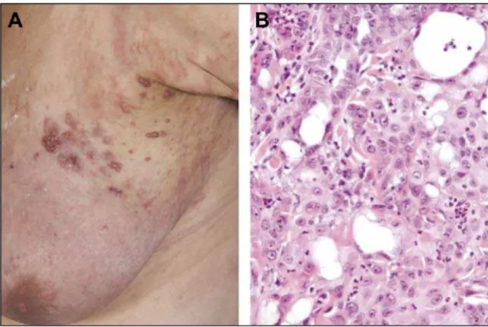
A 68-year-old Caucasian woman was referred to our clinic for therapy of herpes zoster. She had numerous ser-ohematic blister formations in a zosteriform pattern on her left breast, left lateral thorax and left back, on a reddish background. The efflorescences were distributed in a typ-ical zosteriform manner along the left thoracic derma-tomes Th2-4, without crossing the median line (figure 1A). The patient described sensations of pain and itching on and around the efflorescences. The previous history revealed an endometrial cancer of the corpus uteri (pT1b, pNx, G2, FIGO 1b, ER 12/12, PR 9/12) first diag-nosed three years previously. At the time of hospitaliza-tion the patient was not receiving any chemotherapy. She was first considered to have herpes zoster and received a systemic treatment with intravenous acyclovir. The pri-mary vesicular skin changes disappeared and the sur-rounding erythema paled but became infiltrated and chan-ged its distribution. Some vesicles transformed into more solid, indurated papules and nodules. As no therapeutic improvement was achievable by antiviral therapy, we per-formed two skin biopsies. They revealed an extensive infiltration under orthotopic epithelium, of irregular nests of pleomorphic epithelium cells, with many atypical mito-ses. Immunohistopathological stains showed a cytokine expression profile (CK7++, CA125+ and negative for CK20, TTF-1, CDX-2, ER, PR, vimentin), similar to the primary tumor of the corpus uteri. Therefore the diagnosis of cutaneous metastases of an adenocarcinoma with lym-phangiosis carcinomatosa was confirmed (figure 1B). Chemotherapy treatment was not appropriate due to the patient’s deteriorated general condition. The patient was referred to a palliative care unit.
The incidence of cutaneous metastases from all neoplasms reported in literature varies from 0.7 to 10.4 percent [1]. Although metastasis to the skin is not uncommon, a cuta-neous zosteriform distribution is still very rare. Malignan-cies frequently developing skin metastases are malignant melanoma, followed by adenocarcinoma of the breast, lung, colon and ovary [2]. Typical locations are the chest wall, followed by the face and lower extremities [1]. Most skin metastases occurred in the fifth decade of life. In the literature, nearly equal prevalence of cutaneous metastasis is described in both sexes. Two thirds of the
A B
Figure 1. A) Zosteriform distribution of metastases on the left breast. B) Multiple irregular nests of pleomorphic cells with many atypical mitoses. Hematoxylin-eosin stain; origi-nal magnification ×400