Letters to the Editor
Radiol Bras. 2016 Set/Out;49(5):340–346
344
http://dx.doi.org/10.1590/0100-3984.2014.0136 Leiomyoma is a benign tumor composed of smooth muscle
tissue and is considered one of the most common mesenchymal neoplasms in the gastrointestinal tract and uterus(1). Leiomyoma of the breast originates from the stroma of the gland and is ex-tremely rare(2). Mammography and ultrasound studies are com-monly used as screening tools. However, the histopathological evaluation is the definitive diagnostic method. The differential diagnoses include carcinoma, sarcoma, benign tumors and tu-mor-like conditions(3–6). The treatment consists of surgical exci-sion of the leexci-sion, and recurrence is unusual(7).
Smooth muscle tumors are uncommon, especially in the mammary gland. Such tumors account for less than 1% of all breast neoplasms. Deep parenchymal lesions are extremely rare and seem to affect only women. Leiomyomas affect women from 30 to 60 years of age, the mean age being 47.6 years(8). They often occur near the nipple-areola complex, because of the abun-dance of smooth muscle cells in that area(9). Smooth muscle is a component that can be present in other lesions, such as fibroad-enomas and hamartomas. Leiomyomas located in the parenchyma (as in the case reported here) are circumscribed and 1.0–14.0 cm in diameter(1,2).
There are no radiological criteria for making the diagnosis with certainty, histopathological and immunohistochemical studies of the lesion being necessary in order to make the definitive diag-nosis(7–10). The histopathological differential diagnosis is estab-lished with fibroadenoma, phyllodes tumor, adenomyoepithelioma, and leiomyosarcoma of the breast. On histopathology, leiomyo-sarcoma of the breast shows pronounced cell atypia, atypical mi-tosis, vascular invasion, and necrosis(11). Although patients are typically asymptomatic, there can be pruritus, increased breast volume, pain, and hardening of the nipple or nodule(2).
REFERENCES
1. Sidoni A, Lüthy L, Bellezza G, et al. Leiomyoma of the breast: case re-port and review of the literature. Breast. 1999;8:289–90.
Giorge Pereira Sampaio1, Melissa Vieira Koch2, Márcia Boechat2, Viviane Esteves Matos2, Alair Augusto Sarmet Moreira Damas dos Santos3
1. Complexo Hospitalar de Niterói, Niterói, RJ, Brazil. 2. Instituto Fer-nandes Figueira, Rio de Janeiro, RJ, Brazil. 3. Universidade Federal Fluminense (UFF), Niterói, RJ, Brazil. Mailing address: Dr. Giorge Pereira Sampaio. Complexo Hospitalar de Niterói – Radiologia. Rua La Salle, 12, Centro. Niterói, RJ, Brazil, 24020-096. E-mail: giorgesampaio@ hotmail.com.
2. Minami S, Matsuo S, Azuma T, et al. Parenchymal leiomyoma of the breast: a case report with special reference to magnetic resonance im-aging findings and an update review of literature. Breast Cancer. 2011; 18:231–6.
3. Valentim MH, Monteiro V, Marques JC. Primary neuroendocrine breast carcinoma: a case report and literature review. Radiol Bras. 2014;47: 125–7.
4. Bitencourt AGV, Lima ENP, Chojniak R, et al. Correlation between PET/CT results and histological and immunohistochemical findings in breast carcinomas. Radiol Bras. 2014;47:67–73.
5. Pinheiro DJPC, Elias S, Nazário ACP. Axillary lymph nodes in breast cancer patients: sonographic evaluation. Radiol Bras. 2014;47:240–4. 6. Campos GCP, Castro MVK, Mattos VFE, et al. Lymphocytic mastopathy mimicking breast malignancy: a case report. Radiol Bras. 2014;47: 256–8.
7. Heyer H, Ohlinger R, Schimming A, et al. Parenchymal leiomyoma of the breast – clinical, sonographic, mammographic and histological fea-tures. Utraschall Med. 2006;27:55–8.
8. Vecchio GM, Cavaliere A, Cartaginese F, et al. Intraparenchymal lei-omyoma of the breast: report of a case with emphasis on needle core biopsy-based diagnosis. Pathologica. 2013;105:122–7.
9. Rad FS, Zangivand AA. Breast leiomyoma: a case report and review of the literature. Comp Clin Pathol. 2014;23:483–5.
10. Yalta T, Bekar E, Balaban F. Leiomyoma of the breast: a case report. Dicle Medical Journal. 2012;39:283–5.
11. Munitiz V, Rios A, Canovas J, et al. Primitive leiomyosarcoma of the breast: case report and review of the literature. Breast. 2004;13:72–6.
Breast cancer with splenic metastasis in a male patient
Dear Editor,
Here, we report the case of a 53-year-old male patient who was admitted to the Hospital Alemão Oswaldo Cruz in 2014 with a three-month history of intense, progressively worsening lum-bosacral pain. Computed tomography (CT) showed bone lesions in the spine and pelvis, consistent with secondary involvement. We performed a CT-guided pelvic biopsy, which revealed meta-static adenocarcinoma. In an immunohistochemical study, the
biopsy sample tested positive for estrogen and progesterone re-ceptors, indicating that the primary site was in the breast.
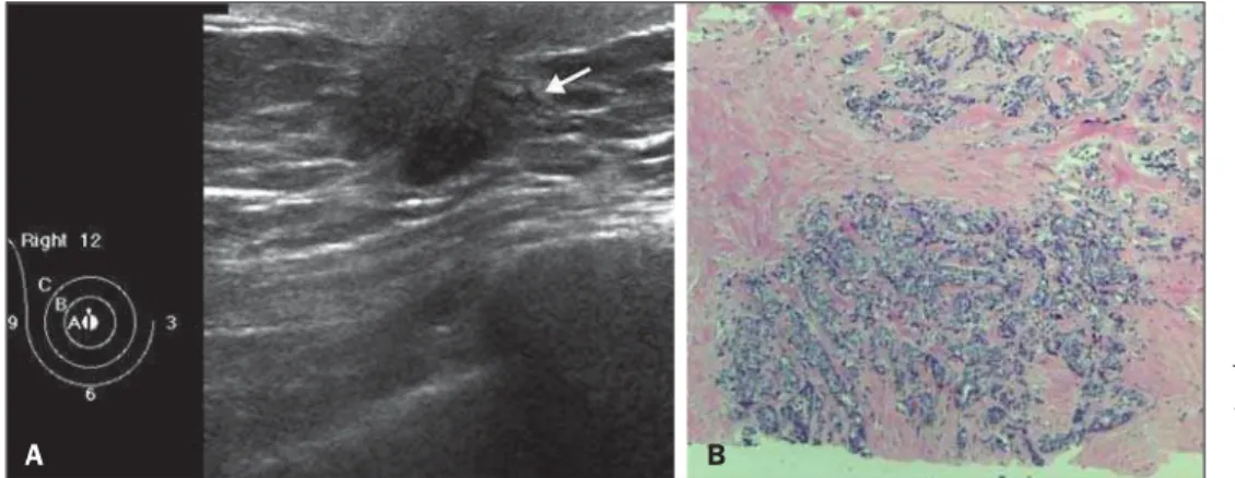
The patient reported having detected a hard, palpable lump, measuring 2.0 cm, in the right breast, three years prior. Ultra-sound showed a solid, hypoechoic, spiculated nodule in the retroareolar region, adjacent to the papilla (Figure 1A), classified as BI-RADS category 5(1), a core biopsy of which showed invasive carcinoma of no special type (invasive ductal carcinoma), as de-picted in Figure 1B, showing positivity for hormone receptors and negativity for HER2.
Figure 1. A: Ultrasound showing a solid, hypoechoic, irregular spiculated nodule, ad-jacent to the papilla of the right breast. B: Right breast biopsy showing massive infil-tration by grade III invasive carcinoma of no special type. Hematoxylin and eosin stain-ing.
Letters to the Editor
Radiol Bras. 2016 Set/Out;49(5):340–346
345
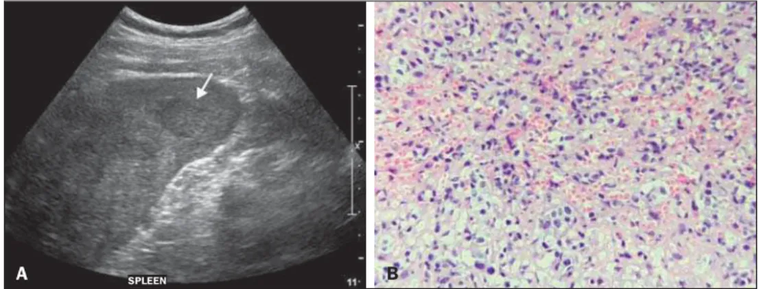
The breast tumor was classified as clinical stage IV, with metastasis to the lungs (lymphangitis carcinomatosa identified on CT) and bones, and surgery for the breast lesion was therefore not indicated. Chemotherapy followed by endocrine therapy was the treatment strategy elected. After a year, the cancer was re-staged. A CT scan of the upper abdomen showed parenchymal nodules suggestive of secondary implants in the spleen, which were also seen on ultrasound (Figure 2A). On the basis of an ultra-sound-guided percutaneous biopsy, the patient was diagnosed with splenic metastasis of breast carcinoma (Figure 2B), and a new chemotherapy regimen was started exclusively for the splenic pro-gression.
Male breast cancer is rare, accounting for 0.6% of all cases of breast cancer and less than 1% of all carcinomas in men. The average age at diagnosis is 65 years(2). The most common com-plaint at diagnosis is of a palpable nodule, typically > 2.0 cm(3). Mammography and ultrasound are used in making the diagno-sis, following the criteria for malignancy in female breast can-cer(2,4–6). The most common histological subtype is invasive duc-tal carcinoma, which often tests positive for estrogen and proges-terone(2).
The treatment of choice is mastectomy and, if necessary, ipsilateral axillary drainage, lymph node involvement being seen in 50–60% of cases(3). The success of chemotherapy and radio-therapy, as well as hormone therapy (tamoxifen being the drug of choice), in the treatment of female breast cancer, allows us to extrapolate that they can also be used in cases of male breast cancer(3).
The metastatic pattern of male breast cancer follows that of female breast cancer in that the bones, lungs, and liver are the most common sites. Splenic metastasis of breast cancer, as shown in this case, is rare in the literature, and the few cases reported have all been in women. Metastases to the spleen are fairly uncom-mon, can be single or multiple, and often occur in the context of multi-organ metastatic carcinoma, usually without clinical sig-nificance, splenectomy being palliative in symptomatic patients.
Bruna Maria Thompson1, Flávio Ferrarini de Oliveira Pimentel1, Jaime Afonso Coelho Nogueira Diógenes1, Marcelo Hajime Kohayagawa2, Maria Regina Vianna2
1. Grupo Fleury/Hospital Alemão Oswaldo Cruz, São Paulo, SP, Brazil. 2. Hospital Alemão Oswaldo Cruz, São Paulo, SP, Brazil. Mailing address: Dra. Bruna Maria Thompson. Rua Afonso de Freitas, 78, ap. 71, Pa-raíso. São Paulo, SP, Brasil, 04006-050. E-mail: thompsonbruna@ gmail.com.
Such metastases are incidental findings on imaging studies for follow-up of the primary tumor (melanoma is the main underly-ing diagnosis) and are radiologically indistunderly-inguishable from pri-mary lesions. The clinical significance of these metastases is not well established in the literature. When occurring in isolation, 60% of splenic metastases are asymptomatic. However, some patients present with fatigue, splenomegaly, or other symptoms. There have been no studies discussing the preferred approach when a single splenic metastasis is identified. The diagnosis can be made by percutaneous biopsy, which has a low (0–2%) complication rate(7).
REFERENCES
1. American College of Radiology. ACR BI-RADS® Atlas, 2013. Reston,
VA: American College of Radiology; 2013.
2. Oger AS, Boukerrou M, Cutuli B, et al. Male breast cancer: prognostic factors, diagnosis and treatment: a multi-institutional survey of 95 cases. Gynecol Obstet Fertil. 2015;43:290–6.
3. Harlan LC, Zujewski JA, Goodman MT, et al. Breast cancer in men in the United States: a population-based study of diagnosis, treatment, and survival. Cancer. 2010;116:3558–68.
4. Valentim MH, Monteiro V, Marques JC. Primary neuroendocrine breast carcinoma: a case report and literature review. Radiol Bras. 2014;47:125– 7.
5. Bitencourt AGV, Lima ENP, Chojniak R, et al. Correlation between PET/ CT results and histological and immunohistochemical findings in breast carcinomas. Radiol Bras. 2014;47:67–73.
6. Pinheiro DJPC, Elias S, Nazário ACP. Axillary lymph nodes in breast cancer patients: sonographic evaluation. Radiol Bras. 2014;47:240–4. 7. Compérat E, Bardier-Dupas A, Camparo P, et al. Splenic metastases: clinicopathologic presentation, differential diagnosis, and pathogenesis. Arch Pathol Lab Med. 2007;131:965–9.
http://dx.doi.org/10.1590/0100-3984.2015.0109
Unusual intrathoracic foreign body: tree branch
Dear Editor,
We report the case of a 46-year-old male who was admitted to the emergency room 4 hours after suffering trauma to the left lateral chest wall, which was penetrated by tree branch during a fall from a bicycle. At the time of the examination, the patient was bleeding from the entrance wound and complaining of severe lo-cal pain. His vital signs were normal. Computed tomography
showed laceration of the left upper lung lobe, with areas of pul-monary contusion and ipsilateral pleural effusion. We also observed a tubular image, with a density of –136 HU, of which the proxi-mal end was in the soft tissues of the chest wall and the distal end was in the lung parenchyma (Figure 1). The patient underwent surgery on the same day, and a piece of tree branch was removed from the chest cavity. A chest tube was inserted, and approximately 1.5 L of blood, mixed with clots, were drained from the pleural space. There was no vascular or mediastinal lesion.
Figure 2. A: Ultrasound showing multiple, hypoechoic splenic nodules, one of which is indicated by the arrow. B: Biopsy demon-strating splenic infiltration by breast