Hereditas 139: 228 – 231 (2003)
Brief report
5S rDNA organization in the fish
Synbranchus marmoratus
(Synbranchidae, Synbranchiformes)
LUCIANO HENRIQUE VIEIRA MESSIAS, DANIELA CRISTINA FERREIRA,
ADRIANE PINTO WASKO, CLAUDIO OLIVEIRA, FAUSTO FORESTI and CESAR MARTINS Departamento de Morfologia,Instituto de Biocieˆncias, Uni6ersidade Estadual Paulista, 18618-000, Botucatu, Sa˜o Paulo, Brazil. E-mail: [email protected]
Received July 14, 2003. Accepted October 29, 2003
Studies on ribosomal RNA genes have gained promi-nence in a broad range of animals and plants, espe-cially in relation to species or population characterization, evolutionary relationships and genome structuring. The 5S rDNA array consists of multiple copies of a highly conserved 120 base pair (bp) coding sequence, separated by a variable non-transcribed spacer (NTS) (LONG and DAVID 1980).
While the 5S rRNA gene is conserved even among non-related taxa, the NTS shows extensive length and sequence variation which can give an accentuated dynamism to these genes (WILLIAMSand STROBECK
1985).
The fish Synbranchus marmoratus BLOCH 1795 (Synbranchidae, Synbranchiformes) is widely dis-tributed in freshwater river systems of different hy-drographic basins of South America. Previous cytogenetic and biochemical studies have been carried out in several populations of the species and detected extensive levels of variation (NAKAMOTOet al. 1986; FORESTI et al. 1992; MELILLO et al. 1996; TORRES
2000). These data strongly support the hypothesis that S. marmoratus represents a complex of species (FORESTIet al. 1992) and that further biological and
genetic studies are required for a better characteriza-tion of the species. To further understand the genetic structure ofS.marmoratusand the 5S rDNA organi-zation among fishes, we have studied the nucleotide sequence and genome organization of 5S rDNA tandem repeats in this species.
MATERIAL AND METHODS
Seven wild adult specimens (4 females and 3 males) of Synbranchus marmoratus were collected from the Ti-eteˆ River, municipality of Pena´polis, Sa˜o Paulo State, Brazil. DNA was extracted from the liver according to SAMBROOKand RUSSEL(2001) and PCR amplifi-cations of the 5S rDNA were performed as described by MARTINS and GALETTIJR. (2001a). Primers 5SA (5%-TAC GCC CGA TCT CGT CCG ATC-3%) and
5SB (5%-CAG GCT GGT ATG GCC GTA AGC-3%)
were designed to amplify the 5S genes and their NTSs. The PCR-amplified products were cloned in the plasmid pGEM-T (Promega) and used to trans-form competent cells of the strain DH5a E. coli
(Invitrogen). Clones were sequenced on an ABI PRISM 377 DNA Sequencer (Applied Biosystems) and the alignment of the sequences was performed using ClustalW (THOMPSONet al. 1994). Nucleic acid
sequences were subjected to Blastn (ALTSCHULet al.
1990) searches at National Center for Biotechnology Information (USA), website (http://www.ncbi.nlm. nih.gov/blast).
The genomic organization of 5S rDNA was deter-mined by Southern-blot hybridization. Genomic DNA (10 mg) was partially to completely digested
with 30U ofHindIII orSacI at 37oC and the digested products were subjected to gel-electrophoresis in 1 % agarose and Southern-transferred to a Hybon-N ny-lon membrane (Amersham Bioscience) according to SAMBROOK and RUSSEL (2001). Inserts of the 5S clones were labeled, hybridized, and detected by the ECL-Direct Nucleic Acid Labelling and Detection System (Amersham Bioscience), following the instruc-tions of the manufacturer.
RESULTS AND DISCUSSION
iden-Brief report 229
Hereditas 139 (2003)
Fig. 1. Consensus sequence of the 5S rDNA repeats ofS.marmoratus. The 5S rRNA gene coding sequence is in boldface type, the primer (5SA and 5SB) regions are underlined and TATA-like elements are in lower case. The
restriction sites forHindIII (AAGCTT) andSacI (GAGCTC) are underlined and in italics. The sequence is deposited
in GenBank under the accession number AY271269.
tified low divergences between the 5S rRNA gene sequence described here and from those of other fish species previously studied (average similarity of 95 %). On the other hand, no relationship could be established between the NTS of the 5S rDNA of S. marmoratus and any sequence of other fish or verte-brate species, suggesting that this spacer region evolves rapidly. The NTS regions are presumed to be free to vary since these variants are selectively neutral or nearly neutral and can become either fixed or lost, thereby causing accentuated differences even between close related species. In contrast, most mutations in the 5S rRNA gene are presumed to be selectively neutral or nearly neutral only when they occur in a subcritical proportion (CRONNet al. 1996). Although
little is known about the NTS sequence among fishes, a TATA-like sequence has been observed about 25 – 30 bp upstream from the 5S rRNA gene of some species (PENDA´ S et al. 1994; SAJDAK et al. 1998;
MARTINS and GALETTI JR. 2001a; WASKO et al.
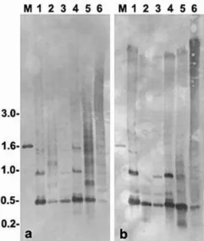
2001). Although the TATA-like elements of S. mar-moratus were located around 180 – 200 bp upstream to the 5S rRNA gene (Fig. 1), such sequence may be involved with the transcription control of these genes. To determine if the genomic organization of the 5S rDNA ofS.marmoratuswas consistent with the PCR products and sequences obtained, Southern-blot hy-bridization experiments were performed. Complete genomic DNA digestion with HindIII or SacI, that cleave once in the 5S rDNA sequence (Fig. 1), showed that most of the 5S rRNA genes are orga-nized in monomers around 460 base pairs in six analyzed specimens (Fig. 2). Faint hybridization bands around 550 bp (samples 1 and 4, Fig. 2a and 2b), 600 bp (samples 1 – 4, Fig. 2b) and 700 bp (samples 2 and 5, Fig. 2a) give support for the presence of 5S rDNA variant copies in the genome of S.marmoratus. Although a complete DNA digestion
was carried out withHindIII and SacI, some of the samples gave partially digested results showing the presence of multimeric units of the 5S rDNA. The membrane hybridization experiments also detected a polymorphism in the 5S rDNA monomer size among the sampled animals. This is clearly visible in the samples 3 and 4 of Fig. 2a and 2b. Such polymor-phism can be useful for population studies in this species.
Fig. 2. Genomic organization of 5S rDNA ofS.
marmora-tusdetermined by Southern-blot and hybridization to a 5S
rRNA gene probe. Aliquots of 10 mg of genomic DNA
L. H. V. Messias et al.
230 Hereditas 139 (2003)
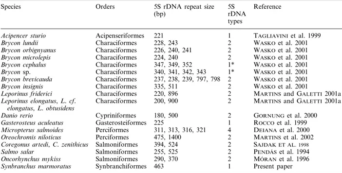
Table 1. 5S rDNA repeat characteristics among fish species.
Species Orders 5S rDNA repeat size 5S Reference
rDNA (bp)
types
221
Acipencer sturio Acipenseriformes 1 TAGLIAVINIet al. 1999
228, 243 2
Characiformes WASKOet al. 2001
Brycon lundii
226, 240, 241 2 WASKOet al. 2001
Brycon orbignyanus Characiformes
224, 240 2
Characiformes WASKOet al. 2001
Brycon microlepis
347, 349, 352 1*
Brycon cephalus Characiformes WASKOet al. 2001
340, 341, 342, 343 1*
Characiformes WASKOet al. 2001
Brycon sp.
237, 238, 239, 797, 798 2 WASKOet al. 2001
Brycon bre6icauda Characiformes
335, 511 2
Characiformes WASKOet al. 2001
Brycon insignis
220, 896 2 MARTINSand GALETTI 2001a
Leporinus friderici Characiformes
Characiformes 200, 900 2 MARTINSand GALETTI 2001a
Leporinus elongatus,L.cf. elongatus,L.obtusidens
180, 500 2 GORNUNG et al. 2000
Danio rerio Cypriniformes
225 1
Gasterosteiformes ROCCO et al. 1999
Gasterosteus aculeatus
311, 313, 316, 321
Micropterus salmoides Perciformes 4 DEIANAet al. 2000
475, 1400 2
Perciformes MARTINSet al. 2002
Oreochromis niloticus
Salmoniformes
Coregonus artedi,C.zenithicus 394, 524 2 SAJDAK ET AL. 1998 Salmoniformes
Salmo salar 255, 525 2 PENDA´ S et al. 1994
290, 370 2
Salmoniformes MO´ RANet al. 1996
Oncorhynchus mykiss
Synbranchiformes 463 1 Present paper
Synbranchus marmoratus
*There are evidences for the presence of a second 5S rDNA type.
5S rDNA variants have been reported for several mammals and amphibians (KOMIYA et al. 1986; L IT-TLE and BRAATEN 1989; LEAH et al. 1990; F RED-ERIKSEN et al. 1997), suggesting that 5S rDNA
variant types have originated early in the evolution-ary process of vertebrates. Variant types of 5S rDNA also have a common occurrence in fish groups (Table 1). However, S. marmoratus (Synbranchiformes), Gasterosteus aculeatus (Gasterosteiformes) and Acipencer sturio (Acipenseriformes) seem to present just one major 5S rDNA type. Synbranchiformes and Gasterosteiformes represent evolved lineages within the Actinopterygii and Acipenseriformes represents a very ancient group of fishes (LAUDER and LIEM
1983). Although variant types of 5S rDNA are com-mon acom-mong fishes, the evolution might have main-tained just one major class of 5S tandem repeats in some groups.
Particularly among fishes, variant classes of 5S rDNA within a single genome that differ extensively in their NTSs, have been identified in Salmo salar (PENDA´ S et al. 1994), Oncorhynchus mykiss (MO´ RAN
et al. 1996), species of Coregonus (SAJDAK et al. 1998),Leporinus(MARTINS and GALETTIJR. 2001a), Brycon(WASKO et al. 2001) and Oreochromis
niloti-cus (MARTINS et al. 2002) (Table 1). Although the
presence of 5S rDNA variant classes seems to be a common feature for fishes (MARTINS and GALETTI
JR. 2001b) (Table 1), the present results of membrane
hybridization show that there is just one major class of 463 bp-tandem repeats of 5S rDNA in the genome
ofS.marmoratus. However, in addition to the 463-bp strong band, faint bands were detected after mem-brane hybridization, showing that the existence of few copies of other types of 5S rDNA-tandem repeats in the genome of S. marmoratus cannot be elimi-nated.
Acknowledgements – This work was supported by grants from FAPESP (Fundac¸a˜o de Amparo a` Pesquisa do Es-tado de Sa˜o Paulo) and CNPq (Conselho Nacional de Desenvolvimento Cientı´fico e Tecnolo´gico).
REFERENCES
Altschul, S. F., Gish, W., Miller, W. et al. 1990. Basic local alignment search tool. – J. Mol. Biol. 215: 403 – 410. Cronn, R. C., Zhao, X., Paterson, A. H. et al. 1996.
Polymorphism and concerted evolution in a tandemly repeated gene family: 5S ribosomal DNA in diploid and allopolyploid cottons. – J. Mol. Evol. 42: 685 – 705. Deiana, A. M., Cau, A., Salvadori, S. et al. 2000. Major
and 5S ribosomal sequences of the largemouth bass Micropterus salmoides (Perciformes, Centrarchidae) are localized in GC-rich regions of the genome. – Chromo-some Res. 8: 213 – 218.
Foresti, F., Oliveira, C. and Tien, O. S. 1992. Cytogenetic studies in fishes of the genusSynbranchus (Pisces, Syn-branchiformes, Synbranchidae). – Naturalia 17: 129 – 138.
Frederiksen, S., Cao, H., Lomholt, B. et al. 1997. The rat 5S rRNA bona fide gene repeat maps to chromosome
19q12qter and the pseudogene repeat maps to 12q12.
– Cytogenet. Cell Genet. 76: 101 – 106.
Brief report 231
Hereditas 139 (2003)
Komiya, H., Hasegawa, M. and Takemura, S. 1986. Differ-entiation of oocyte- and somatic-type 5S rRNAs in animals. – J. Biochem. 100: 369 – 374.
Lauder, G. V. and Liem, K. F. 1983. The evolution and interrelationships of the actinopterygian fishes. – Bull. Mus. Comp. Zool. 150: 95 – 197.
Leah, R., Frederiksen, S., Engberg, J. et al. 1990. Nucle-otide sequence of a mouse 5S rRNA variant gene. – Nucleic Acids Res. 18: 7441 – 7441.
Little, R. and Braaten, D. 1989. Genomic organization of human 5S rDNA and sequence of one tandem repeat. – Genomics 4: 376 – 383.
Long, E. O. and David, I. D. 1980. Repeated genes in eukaryotes. – Annu. Rev. Biochem. 49: 727 – 764. Martins, C. and Galetti Jr., P. M. 2001a. Organization of
5S rDNA in species of the fishLeporinus: two different
genomic locations are characterized by distinct nontran-scribed spacers. – Genome 44: 903 – 910.
Martins, C. and Galetti Jr., P. M. 2001b. Two 5S rDNA arrays in Neotropical fish species: is it a general rule for fishes? – Genetica 111: 439 – 446.
Martins, C., Wasko, A. P., Oliveira, C. et al. 2002. Dynam-ics of 5S rDNA in the tilapia (Oreochromis niloticus) genome: repeat units, inverted sequences, pseudogenes and chromosome loci. – Cytogenet. Gen. Res. 98: 78 – 85.
Melillo, M. I. F. M., Foresti, F. and Oliveira, C. 1996. Additional cytogenetic studies on local populations of Synbranchus marmoratus (Pisces, Synbranchiformes, Synbranchidae). – Naturalia 21: 201 – 208.
Mo´ran, P., Martı´nez, J. L., Garcia-Va´squez, E. et al. 1996. Sex linkage of 5S rDNA in rainbow trout (Oncorhynchus mykiss). – Cytogenet. Cell Genet. 75: 145 – 150. Nakamoto, W., Machado, P. E. and Foresti, F. 1986.
Hemoglobin patterns in different populations of
Syn-branchus marmoratus, Bloch 1795 (Pisces, Synbranchi-dae). – Comp. Biochem. Physiol. 84: 377 – 381. Penda´s, A. M., Mo´ran, P., Freije, J. P. et al. 1994.
Chro-mosomal location and nucleotide sequence of two tanden repeats of the Atlantic salmon 5S rDNA. – Cytogenet. Cell Genet. 67: 31 – 36.
Rocco, L., Russo, C., Stingo, V. et al. 1999.
Characteriza-tion of 5S rDNA in Gasterosteus aculeatus (Teleostei,
Gasterosteidae). – Ital. J. Zool. 66: 285 – 289.
Sajdak, S. L., Reed, K. M. and Phillips, R. B. 1998. Intraindividual and interspecies variation in the 5S rDNA of coregonid fish. – J. Mol. Evol. 46: 680 – 688. Sambrook, J. and Russel, D. W. 2001. Molecular cloning.
A laboratory manual, 3rd edn. – Cold Spring Harbor Laboratory Press.
Tagliavini, J., Williot, P., Congiu, L. et al. 1999. Molecular cytogenetic analysis of the karyotype of the European Atlantic sturgeon,Acipenser sturio. – Heredity 83: 520 – 525.
Thompson, J. D., Higgins, D. G. and Gibson, T. J. 1994. Clustal W: improving the sensitivity of progressive mul-tiple sequence alignment through sequence weighting, position-specific gap penalties and weight matrix choice.
– Nucleic Acids Res. 22: 4673 – 4680.
Torres, R. A. 2000. O geˆnero Synbranchus (Pisces,
Syn-branchiformes, Synbranchidae): interrelac¸o˜es
citotaxo-noˆmicas, evolutivas e natureza da variabilidade
cariotı´pica. PhD. Thesis. – Instituto de Biocieˆncias, UNESP, Brazil.
Wasko, A. P., Martins, C., Wright, J. M. et al. 2001. Molecular organization of 5S rDNA in fishes of the
genusBrycon. – Genome 44: 893 – 902.