Infection of silkworm larvae by the entomopathogenic fungus Metarhizium anisopliae
Infecção de lagartas do bicho-da-seda pelo fungo entomopatogênico Metarhizium anisopliae
Lucineia de Fátima Chasko Ribeiro1 Jeronimo Tavares2 Sóstenez Alexandre Vessaro Silva1
Luis Francisco Angeli Alvez2 Eduardo Alexandre Loth2 Rose Meire Costa Brancalhão1*
ISSNe 1678-4596
Metarhizium anisopliae is an entomopathogenic fungus that is capable of adapting to heterogeneous media and it infects a wide variety of insects primarily due to the evolution of genes which cause the degradation of the host cuticle,
detoxification and biosynthesis of toxins (GAO et
al., 2011). M. anisopliae naturally infects over 300
species of insects (ALVES, 1998), as well as the
silkworm Bombyx mori (Lepidoptera: Bombycidae), which is an insect model in scientific studies and has
economic importance due to its role in silk production
(STAYKOVA et al., 2012).
Entomopathogenic fungi act via contact
and they require some environmental conditions to germinate on insects. Such conditions are reported in the B. mori creation room, which increases the infection and favours the multiplication and spread of the pathogen. Some previous studies conducted
in sericulture regions of Brazil and other countries
have shown fungal incidence in B. mori larvae
(ALVES, 1998; KUMARI et al., 2011). However,
data that correlate symptoms and histopathology of infection are scarce.
Considering the economic importance of B. mori, a better understanding and description of fungal infection process may help to manage the risks of disease spreading in silk production. Silkworms are widely used as a model for cellular immunity response studies in insect-pathogen interactions, even though there is a lack of information about histopathological studies. Consequently, this studied analysed the symptomatology and histopathology of B. mori
larvae inoculated experimentally by M. anisopliae.
The fungus M. anisopliae, E9 strain,
was obtained from Deois flavopicta (Hemiptera:
Cercopidae) and supplied by EMCAPA - Capixaba
Research Company Agropecuaria. It was cultured in Petri dishes with a complete medium for conidial production (agar 20g, yeast extract 5g, KH2PO4
0.36g, Na2HPO4. 7H20 1.05g, MgSO4.7H2O 0.60g,
KCl 1.00g, NaNO3 1.58g, glucose 10g and distilled water 1000mL), which was previously autoclaved
and incubated at 26°C with a 12 hour photoperiod
for 10 days (ALVES, 1998). The conidia were then
collected by scraping the surface of the medium
1Mestrado em Biociências e Saúde, Universidade Estadual do Oeste do Paraná (UNIOESTE), 85819-110, Cascavel, PR, Brasil. E-mail: rosecb@gmail.com. *Corresponding author.
2Centro de Ciências Biológicas e da Saúde, Universidade Estadual do Oeste do Paraná (UNIOESTE), Campus de Cascavel, Cascavel, PR, Brasil.
ABSTRACT: The isolate E9 of Metarhizium anisopliae was used in commercial hybrids of Bombyx mori larvae to evaluate its biological effect. Symptomatological analyses showed typical signs of fungal infection. Histopathology revealed the presence of large numbers of hemocytes in the hemocoel, and on the sixth dpi the bodies of the insects appeared to be colonised by the fungus. The isolate E9 is pathogenic to larvae B. mori and; therefore, death of the insects was caused by the colonization of fungus in the epidermal and mesodermal tissues. Key words: Bombyx mori, histopathology, Metarhizium anisopliae, fungal infection.
RESUMO: O isolado E9 de Metarhizium anisopliae foi usado em larvas híbridas de Bombyx mori para avaliar seu efeito biológico. A
sintomatologia revelou sinais típicos de infecção por fungos. Na histopatologia foi verificado um aumento no número de hemócitos, sendo que, no 6dpi, todo o corpo do inseto se apresentou colonizado pelo fungo. O isolado E9 é patogênico para lagartas de B. mori, causando sua morte pela colonização dos tecidos de origem epidérmica e mesodérmica.
Palavras-chave: Bombyx mori, histopatologia, Metarhizium anisopliae, infecção fúngica.
and suspended in a solution of distilled water and
0.01% Tween 80®. After quantification, using a
Neubauer chamber, a suspension was prepared at a concentration of 1.5×10⁸ conidia mL-1.
Third instar B. mori larvae were obtained
from the BRATAC S/A silk company; they were
kept in polyethylene boxes at room temperature and humidity and fed with fresh mulberry leaves twice
daily until the fifth instar.
At the beginning of the fifth instar, 40
larvae were quickly immersed for 5 seconds in the
conidia suspension. An identical control group was
immersed in a solution of distilled water plus 0.01%
Tween 80®. Larvae were subsequently placed in
boxes and fed with mulberry leaves, as described
above. The signs and symptoms of the disease were assessed daily, based on ALVES (1998), from the first to the eleventh day post-inoculation (dpi) and the dead larvae were transferred to a Petri dish with its bottom covered with filter paper (moist chamber) to promote the extrusion of the fungus, which confirmed the mortality of the larvae. The experiment was
conducted in triplicate.
Two larvae from each group (control and inoculated) were anesthetized with ether and fixed in Bouin for 24 hours. This procedure was performed daily from the first to the seventh dpi. After fixation, the larvae were rinsed in 70%
alcohol, processed for routine histological, and stained with hematoxylin and eosin for analysis
of general morphology, and Gomori methenamine technique (GROCOTT, 1955) for fungi.
Evidence of infection of B. mori larvae by M. anisopliae was observed externally from the
second dpi. The infected larvae decreased the size of
the food that was available, had smaller bodies than insects in the control group, and began to present
darkened spots in the integument. On the third dpi
decreased activity was evident with dry stools and
integument with a wrinkled appearance. Between the fourth and fifth dpi browning was evident with the
appearance of dark spots on the surface, especially in the regions of the intersegmental membranes and
spiracles. On the sixth dpi the larvae began to die and on the seventh dpi all the larvae were dead. These dark spots corroborated previous studies (ALVES, 1998; TOLEDO et al., 2010) and confirmed what is
known about the integument of insects being the main route of entomopathogenic fungal entry, including M. anisopliae; the dark spots were the result of fungal
germination and penetration in the cuticle through
the action of extracellular enzymes and mechanical
pressure exerted by hyphae.
In the hemocoel, hyphal bodies grow profusely and multiply by budding of pre-existing
cells, the blastospores, as observed by KUMAR et al. (2004) in B. mori infected with A. flavus, and by GAO et al. (2011) in a comparative analysis of the
genome and the broad-spectrum insect pathogen of
M. anisopliae and M. acridum.
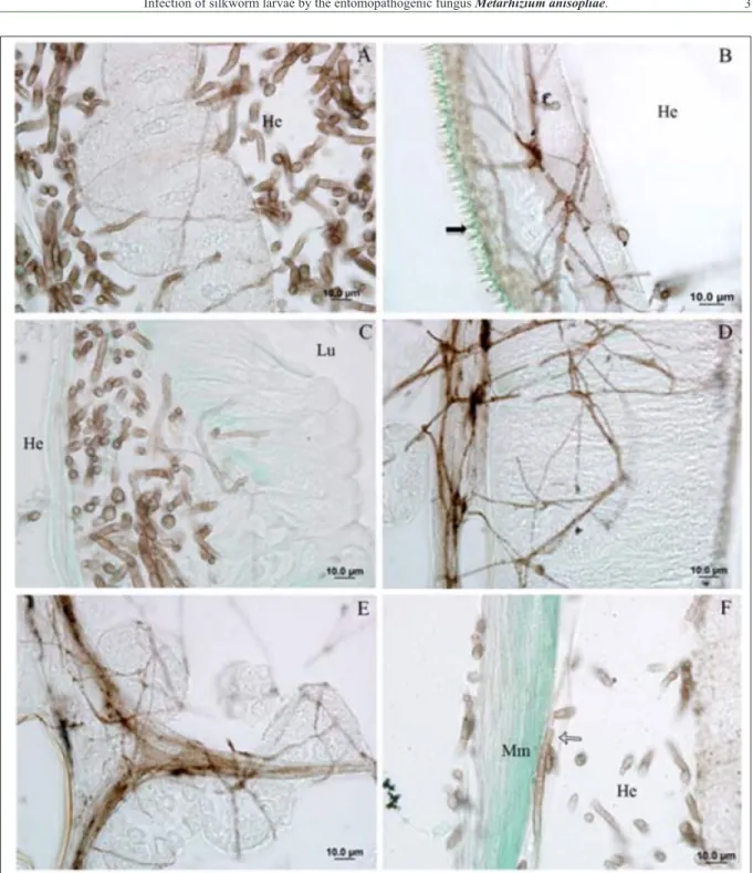
On the sixth dpi, hyphae forming a mycelia complex in the fatty tissue, integument, digestive tract, silk gland, reproductive organs, muscle and
trachea (Figure 1A-F). Furthermore, during this
period microscopic image showed little injury to the
internal tissues, as reported by KUMAR et al. (2004). A large development of mycelium was observed in the mid gut. This was related to the
availability of nutrients in tissues because, as pointed
out by TOLEDO et al.(2010), nutrient limitation
leads to the formation of hyphae with a smaller
diameter. Fatty tissue is another preferential target. Figure 1F shows that we detected the presence of M. anisopliaehyphae and blastospores in the lumen
of the tracheal system. Tracheae are responsible for aeration and; therefore, form channels of
oxygen transport which, in this case, can act as an important distribution system of the pathogen since the spiracles are also sites of pathogen penetration
(TOLEDO et al., 2010). Furthermore, SCHRANK & VAINSTEIN (2010) state that the blastospores
facilitate dispersal of the fungus, thereby causing
generalized infection.
After the seventh dpi all the B. mori
larvae were dead, mainly because of the action of mycotoxins and mechanical blockage of the digestive
tract. Death of insects was following by major
branches of the fungus through the B. moriepidermal and mesodermal tissues, with saprophytes growing on the corpse. Mycelium takes the place of the internal
organs and; therefore, soon after death B. mori has a
normal appearance (GAO et al., 2011).
The mummification of bodies of B. mori occurred between the eighth and ninth dpi; during
this period M. anisopliae emerged through the integument of the larvae, by mechanical pressure, especially in the intersegmental regions, which
offered less resistance to the fungus (KUMAR et
al., 2004). Subsequently, aerial hyphae were formed that originated branched conidiophores which were
disseminated in the environment (ALVES, 1998). In the colonization of the B. moriintegument, hyphae of M. anisopliae passed through the basal lamina and reached the basal surface of epithelial cells
formation of papillae on the edge of the epithelial layers
and cuticle. As the infectious process advanced from the seventh dpi (Figure 2B) there was separation of the
layers and lysis of the epithelial cells, and the area was fully occupied by fungal hyphae.
M. anisopliae intensified growth after
the death of insects at the tenth and eleventh
dpi, forming a green roof on the integument,
which confirmed fungus infection. No other
microorganism was observed in the microscopic analysis, probably because the fungus secretes antibiotic substances, which prevent their
proliferation (ALVES, 1998). It is noteworthy that although no quantification was performed, it was
Figure 1 - Photomicrographs of B. mori tissues infected with M. anisopliae, Gomori staining. Hyphae images, in brown, on fat tissue(A),
integument (B), mid gut epithelium (C), silk gland (D), testicle (E), muscle (Mm) and trachea (white arrow) (F). Hemocoel
evident that there was an increase in the number of hemocytes in the hemocoel, on the second dpi, compared to the control, which was also reported
by SEWIFY and HASHEM (2001) in relation to Galleria mellonella (Lepidoptera: Pyralidae). Hemocytes recognise and remove fungal conidia by phagocytosis and encapsulation (CHOUVENC
et al., 2009) and on the third dpi it was possible to visualise hemocytes surrounding the hyphal
structures. However, this protection is compromised
when forming the hyphal bodies, which, after 20 minutes of contact with the hemolymph, express a gene encoding the N-terminal domain of the protein
similar to collagen, MCL1. This protein forms a
collagenous protective coat in hyphae and enables
M. anisopliae to evade insect immune responses
(WANG &St. LEGER, 2006). Contributing to the
infective capacity, M. anisopliae has a number
of protein families, such as ABC transporters, and cytochrome P450, which act in defence against secondary metabolites and detoxification,
respectively, and which are produced by the host
(GAO et al., 2011). CHOUVENC et al. (2009), SCHRANK & VAINSTEIN (2010) stress the role
of toxins, such as destruxins, which weaken the immune defences of the host, cause damage in the muscle system and Malpighian tubules, affect
excretion and lead to difficulties in feeding and mobility. This was corroborated in the present
study, where a loss in motor coordination was
observed between the fourth and fifth dpi.
The combined analysis of symptoms
and histopathology revealed that B. mori larvae
are highly susceptible to M. anisopliae E9 isolate and that death is caused by fungal colonization in
the silkworm tissues, resulting in the characteristic
symptoms of infection and the mummification of
the insect’s body.
ACKNOWLEDGEMENTS
Araucaria Foundation (award No. 546/2014), the MA program of the Univerisdade Estadual do Oeste do Paraná (UNIOESTE), and the BRATAC sericulture producer.
REFERENCES
ALVES, S. B. Fungos entomopatogênicos. In: ALVES, S. B.
Controle microbiano de insetos. Piracicaba: FAELQ, 1998. p.289-370.
CHOUVENC, T. et al. Cellular encapsulation in the eastern
subterranean termite, Reticulitermes flavipes (Isoptera), against infection by the entomopathogenic fungus Metarhizium anisopliae.
Journal of Invertebrate Pathology, v.101, p.234-241, 2009.
Available from: <http://www.ncbi.nlm.nih.gov/pubmed/19463828>. Accessed: may, 29, 2014. doi: 10.1016/j.jip.2009.05.008.
GAO Q. et al. Genome sequencing and comparative transcriptomics
of the model entomopathogenic fungi Metarhizium anisopliae and
M. acridum. PLoS Genetics, v.7, p.1-18, 2011. Available from:
<http://www.ncbi.nlm.nih.gov/pubmed/21253567>. Accessed: jun,
10, 2015. doi: 10.1371/journal.pgen.1001264.
GROCOTT, R. G. A stain for fungi in tissue sections and smears using Gomori’s methenamine–silver nitrate technic.
American Journal of Clinical Pathology, v.25,
p.975-979, 1955. Available from: <http://www.ncbi.nlm.nih.gov/ pubmed/14398663>. Accessed: jun, 10, 2015.
KUMAR, V. et al. Surface ultrastructural studies on the germination,
penetration and conidial development of Aspergillus flavus Link:
Fries infecting silkworm, Bombyx mori Linn. Mycopathologia,
v.157, p.127-135, 2004. Available from: <http://www.ncbi.nlm. nih.gov/pubmed/15008355>. Accessed: may, 10, 2015.
KUMARI, S. S. et al. Antifungal activity of Turbinaria conoides
and evaulation for the effective concentration against the infection
of Beaveria bassiana in Silkworm larvae. Resarch Journal of
Microbiology, v.6, p.115-123, 2011. Available from: <http://
scialert.net/abstract/?doi=jm.2011.115.123>. Accessed: may, 10,
2014. doi: 10.3923/jm.2011.115.123.
SCHRANK, A.; VAINSTEIN, M. H. Metarhizium anisopliae enzymes and toxins. Toxicon, v.56,
p.1267-Figure 2 - Photomicrographs integument of B. mori infected with M. anisopliae. In (A) and (B) changes in integument, in 5th and 8th dpi,
respectively. Fungus hyphae (hollow arrow) and papillae (Pi), visible at the edge of epithelial and cuticular layers. In (B), complete
1274, 2010. Available from: <http://www.ncbi.nlm.nih.gov/ pubmed/20298710>. Accessed: may, 10, 2015. doi: 10.1016/j.
toxicon.2010.03.008.
SEWIFY, G. H.; HASHEM, M.Y. Effect of the
entomopathogenic fungus Metarhizium anisopliae (Metsch.) Sorokin on cellular defence response and oxygen uptake of the wax moth Galleria mellonella L. (Lepidoptera-Pyralidae).
Journal of Applied Entomology, v.125, p.533-536, 2001.
Available from: <http://onlinelibrary.wiley.com/doi/10.1046/ j.1439-0418.2001.00564.x/full>. Accessed: may, 10, 2015. doi:
10.1046/j.1439-0418.2001.00564.x.
STAYKOVA, T. et al. Genetic variability in Silkworm (Bombyx mori L.) strains with Different Origin. Acta Zoologica Bulgarica,
v.4, p.89-94, 2012. Available from:
<http://www.acta-zoologica-bulgarica.eu/downloads/acta-zoologica-bulgarica/2012/ supplement-4-089-094.pdf>. Accessed: may, 10, 2015.
TOLEDO, A. V. et al. Histopathology caused by the entomopathogenic
fungi, Beauveria bassiana and Metarhizium anisopliae, in the adult planthopper, Peregrinus maidis, a maize virus vector. Journal of
Insect Science, v.10, p.1-10, 2010. Available from: <http://www.
ncbi.nlm.nih.gov/pubmed/20578956>. Accessed: may, 10, 2015.
doi: 10.1673/031.010.3501.
WANG C.; St.LEGER, R. J. A collagenous protective coat enables
Metarhizium anisopliae to evade insect immune responses.
Proccedings of the National Academy Science of the United States of America, v.103, p.6647-6652, 2006. Available from: