Methicillin-resistant coagulase-negative staphylococci from healthy dogs
in Nsukka, Nigeria
Kennedy F. Chah
1, Elena Gómez-Sanz
2, John A. Nwanta
3, Brendan Asadu
3,
Ifeoma C. Agbo
1, Carmen Lozano
2, Myriam Zarazaga
2, Carmen Torres
21
Department of Veterinary Pathology and Microbiology, University of Nigeria, Nsukka.
2
Biochemistry and Molecular Biology, University of La Rioja, Logroño, Spain.
3
Department of Veterinary Public Health and Preventive Medicine, University of Nigeria, Nsukka.
Submitted: December 18, 2012; Approved: April 1, 2013.
Abstract
The occurrence, resistance phenotype and molecular mechanisms of resistance of methicillresistant staphylococci from groin swabs of 109 clinically healthy dogs in Nsukka, Nigeria were in-vestigated. The groin swab samples were cultured on mannitol salt agar supplemented with 10mg of cloxacillin. Sixteen methicillin-resistant coagulase negative staphylococci (MRCoNS), all harbour-ing themecAgene were isolated from 14 (12.8%) of the 109 dogs studied. The MRCoNS isolated were:S. sciurisubspeciesrodentium,S. lentus,S. haemolyticus, andS. simulanswithS. sciuri sub-species rodentium(62.5%) being the predominant species. Thirteen (81.3%) of the MRCoNS were resistant to tetracycline while 12 (75%) and 10 (62.5%) were resistant to kanamycin and trimtho-prim-sulphamethoxazole respectively. None of the isolates was resistant to fusidic acid, linezolid and vancomycin. Thirteen (81.3%) of the MRCoNS were multi-drug resistance (MDR). Other anti-microbial genes detected were:blaZ,tet(K),tet(M),tet(L),erm(B),lnu(A),aacA-aphD, aphA3,str,
dfr(G),catpC221,and catpC223. Methicillin-resistant staphylococci are common colonizers of healthy dogs in Nigeria with a major species detected being S. sciuri subsp. rodentium.
Key words:methicillin-resistant,S. sciuri, resistant genes, dogs.
Introduction
Staphylococci are Gram-positive cocci usually found as transient normal flora of the skin and mucous mem-branes of mammals and birds and can easily spread to hu-mans by contact and through formites (Fusi Ngwaet al., 2007). According to Bergeronet al.(2011) the Staphylo-coccusgenus groups together 45 species and 21 subspecies which are classified in two groups based on their ability to produce the enzyme coagulase: coagulase-positive staphy-lococci (CoPS) and coagulase-negative staphystaphy-lococci (CoNS).
Thirty-eight different species are found within the CoNS group, including theS. sciurisubgroup, which con-sists of 3 species and 3 subspecies (S. sciuri subspecies
rodentium, S. sciurisubspeciessciuri, S. sciuri subspecies carnaticus; S. lentus, and S. vitulinus) (Kloos and
Ban-nerman, 1994). Staphylococcus sciuri, first described in 1976 by Klooset al.(1976), is the most abundant member of the genus Staphylococcus with a wide habitat range (Kloos and Bannerman, 1994; Kloos et al., 1997). Al-though CoNS are saprophytic and rarely pathogenic (Kloos
et al., 1997), multi-drug resistant strains have been associ-ated with severe cases of difficult to treat infections, espe-cially in immune compromised individuals (Zell et al., 2008).
Companion animals, particularly dogs and cats, are frequently implicated as potential reservoirs of methi-cillin-resistant staphylococci (Saleha and Zunita, 2010). Prevalence of staphylococcal species in companion ani-mals is important because of the potential for zoonotic in-fections and possibility of resistance genes transfer (Rachal
et al., 2009). Human colonization and infections with
Send correspondence to K.F. Chah. Department of Veterinary Pathology and Microbiology, University of Nigeria, Nsukka. E-mail: ken-nedy.chah@unn.edu.ng.
methicillin-resistant staphylococci (MRS) have been re-ported in several parts of Nigeria (Fusi Ngwaet al., 2007; Oloweet al., 2007; Ghebremedhinet al., 2009). However, no information exists on the occurrence and molecular characteristics of MRS from dogs in Nigeria. Thus, the ob-jective of this study was to determine the occurrence, resis-tance phenotypes and molecular mechanisms of resisresis-tance of MRS in healthy dogs in Nsukka, Nigeria.
Material and Methods
Sample collection and processing
Dogs from the university town of Nsukka and four other surrounding communities (Ibagwa Aka, Obollo Afor, Orba, and Opi) were used for the study. A total of 109 clini-cally healthy dogs consisted of 20 individual household-dogs from the university town, 50 household-dogs displayed for sale in the local markets in the four communities, and 39 dogs pre-sented to the University of Nigeria Veterinary Teaching Hospital, Nsukka for routine vaccination were sampled. In-formed consent of the dog owners was obtained prior to sample collection. The cotton tip of each swab stick was moistened in sterile normal saline and gently rolled over the groin area for about 10 seconds. Each swab sample was streaked on mannitol salt agar (MSA; Oxoid, Basingstoke) supplemented with 10 mg/mL of cloxacillin. Inoculated plates were incubated at 37 °C for 24 to 48 h. On each plate that produced growth, three colonies were randomly se-lected and purified on nutrient agar. Purified colonies were subjected to Gram staining and catalase test and presump-tive staphylococcal colonies (Gram-posipresump-tive cocci in bunches with positive catalase test) were further evaluated for coagulase production (tube coagulase using rabbit plas-ma) and haemolysis on 5% sheep blood agar using standard procedures. Representative colony/colonies (if different morphology was shown) from each sample was/were se-lected for further analysis, including molecular identifica-tion.
Identification of staphylococci and methicillin-resistant staphylococci
Coagulase-negative staphylococci were identified by PCR amplification followed by sequencing ofsodAgene (Poyartet al., 2001). The presence ofmecA in all recovered isolates was investigated by PCR (Gómez-Sanz et al., 2011).
Antimicrobial resistance pheno- and genotype
Antimicrobial susceptibility of the methicilin-resistant staphylococcal isolates to 17 antimicrobial agents was determined by the agar disk-diffusion method. Antimicrobial agents tested were as follows: penicillin (10 U), oxacillin (1mg), cefoxitin (30 mg) erythromycin (15 mg), clindamycin (2 mg), gentamicin (10 mg),
kana-mycin (30mg), streptomycin (10 U), tobramycin (10mg), tetracycline (30 mg), trimethoprim-sulphamethoxazole (25 mg), chloramphenicol (30 mg), ciprofloxacin (5 mg), mupirocin (200 mg), fusidic acid (10 mg), vancomycin (30mg), and linezolid (30mg). The disk-diffusion method and breakpoints recommended by Clinical and Laboratory Standards Institute (2010) were employed for all antimi-crobials except for streptomycin and fusidic acid, where the methods and breakpoints recommended by the Société Française de Microbiologie (http://www.sfm.asso.fr) were used. The double-disk diffusion test (D-test) was per-formed on all isolates to detect inducible clindamycin resis-tance.
The presence of the following 30 antimicrobial resis-tance genes was investigated by PCR:mecA,blaZ,tet(K),
tet(M),tet(L), erm(A),erm(B), erm(C),erm(T), mph(C),
msr(A), msr(B), lnu(A), vga(A), vga(C), aacA-aphD,
aphA-3, aadD, aadE, aadA, str, dfr(A), dfr(D), dfr(G),
dfr(K),fexA,cfr,catpC194,catpC221, andcatpC223(Schwarzet al., 2001; Hanselmanet al., 2008). Positive controls from the collection of the University of La Rioja were included in each PCR reaction.
Results
Occurrence of methicillin-resistant staphylococcal species
Sixteen methicillin-resistant coagulase negative staphylococci (MRCoNS), all harbouring themecA gene were isolated from 14 (12.8%) of the 109 dogs sampled. The MRCoNS isolated belonged to four different species namely: S. sciuri subspecies rodentium, S. lentus, S. haemolyticus,and S. simulans, with S. sciuri subspecies
rodentium(62.5%) being the predominant species detected (Figure 1).
Resistance pheno- and genotype of the MRS to other antimicrobials
Resistance phenotype of the staphylococcal isolates investigated as well as their corresponding molecular mechanisms of resistance are shown in Table 1. Thirteen (81.3%) of the MRCoNS were resistant to tetracycline while 12 (75%) and 10 (62.5%) were resistant to kana-mycin and trimethoprim-sulphamethoxazole respectively. None of the isolates was resistant to fusidic acid, linezolid and vancomycin. High rate of multi-drug resistance (MDR) was observed among the MRCoNS as 13 (81.3%) of them were resistant to more than 3 classes of antimicrobial agents.
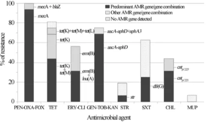
The percentage of each antimicrobial resistance gene and/or gene combination detected among the MRCoNS isolates is presented in Figure 2. The nine (56.3%) erythro-mycin resistant isolates were also resistant to clindaerythro-mycin. Constitutive clindamycin resistance was observed in all but 3 MRCoNS isolates. These 3 isolates revealed the typical D-shaped halo around clindamycin disk, characteristic of inducible clindamycin resistance. Table 1 and Figure 2 show the different resistance genes detected among iso-lates. In some cases, the resistance mechanism implicated could not be detected even though a wide variety of resis-tance genes were tested. Six of the 10 MRCoNS that were also resistant to trimethoprim-sulphamethoxazole lacked any of the so far described genes encoding trimethoprim re-sistance [dfr(A),dfr(D),dfr(G),dfr(K)]. Based on these re-sults, trimethoprim (5mg/mL) and sulfonamide (20mg/mL) antimicrobial agents were tested separately (CLSI 2010) and the results revealed that all trimethoprim-sulpha-methoxazole resistant strains were also trimethoprim resis-tant. One methicillin-resistant S. sciuri subspecies
rodentium(C2867) was resistant to mupirocin butmupA
gene was not detected.
Discussion
Resistance of staphylococci to methicillin and other antimicrobials is a global problem in the chemotherapy of staphylococcal infections. As pointed out by Huebner and Goldmann (1999), this resistance has underscored the need for species identification which is an important step for monitoring the reservoir and distribution of these bacteria. In the present study, the major MR staphylococcal group present in the groin area of dogs in Nsukka was CoNS, with
S. sciurisubspeciesrodentium,being the predominant spe-cies and subspespe-cies identified. Although CoNS are sapro-phytic and rarely pathogenic (Kloos and Bannerman, 1994), they have been associated with opportunistic infec-tions, especially in immunocompromised individuals (Zell
et al., 2008). Previous studies have shownS. sciuri to be present not only as part of the skin, nasal and oral micro-flora of healthy dogs, but also as a causative agent of infec-tions, although at lower rates than S. pseudintermedius Table
(Stepanovic et al., 2001; Hauschild and Wójcik, 2007).
Staphylococcus sciuri was among the staphylococcal strains isolated from cases of canine pyoderma in Natal City, Brazil (Limaet al., 2012). A case of human wound in-fection by a multidrug resistant strain ofS. sciurihas been reported (Coimbraet al., 2011). Thus, this staphylococcal species could constitute a health risk to both humans and animals especially when the natural mucocutaneous barrier is breached.Staphylococcus sciurihas been described as a natural reservoir of the methicillin resistance gene,mecA (Klooset al., 1997; Coutoet al., 2000), which may explain the high rate ofmecA positiveS. sciurirecorded in the pres-ent study. The other MRCoNS isolated in this study, al-though at low rates, have also been reported in dogs as commensal and/or pathogens (Hauschild and Wójcik, 2007).
The absence of MRCoPS colonization among the tested dog population is similar to the findings of Vengust
et al.(2006) and Baptisteet al.(2005) who failed to detect the organism in healthy dogs in Slovenian and a community in United Kingdom, respectively. However, low MRCoPS carriage rates in healthy dogs have been reported by other authors (Hanselmanet al., 2008; Loeffleret al., 2011), with
S. pseudintermedius as the prominent MRCoPS species, even though their occurrence is still low (0-4.5%) (Gómez-Sanzet al., 2011; van Duijkerenet al., 2011). Fail-ure to detect methicillin-resistant S. aureus and S. pseudintermediusamong the sampled dogs suggests that these species are not predominant methicillin-resistant staphylococci in the study area. Alternatively, groin swabs from healthy dogs were used in the present study whereas most reports on S. pseudintermediusin dogs used nasal, skin, perineal or combined body-site samples (Rubin and Chirino-Trejo, 2011; van Duijkerenet al., 2011). It could therefore appear that the groin area of dogs is less colonized
by methicillin-resistantS.pseudintermediusthan the mu-cous or skin.
High rate of MDR was observed among the MRCoNS as 81.3% of them were resistant to more than 3 classes of antimicrobial agents. Cross resistance to other antimicrobials is most common in methicillin-resistant than in methicillin-sensitive staphylococcal isolates (Orrett and Land, 2006; John and Harvin, 2007). In our MRCoNS iso-lates, cross-resistance was due in most cases to tetracy-clines, aminoglucosides, trimethoprim-sulphamethoxazole and/or macrolides-lincosamides. Interestingly, our isolates were recovered from healthy animals whereas nosocomial strains are more likely to be MDR than commensal (John and Harvin, 2007).
Only 2 of the 16 MRCoNS (both beingS. sciuri sub-speciesrodentium) had the same resistance phenotype; in-dicating a high diversity of antimicrobial resistance profiles among the strains. Interestingly, no tetracycline resistance gene was detected in 1 tetracycline-resistant MRS. sciuri
in veterinary practice in Nigeria, thus, mupirocin resistance in one of theStaphylococcusstrain in the present study may not be attributed to mupirocin usage. These data altogether suggest the potential presence of novel characteristics and presents S. sciuri as a potential reservoir of novel anti-microbial resistant properties.
The findings in this study highlight the existence of MDR strains of MRCoNS, particularlyS. sciurisubspecies
rodentium,in healthy dogs in Nsukka, Nigeria. Since dogs are in close contact with their owners, the risk of transmis-sion of MDR staphylococci between animals and humans as well as the possibility of transfer ofmecA and other re-sistance genes from the CoNS to human pathogenic S. aureusare serious public health concerns.
Acknowledgments
This work was partially supported by Project SAF2009-08570 from the Ministry of Education and Sci-ence of Spain and FEDER. E. Gómez-Sanz has a fellow-ship from the Gobierno de La Rioja of Spain; and C. Lozano has a fellowship from the Ministerio de Ciencia e Innovación of Spain.
References
Baptiste KE, Williams K, Williams NJ, Wattret A, Clegg PD, Dawson S, Corkill JE, O’Neill T, Hart CA (2005) Methi-cillin-resistant staphylococci in companion animals. Emerg Infect Dis 11:1942-1944.
Bergeron M, Dauwalder O, Gouy M, Freydiere AM, Bes M, Meugnier H, Benito Y, Etienne J, Lina G, Vandenesch F, Boisset S (2011) Species identification of staphylococci by
amplification and sequencing of thetufgene compared to
thegapgene and by matrix-assisted laser desorption
time-of-flight mass spectrometry. Eur J Clin Microbiol Infect Dis 30:343-354.
Clinical and Laboratory Standards Institute (CLSI) (2010) Perfor-mance standards for antimicrobial susceptibility testing; Eighteenth informational supplement. CLSI Document M100-S20. Wayne, PA.
Coimbra DG, Almeida AGCS, Junior JBO, da Silva LAF, Pi-mentel BJ, Gitai DLG, Moreina LS, Silva-Filho EA, de
An-drade TG (2011) Wound infection by multiresistant
Staphy-lococcus sciuri identified by molecular methods. New
Microbiologica 34:425-427.
Couto I, Sanches IS, Sá-Leão R, de Lencastre H (2000) Molecular
characterization of Staphylococcus sciuri strains isolated
from humans. J Clin Microbiol 38:1136-1143.
Foster TJ (2005) Immune evasion by staphylococci. Nat Rev Microbiol 3:948-958.
Fusi Ngwa CN, Egri-Okwaji MT, Odugbemi T, Iroha E (2007) A study on pediatric MRSA in Lagos, Nigeria. Int J Biol Chem Sci 1:54-60.
Ghebremedhin B, Olugbosi MO, Raji AM, Layer F, Bakare RA, Konig B, Konig W (2009) Emergence and commu-nity-associated MRSA strain with a unique resistance pro-file in Southwest Nigeria. J Clin Microbiol 47:2975-2980. Gómez-Sanz E, Torres C, Lozano C, Saenz Y, Zarazaga M (2011)
Detection and characterization of methicillin-resistant
Staphylococcus pseudintermediusin healthy dogs in La
Rio-ja, Spain. Comp Immun Microbiol Infect Dis 34:447-453. Hanselman BA, Kruth S, Weese JS (2008) Methicillin-resistant
staphylococcal colonization in dogs entering a veterinary teaching hospital. Vet Microbiol 126:277-281.
Hauschild T, Wójcik A (2007) Species distribution and properties of staphylococci from canine dermatitis. Res Vet Sci 82:1-6. Huebner J, Goldmann DA (1999) Coagulase-negative
staphylo-cocci: role as pathogens. Annu Rev Med 50:223-236. John JF, Harvin AM (2007) History and evolution of antibiotic
re-sistance in coagulase-negative staphylococci: Susceptibility profiles of new anti-staphylococcal agents. Therap Clin Risk Manag 3:1143-1152.
Kloos WE, Schleifer KH, Smith RF (1976) Characterization of
Staphylococcus sciurisp. nov. and its subspecies. Int J Syst
Bacteriol 26:22-37.
Kloos WE, Bannerman TL (1994) Update on clinical significance of coagulase negative staphylococci. Clin Microbiol Rev 7:117-140.
Kloos WE, Ballard BN, Webster JA, Hubner RJ, Tomasz A, Couto I, Sloan GL, Dehart HP, Fiedler F, Schubert K, de Lencastre H, Sanches IS, Heath HE, Leblanc PA, Ljungh A
(1997) Ribotype delineation and description of
Staphylococ-cus sciuri subspecies and their potential as reservoirs of
methicillin resistance and staphylolytic enzyme genes. Int J Syst Bacteriol 47:313-323.
Lima LF, Lira AC, Coutinho HDM, Junior JP, Barreto HM (2012) Antimicrobial resistance in staphylococci isolated from ca-nine pyoderma. Comunicata Scientiae 3:181-185.
Loeffler A, Pfeiffer DU, Lindsay JA, Magalhães RJ, Lloyd DH (2011) Prevalence of and risk factors for MRSA carriage in companion animals: a survey of dogs, cats and horses. Epidemiol Infect 139:1019-1028.
Olowe OA, Eniola KIT, Olowe RA, Olayemi AM (2007) Anti-microbial susceptibility and betalactamase detection of MRSA in Osogbo, S.W. Nigeria. Nat Sci 5:44-48.
Orrett FA (2008) The emergence of mupirocin resistance among
clinical isolates of methicillin-resistant Staphylococcus
aureusin Trinidad: a first report. Jap J Infect Dis
61:107-110.
Orrett FA, Land M (2006) Methicillin-resistantStaphylococcus
aureusprevalence: current susceptibility patterns in
Trini-dad. BMC Infect Dis 6: doi: 10.1186/1471-2334-6-83. Poyart C, Quesne G, Boumaila C, Trieu-Cuot P (2001) Rapid and
accurate species-level identification of coagulase-negative
staphylococci by using the sodAgene as a target. J Clin
Microbiol 39:4296-4301.
Rachal T, Leonard K, Martinez L, Breaux JG, Corbin A, Natha-niel R (2009) Prevalence of SCCmec types in methicillin re-sistant Staphylococcus intermedius in healthy pets from Southeastern United States. J Infect Dis Immun 1:006-010. Rubin JE, Chirino-Trejo M (2011) Prevalence, sites of
coloniza-tion, and antimicrobial resistance among Staphylococcus
pseudintermediusisolated from healthy dogs in Saskatoon,
Canada. J Vet Diagn Invest 23:351-354.
Saleha A, Zunita Z (2010) Methicillin resistantStaphylococcus
aureus(MRSA): An emerging veterinary and zoonotic
pa-thogen of public health concern and some studies in Malay-sia. J Anim Vet Adv 9:1094-1098.
Pres-ence of newmecA andmph(C) variants conferring antibiotic
resistance inStaphylococcusspp. isolated from the skin of
horses before and after clinic admission. J Clin Microbiol 44:4444-4454.
Schwarz S, Chaslus-Dancla E (2001) Use of antimicrobials in vet-erinary medicine and mechanisms of resistance. Vet Res 32:201-225.
Stepanovic S, Dimitrijevic V, Vukovic D, Dakic I, Savic B,
Svabic-Vlahovic M (2001)Staphylococcus sciurias a part
of skin, nasal and oral flora in healthy dogs. Vet Microbiol 82:177-185.
van Duijkeren E, Kamphuis M, van der Mije IC, Laarhoven LM, Duim B, Wagenaar JA, Houwers DJ (2011) Transmission of
methicillin-resistant Staphylococcus pseudintermedius
be-tween infected dogs and cats and contact pets, humans and the environment in households and veterinary clinics. Vet Microbiol 150:338-343.
Vengust M, Anderson MEC, Rousseau J, Weese JS (2006) Methi-cillin-resistant staphylococcal colonization in clinically healthy dogs and horses in the community. Lett App Microbiol 43:602-606.
Zell C, Resch M, Rosenstein R, Albrecht T, Hertel C, Gotz F (2008) Characterization of toxin production of coagulase-negative staphylococci isolated from food and starter cul-tures. Int J Food Microbiol 127:246-251.