903 903 903 903 903 Mem Inst O swaldo Cruz, Rio de Janeiro, Vol. 100(8): 903-908, D ecem ber 2005
Basic biology of
Pneumocystis carinii
- A M ini Review
Wanderley de Souza/+, M arlene Benchimol*
Laboratório de Ultraestrutura Celular Hertha Meyer, Instituto de Biofísica Carlos Chagas Filho, Universidade Federal do Rio de Janeiro, CCS-Bloco G, 21941-900 Rio de Janeiro, RJ, Brasil *Laboratório de Ultra-estrutura Celular, Universidade Santa Úrsula,
Rio de Janeiro, RJ, Brasil
Basic aspects of cell biology of Pneumocystis carinii are reviewed with major emphasis on its life cycle and the structural organization of the trophozoites and cyst forms. Initially considered as a protozoan it is now established that Pneumocystis belongs to the Fungi Kingdom. Its life cycle includes two basic forms: (a) trophozoites, which are haploid cells that divide by binary fission and may conjugate with each other forming an early procyst and (b) cysts where division takes place through a meiotic process with the formation of eight nuclei followed by cytoplasmic delimitation and formation of intracystic bodies which are subsequently released and transformed into trophozoi-tes. Basic aspects of the structure of the two developmental stages of P. carinii are reviewed.
Key words:Pneumocystis carinii - life cycle - trophozoites - cyst form - fine structure
At the beginning of the XX century several parasi-tologists dedicated most of their time looking for new parasites in the bloodstream, tissues, and faeces of nor-mal as well as experimentally infected aninor-mals. While car-rying out studies on experimentally infected guinea pigs and in humans with a disease later on designated as Chagas disease or American trypanosomiasis, Chagas (1909), using a light microscope, observed the presence of cystic forms in histological sections of lungs. It is im-portant to remember that the description of the schizonts as intracellular dividing forms of malaria parasites had been described a few years before. This fact probably influenced Chagas to consider the cystic forms as tissu-lar schizonts occurring during the life cycle of a new try-panosome species designated as Schizotrypanum cruzi. Five years later, Delanoe and Delanoe (1914) examining rats collected in Paris described the same forms and con-sidered them as representative of a new protozoan spe-cies which was designated as Pneumocystis carinii as a honour to Antonio Carini, an Italian biologist who de-scribed the same microorganism in the lungs of rats col-lected in Brazil and which were simultaneously infected with Trypanosoma lewisi.
During many years P. carinii was considered as a pro-tozoan without any special medical importance. However, two groups of observations were responsible for the in-clusion of P. carinii among the most important microor-ganisms studied in the past years. The first one, was the conclusion based on comparative analysis of the 16 S ribosomal RNA, mitochondrial genomic gene sequences, aminoacid sequences of peptides and proteins, that P. carinii belongs to the Fungi Kingdom rather than to Pro-tozoa, as considered since its original description (Ypma-Wong et al. 1992, Cushion et al. 1994). According to the
Financial support: CNPq, Faperj, Pronex, Capes, Ausu
+Corresponding author. E-mail: [email protected]
Received 13 July 2005 Accepted 14 December 2005
molecule used for comparative phylogenetic analysis, P. carinii has been included in different groups such as Chytridomycota, Zygomycota, Ascomycetons or Usto-mycetons red yeasts. At present, most authors include P. carinii as a fungus related to ascomycetous yeasts where the well known Sacharomyces cerevisae is located. Based on the analysis of the gene sequences obtained in the genome project, affinity of P. carinii with Schizosac-charomyces pombe and Neurospora crassa has been sug-gested (review in Cushion et al. 1991, Cushion 2004). How-ever, P. carinii presents unique morphological and life cycle characteristics as will be described below.
Biochemical and molecular studies have shown that there is significant genetic diversity in the natural P. carinii population. In the same infected animal several strains may co-exist. P. carinii has been isolated from humans, monkeys, rats, mice, ferrets, sloths, dogs, cats, sheeps, marmosets, and voles. The available information indicates that there are distinct species of Pneumocystis. Analysis based on nucleotide variations of the rRNA genes revealed the existance of several genotypes (Siripattanapipong et al. 2005). During the international workshop for Pneumocystis held in 2001 it was suggested that the spe-cies described in humans should be named P. jirovecii. However, several authors did not follow this recommen-dation. Different host species are infected with geneti-cally different protozoan populations indicating the exist-ence of multiple strains and/or species. Cross infection is also observed.
in-904 904 904 904
904 Basic biology of P. carinii • W de Souza, M Benchimol
fection dramatically increased in the 1980s with the ap-pearance of the acquired immunodeficiency syndrome (Aids) where this organism was the major cause of oppor-tunistic infection and mortality. Indeed, up to 90% of Aids patients developed pneumocystosis, characterized by in-tense organism proliferation with little or no inflammatory response (reviews in Frenkel et al. 1966, Mills 1986).
Due to all these factors the interest in studying P. carinii increased and the Society of Protozoologists or-ganizes every two years a special scientific meeting on opportunistic organisms where papers dealing with P. carinii and Toxoplasma gondii predominate. The papers presented in these meetings are published in special is-sues of the Journal of Eukaryotic Microbiology (http:// www.blackwellpublishing.com/journals).
In this short review we intend to analyze some basic aspects of the biology of P. carinii emphasizing its life cycle, and its cell biology, with special emphasis on its morphology, as observed by electron microscopy.
The life cycle
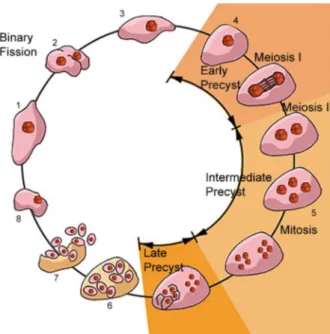
There are several reports on the life cycle of P. carinii, each one presenting different views and various devel-opmental stages. We will consider here a life cycle ac-cepted by most of the authors and which incorporates data obtained using electron microscopy (Fig. 1).
It is not yet clear which is the main infective form re-sponsible for the primary infection. However, it is well established that airbone transmission is the most impor-tant one. For instance, corticosteroid-treated rats develop P. carinii infection when housed with infected rats. Infec-tion by drinking water or food was excluded in these ex-periments. One way to start working with P. carinii is to administer corticosteroids to normal laboratory rodents, thus indicating that highly infective forms are present in the environment.
Fig. 1 shows a schematic view of the life cycle of P. carinii. Two developmental stages are well characterized: the mature cyst and the trophozoite. The trophozoite form is variable in shape, measuring about 0.3 µm in diameter, and they usually form clusters. Some authors consider them to have ameboid characteristic, with the presence of cytoplasmic projections similar to filopodia. However, no such type of cell motility has been reported in living samples.
Trophozoites originate directly from the cyst. Each mature cyst may contain up to eight spherical intracystic bodies, which give rise to eight trophozoites. It has been reported that trophozoites may originate from cysts con-taining spherical, banana-shaped or ameboid intracystic bodies. The initial trophozoite is haploid and divides by binary fission or endogeny. Two trophozoites may conju-gate giving rise to a diploid cell which then divides, as described above for the haploid trophozoites or begin a meiotic process of division with two meiotic cycles where three nuclear divisions takes place, forming a large spheri-cal cell with eight nuclei. Subsequently, there is a process of cell delimitation, forming eight intracystic bodies. Im-portant evidence for the presence of meiosis during the P. carinii life cycle is the presence of a synaptonemal com-plex (Matsumoto & Yoshida 1984). Preliminary analysis of the partial genome of P. carinii genes coding for pro-teins similar to those involved in the mating process in other fungi have been detected (Smulian et al. 2001).
It is important to point out that all these forms have been identified based on the observation of infections in animals and, in a few cases, in cell cultures. Further stud-ies are still necessary to reproduce in vitro the complete life cycle of P. carinii.
Attempts have been made to cultivate the protozoan in different cell lines, using an approach typical for proto-zoa. Samples isolated from the lung of infected animals were inoculated into cell cultures in which they prolifer-ated. Cysts and trophozoites have been observed al-though the latter predominates, growing as clusters in the supernatant. However, many organisms attached to portions of the cells. In some cases the morphology of the attachment resembles that observed in vivo where P. carinii attaches to type I pneumocytes (Bartlett et al. 1994). Attempts to cultivate the organism in cell free media, as usual for fungi, succeeded in ten-fold amplifications of the number of cells. However, continuous axenic cultiva-tion of P. carinii is difficult, and has been obtained only by few groups (Merali et al. 1999).
Cell biology of the trophozoite
Trophozoites are pleomorphic and usually associate with each other forming clusters. They are easily identi-fied in the lung of infected animals due to their irregular shape and close association with pneumocytes. Due to its pleomorphism the association of the trophozoites to each other is very irregular, forming complex aggregates with many interdigitations, sometimes making it difficult to distinguish each individual cell (Figs 2-4).
The cytoplasm of the trophozoite is poor in organelles (Vavra & Kucera 1970). Free ribosomes predominate. Gly-cogen particles are also seen. Tubular and ramified
905 905 905 905 905 Mem Inst O swaldo Cruz, Rio de Janeiro, Vol. 100(8), D ecem ber 2005
tures, resembling the endoplasmic reticulum are observed. A small nucleus is seen in variable positions. Usually it appears homogeneous, with an electrondensity similar to that of the cytoplasm. A nucleolus is evident and located centrally or peripherycally to the nucleus. Dividing nuclei have been observed. The mitotic process occurs within an intact nuclear membrane and there are mitochondria with short cristae. Osmiophilic bodies, vacuolar spaces, microtubules and an incipient Golgi complex were also reported, although not frequently seen (Dei-Cas et al. 1989).
For some authors the trophozoite is limited by two unit membranes spanning approximately 20-30 nm. The outer membrane is decorated with an electron dense layer and separated from the inner membrane by a thin electron lucent space (Vossen et al. 1978, Hughes 1987). Accord-ing to our view, one characteristic feature of the tropho-zoite is the presence of a 20-30 nm thick and dense coat, which is found in the whole surface of the cell, including the regions of interdigitations (Figs 2-4). At high magnifi-cation it is clear that the coat is not homogeneous, leav-ing small electron translucid areas (Fig. 4). Cross sections of filopodium-like regions give the impression that some periodicity exists.
In heavily infected animals areas are found where por-tions of the P. carinii surface were released as a shed-ding-like process. Hundreds of longitudinally and transversally sectioned small tubules are observed, al-ways containing the plasma membrane and the character-istic surface coat (Figs 3-4).
Freeze-fracture analysis revealed the presence of only one unit membrane displaying a large number of randomly distributed intramembranous particles on both protoplas-mic and extracellular faces (Yoneda et al. 1982). The par-ticle density was higher on the P than on the E fracture face (Yoshida 1989).
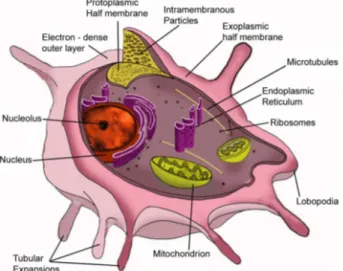
Fig. 5 shows a schematic view of the trophozoite forms displaying various organelles.
What is the nature of the thick surface coat seen only in the trophozoites surface? There are some suggestions that it anchores the organism or is involved in nutrient uptake. Studies using labeled lectins showed that resi-dues of mannose, N-acetyl-glucosamine and galactose/ N-acetyl-galactosamine are exposed on the cell surface (Yoshikawa et al. 1987, Cushion et al. 1988, Pesanti & Stanley 1988). Labeling with lectins recognizing fucose or sialic acids was very light or even absent (De Stefano et al. 1992).
One important surface antigen of P. carinii has 116 kDa in polyacrilamide gel electrophoresis (SDS-PAGE). Under non-reducing conditions it appears to exist as an
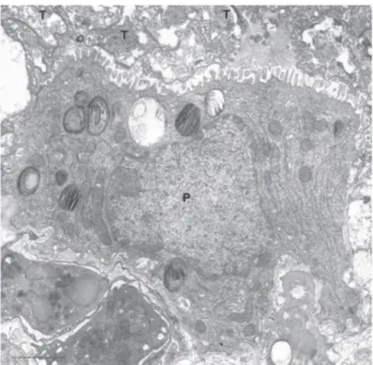
Fig. 2: thin section showing the presence of a large number of trophozoites (T) in the apical portion of a pneumocyte (P). Bar = 2 µm.
906 906 906 906
906 Basic biology of P. carinii • W de Souza, M Benchimol
aggregated form with a molecular weight of > 2. 106 kDa.
This protein is localized in the surface coat and has simi-larities with mucin-type glycoproteins (Radding et al. 1989). Based on the fact that administration of monoclonal anti-bodies recognizing this protein had a benefitial effect on the course of the experimental pneumonia, it has been suggested that it plays some role in pathogenesis (Gigliotti & Hughes 1988). Indeed, the surface of tropho-zoites is covered by the major surface glycoprotein or glycoprotein A which is a family of protein encoded by up to one hundred heterogeneous genes. These genes are localized at the ends, upstream of the subtelomeric and telomeric repeats of all chromosomes. Transcription is lim-ited to a single gene each time and is involved in a pro-cess that resembles the antigenic variation that has been well characterized in Trypanosma brucei (Stringer & Keely 2001).
Trophozoites interact with the surface of pneumocytes (Fig. 2). In most of the cases such interaction occurred through the surface coat that established contact with the microvilli of the epithelial cells. At some points such contact was done through filopodium-like structures. There are evidences that fibronectin is a mediator of the P. carinii attachment to pneumocytes with the participation of fibronectin-binding receptor on the fungus surface and a fibronectin-binding integrin of the host cell surface (Pottratz et al. 1991, 1994, Aliouat et al. 1993).
Part of the P. carinii population is ingested by alveo-lar macrophages in a process mediated by the Dectinn-1 β-glucan receptor, with production of hydrogen peroxide and subsequent killing of the organism (Steele et al. 2003).
Cell biology of the cystic form
The cysts are easily identified due to their typical morphology, being spherical structures with a mean di-ameter of 5-8 µm, containing up to eight intracystic bod-ies (Figs 6-12). Each intracystic body has a mean diameter of 1.2 µm.
Fig. 5: schematic view of the trophozoite form where the main structures described in the text are indicated.
907 907 907 907 907 Mem Inst O swaldo Cruz, Rio de Janeiro, Vol. 100(8), D ecem ber 2005
The cyst wall has two layers and is approximately 50 nm thick (Figs 6-8). The outer layer, with a thickness of about 15 nm, is more electrondense. The inner layer, with a thickness of 35 nm, is less dense and is in contact with the plasma membrane. Some authors described the pres-ence of a membrane-like structure on the outer electrondense layer (DeStefano et al. 1990a). However, this structure was not seen in most of the published elec-tron micrographs. A general schematic view of the cystic form is shown in Fig. 12.
Freeze-fracture studies show the presence of intrabranous particles on the fracture faces of the cyst mem-brane (Yoneda et al. 1982). This number, however, was smaller than that observed in the membrane of the tro-phozoites.
Biochemical analysis has shown that the cyst wall is rich in glucosyl/mannosyl and galactose/N-acetyl-D-ga-lactosamine residues (DeStefano et al. 1990b). The inner portion of the cyst contains two components: a matrix and the intracystic bodies. The matrix contains mitochon-dria, ribosomes, empty vacuoles and membrane debris. Its dimension varies according to the size of the cyst and the number of intracystic bodies. The intracystic body is a spherical to oval shape, with a centrally located nucleus, endoplasmic reticulum, free ribosomes, mitochondria, and glycogen particles.
It is very common to observe cystic bodies with a banana-like shape. These forms only show the matrix con-tent, but no intracystic bodies. They are considered as remnants of the ruptured cysts which have released the intracystic bodies.
The use of the Thiéry technique, which reveals the presence of carbohydrates, showed the presence of gly-cogen particles in the trophozoites, the intracystic bodies and in the cyst matrix. The trophozoite’s plasma mem-brane, the membrane lining the cyst and the intracystic bodies was also labeled, but the trophozoite’s surface coat and the cyst wall were not labeled.
Perspectives
It is clear from the results described above that we know little about the cell biology of Pneumocystis. This is because there is no experimental system in which the or-ganisms can be easily maintained in vitro, that would en-able to its life cycle be studied in detail. Therefore, it is important that such a system is found to open up new possibilities for further biochemical and molecular stud-ies.
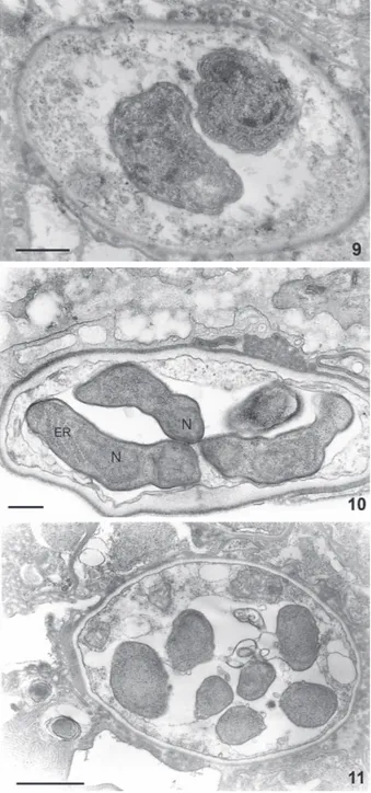
Figs 9-11: evolution of the cyst form. Cysts containing two, four, and eight intracystic bodies are shown. Each intracyst body con-tains all components characteristics of an eukaryotic cell such as the nucleus, endoplasmic reticulum and other organelles. Bars: Fig. 9 = 1.5 µm; Fig. 10 = 250 nm; Fig 11 = 1 µm.
908 908 908 908
908 Basic biology of P. carinii • W de Souza, M Benchimol
REFERENCES
Aliouat EM, Dei-Cas E, Ouaissi A, Palluault F, Soulez B, Camus D 1993. In vitro attachment of Pneumocystis carinii from mouse and rat origin. Biol Cell 77: 209-217.
Bartlett MS, Goheen MP, Lee CH, Shaw MM, Durkin MM, Smith JW 1994. Close association of Pneumocystiscarinii from infected rat lung with culture cells as shown by light and electron microscopy. Parasitol Res 80: 208-215. Chagas C 1909. Nova tripanozomiaze humana. Mem Inst
Oswaldo Cruz 1: 159-181.
Cushion MT 2004. Pneumocystis: unraveling the cloak of ob-scurity. TrendsMicrobiol 12: 243-249.
Cushion MT, De Stefano JÁ, Walzer PD 1988. Pneumocystis carinii: surface reactive carbohydrates detected by lectin probes. Exp Parasito1 67: 137-147.
Cushion MT, Harmsen A, Matsumoto Y 1994. Recent advances in the biology of Pneumocystis carinii. J Med Vet Mycol 32: 217-228.
Cushion MT, Stringer JR, Wa1zer PD 1991. Cellular and mo-lecular biology of Pneumocystis carinii. IntRev Cytol 131: 59-107.
Dei-Cas E, Soulez B, Camus D 1989. Ultrastructural study of Pneumocystis carinii in explants cultures of rabbit lung and cultures with and without feeder cells. J Protozool (Suppl.) 36: 55S-57S.
Delanoe P, Delanoe M 1914. Sur le rapports des kystes du pommon des rats avec le Trypanosoma lewisi. Comp R Acad Sci 155: 658-661.
De Stefano JA, Cushion MT, Sleight RG, Walzer PD 1990a. Analysis of Pneumocsytis carinii cyst wall. I. Evidence for an outer surface membrane. J Protozool 37: 428-435. De Stefano JA, Cushion MT, Puvanesarajah V, Walzer PD 1990b.
Analysis of Pnemocystis carinii cyst wall. II. Sugar compo-sition. J Protozool 37: 436-441.
De Stefano JA, Trink1e LS, Wa1zer PD, Cushion MT 1992. Flow cytometrical analysis of lectin binding to Pneumocystis carinii surface carbohydrates. J Parasitol 78: 271-280. Frenkel JK, Good JF, Shultz JA 1966. Latent Pneumocystis
infection of rats, relapse and chemotherapy. Lab Invest 15: 1559-1577.
Giglioli F, Hughes WT 1988. Passive immunoprophylaxis with specific monoclonal antibodies confers partial protection against Pneumocystis carinii pneumonitis in animal mod-els. J Clin Invest 81: 1666-1668.
Hughes WT 1987. Pneumocystis carinii Pneumonitis, CRC Press, Boca Raton, FL.
Itatani CA 1994. Ultrastructural demonstration of a pore in the cyst wall of Pneumocystis carinii. J Parasitol 80: 644-648. Matsumoto Y, Yoshida Y 1984. Sporogony in Pneumocystis carinii: synaptonemal complexes and meiotic nuclear divi-sions observed in precysts. J Protozool 31: 420-428. Merali S, Frevert U, Williams JH, Chin K, Bryan R, Clarkson
Jr AB 1999. Continuous axenic cultivation of P. cariniii. Proc Natl Acad Sci NY 96: 2402-2407.
Mills J 1986. Pneumocystis carinii and Toxoplasma gondii in-fections in patients with AIDS. Rev Infect Dis 8: 1001-1011.
Pesanti EL, Stanley JD 1988. Glycoproteins of Pneumocystis carinii characterization by electrophoresis and microscopy. J Infect Dis 158: 1353-1359.
Pottratz ST, Paulsrud J, Smith JS, Martin WJ 1991. Pneumocystis carinii attachment to cultured lung cells by pneumocystis gp120, a fibronectin binding protein. J Clin Invest 88: 403-407.
Pottratz ST, Weir AL, Wisniowski PE 1994. Pneumocystis carinii attachment increases expression of fibronectin-binding integrins on cultured lung cells. Infect Immun 62: 5464-5469.
Radding JA, Armstrong MYK, Ullu E, Richards FF 1989. Iden-tification and isolation of a major cell surface glycoprotein of Pneumocystis carinii. Infect Immun 57: 2149-2157. Siripattanapipong S, Worapong J, Mungthin M, Leelayoova S,
Tan-ariya P 2005. Genotypic study of Pneumocystis jirovecii in human immunodeficiency virus-positive pa-tients in Thailand. J Clin Microbiol 43: 2104-2110 Steele C, Marrero L, Swain S, Harmsen AG, Zheng M, Brown
GD, Gordon S, Shellito JE, Kolls JK 2003. Alveolar mac-rophage-mediated killing of Pneumocysts carinii f. sp. muris invoves molecular recognition by the Dectin-1 β-glucan re-ceptor. J Exp Med 198: 1677-1688.
Smulian AG, Sesterhenn T, Tanaka R, Cushion MT 2001. The ste3 pheromone receptor gene of Pneumocystiscarinii is surrounded by a cluster of signal tranduction genes. Genet-ics 157: 991-1002.
Stringer JR, Keely SP 2001. Genetics of surface antigens ex-pression in Pneumocystis carinii. Infect Immun 69: 627-639.
Vavra J, Kucera K 1970. Pneumocystis carinii Delanoe, its ul-trastructure and ultrastructural affinities. J Protozool 17: 463-483.
Vossen MEMH, Beckers PJA, Meuwissen JHET, Stadhouders AM 1978. Developmental biology of Pneumocystis carinii: an alternative view on the life cycle of the parasite. Z Parazitenkd 55: 101-118.
Yoneda K, Walzer PD, Richey CS, Birk MG 1982. Pneumocystis carinii: freeze-fracture study of stages of the organism. Exp Parasitol 53: 68-76.
Yoshida Y 1989. Ultrastructual studies of Pneumocystis carinii. J Protozool 36: 53-60.
Yoshikawa H, Tegoshi T, Yoshida Y 1987. Detection of surface carbohydrates on Pneumocystis carinii by fluorescein-con-jugated lectins. Parasitol Res 74: 43-49.
Ypma-Wong MF, Fonzi WA, Sypherd PS 1992. Fungus-spe-cific translation elongation factor 3 gene present in Pneumocystis carinii. Infect Immun 60: 4140-4145.