www.scielo.br/aabc
Secretory organelles of pathogenic protozoa
WANDERLEY DE SOUZA*
Laboratório de Ultraestrutura Celular Hertha Meyer, Instituto de Biofísica Carlos Chagas Filho CCS-Bloco G, Universidade Federal do Rio de Janeiro, Ilha do Fundão
21949-900 Rio de Janeiro, RJ, Brasil
Manuscript received on September 6, 2005; accepted for publication on February 1, 2006.
ABSTRACT
Secretory processes play an important role on the biology and life cycles of parasitic protozoa. This review focus on basic aspects, from a cell biology perspective, of the secretion of (a) micronemes, rhoptries and dense granules in members of the Apicomplexa group, where these organelles are involved in the process of protozoan penetration into the host cell, survival within the parasitophorous vacuole and subsequent egress from the host cell, (b) the Maurer’s cleft inPlasmodium, a structure involved in the secretion of proteins synthesized by the intravacuolar parasite and transported through vesicles to the erythrocyte surface, (c) the secretion of macromolecules into the flagellar pocket of trypanosomatids, and (d) the secretion of proteins which make the cyst wall ofGiardiaandEntamoeba, with the formation of encystation vesicles.
Key words:parasitic protozoa, cell secretion, Apicomplexa, trypanosomatids, encystation vesicles.
INTRODUCTION
The protozoa kingdom comprises a large number of species, including some which are agents of human and veterinary diseases such as malaria, leishmaniasis, Chagas’ disease, African trypanoso-miasis, amebiasis, trichomoniasis, giardiasis, tox-oplasmosis, coccidiosis, theileriosis, and babesio-sis, to mention only those more important. Some of these protozoa, as is the case ofTrichomonas, present a simple life cycle. For others, however, as occurs with Apicomplexa (which includes Plas-modium, Toxoplasma, Eimeria, etc), and some try-panosomatids, the life cycle is relatively complex, displaying several developmental stages in the ver-tebrate host and, in some cases, in inverver-tebrate hosts. These protozoa are also of interest from the cell biology point of view since they present
spe-*Member Academia Brasileira de Ciências E-mail: [email protected]
cial cytoplasmic structures and organelles, which have been studied in some detail in the last years providing new information of general biological in-terest. In a previous review we analyzed organelles involved in the metabolic pathways (De Souza 2002). Here, we intend to review, from a cell bi-ology perspective, organelles involved in secretory processes. We will not emphasize aspects well cov-ered in a previous review (Becker and Melkonian 1996).
CELL SECRETION IN APICOMPLEXA
(Fig. 1). In addition to cytoskeleton components, such as the conoid-sub-pellicular microtubules com-plex, two organelles were initially recognized and designated as micronemes and rhoptries. Later on another structure, which can be found in other re-gions of the protozoan body, was recognized and designated as dense granules.
Apicomplexan protozoa present a typical en-doplasmic reticulum with associated ribosomes. In T. gondiithe ER is distributed throughout the cell and the nuclear envelope itself provides a signifi-cant fraction of the ER. Vesicles with a fuzzy coat bud off from the nuclear envelope and the ER. In Plasmodiumthe ER is less well developed. Several cisternae of the Golgi complex are observed in the anterior region, just above the nucleus ofT. gondii. Vesicles with a clathrin-like coat bud off from the trans portion of the Golgi complex. Proteins such as COP I and II, Arf 1 and Sar 1, involved in vesi-cle formation at the ER-Golgi complex system, have been found inT. gondii(Review in Joiner and Roos 2002, Ngô et al. 2000).
THEMICRONEMES
Micronemes are small, cigar-shaped organelles that are restricted to the apical third of the protozoan body. Their number varies according to the species and the developmental stages. In some species are hardly seen while in others are so numerous that correspond to the most abundant organelle found in the cell (Fig. 2). The organelle is surrounded by a typical unit membrane and presents an electron dense matrix due to its large protein content. Indeed the organelle is intensely stained when the protozoa are submitted to the ethanolic phosphotungstic acid technique, which reveals basic proteins (De Souza and Souto-Padrón 1978). At present, we still do not have a clear explanation for this labeling pattern since the known micronemal proteins have isolec-tric points lower than 7.0. All proteins found in the micronemes are synthesized with an N-terminal signal sequence that mediates their entrance into the secretory pathway by translocation across the endo-plasmic reticulum membrane. A mutagenesis
anal-ysis of the C-terminal portion of MIC2 showed the presence of two conserved amino acid motifs me-diating the target of this protein to the micronemes (Di Cristina et al. 2000). One motif is a tyrosine-based signal and the other one consists of a stretch of acidic residues.
Many of the micronemal proteins are glycosy-lated as seen by labeling of the micronemes when thin sections ofT. gondiiare incubated in the pres-ence of gold-labeled lectins (Carvalho et al. 1991). It has been shown that when the infective forms of Apicomplexan parasites touch the host cell surface they trigger a process of Ca2+
release and the discharge of the content of the micronemes at the junction between the parasite and the host cell (Carruthers et al. 1999a, Bouchot et al. 1999, Vieira and Moreno 2000) which then mediates par-asite attachment (Carruthers et al. 1999a, b, Car-ruthers and Sibley 1999). This process takes place in a few seconds and the released proteins are not incorporated together with the parasites but instead are capped and released from the posterior end of the protozoan (Carruthers et al. 1999a). During re-distribution on the parasite surface, transmembrane MICs are thought to connect external recep-tors to the submembranous acto-myosin motor that provides the power for parasite motility. Chelat-ing of parasite intracellular Ca2+inhibited both mi-croneme release and invasion of host cells. What is the origin of the Ca2+
used by the protozoan? There is enough data indicating that the Ca2+
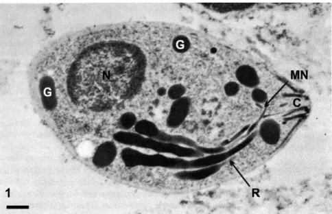
su-Fig. 1 – Transmission electron microscopy of a tachyzoites of Toxoplasma gondiisubmitted to the ethanolic phosphotungstic acid technique, which labels structures containing basic proteins. In addition to the nucleus (N), staining of the dense granules (G), Rhoptries (R), Micronemes (M) and the Conoid (C) is observed. Bar, 0.3µm. After De Souza and Souto-Padrón 1978.
perfamily (Lovett et al. 2002).
It has been shown that isoforms of phospho-glucomutase, a cytosolic enzyme, are implicated in Ca2+-mediated signaling events. One isoform, known as parafusin, plays an important role dur-ing exocytic activity in ciliated protozoa (Zhao and Satir 1998). A protein called parafusin-related pro-tein has been identified inT. gondiiand showed to be localized to an apical subpopulation of mi-conemes and to be redistributed during invasion of the host cell by the protozoan (Matthiesen et al. 2001a, b).
Several micronemal proteins contain one or more adhesive motifs found in mammalian proteins such as Epidermal Growth Factor, integrins, throm-bospondin and kallikrein. Four of them, all desig-nated as MIC (MIC1-4, from micronemal proteins), have been studied in some detail.
MIC1 has a size of 60 kDa and contains two degenerate repeats similar to an adhesive sequence found in thrombospodin and known as type I repeats (TSP-I), and is able to bind to the
consist-Fig. 2 – Routine transmission electron microscopy showing a trophozoite ofCyrilia lignieresi, a haemogregarine found in erythrocytes of a fresh-water fish. Structures such as lipidic in-clusions (Li), amilopectin granules (A), dense granules (D), rhoptries (R) and a large number of micronemes (M) are observed. Spherical bodies are seen within the flagellar pocket (small arrowheads). HCN, host cell nucleus. Bar, 1µm. After Diniz et al. 2002.
ing of threeαβdimmers (Jewett and Sibley 2004). Following discharge MIC2 is proteolitically cleaved by proteases with release of its ectodomain from the parasite surface, a process that seems to be involved on parasite invasion (Carruthers et al. 2000, Brossier et al. 2003). Recently, proteins be-longing to the rhomboid family of intramembrane-cleaving serine proteases, designated as TgROMs, were detected (Brossier et al. 2005, Dowse et al. 2005).
The binding of MIC2 to the host cell surface may establish connection between the host cell sur-face receptor and the cytoskeleton machinery of the parasites activating the gliding process necessary for the penetration of the parasite into the host cell. It has been shown that Plasmodium sporozoites, which do not express a homologue of MIC2, known as TRAP, fail to glide or invade host cells (Sultan et al. 1997).
MIC3 has a size of 90 kDa and possesses five partially overlapping epidermal growth factor (EGF) domains and an NH2terminal chitin
binding-like domain, which probably are also involved in the process of parasite association to the host cell surface (Fourmaux et al. 1996, Soldati et al. 2001). It is a disulfide-linked heterodimer comprised of two 38 kDa isoforms (Achbarou et al. 1991). Sin-gle substitution of two critical amino acids in the chitin binding-like domains of this protein abolished its binding to cells and decrease parasite virulence (Cérède et al. 2005).
molecule to adopt a protease-resistant, rigid struc-ture that could favor its interaction with host cell ligands (Periz et al. 2005).
MIC 6, which is a transmembrane protein, forms trimeric complexes with soluble micronemal proteins such as MIC1 and MIC 4, functioning as an escort protein. Deletion of MIC 6 prevents targeting of these two proteins to the microneme (Reiss et al. 2001). MIC 8 also seems to work as a escort protein to MIC 3 (Meissner et al. 2002).
InPlasmodium falciparuma micronemal pro-tein, known as EBA 175, has been shown to bind to sialic acid (Sim 1995) a molecule, which plays a fundamental role on the process of parasite-erythro-cyte interaction.
More recently several other proteins which do not present recognizable adhesive motifs have been identified in the micronemes ofT. gondii. TgMIC 5 has homology to the parvulin family of peptidyl prolyl cis-trans isomerases and may assist in the folding of other micronemal proteins (Brydges et al. 2000). Other proteins, including Tg MIC10 entirely devoid of cysteines, were recently identified (Hoff et al. 2001). MIC 11, a 16 kDa protein, was recently identified in several coccidian parasites. During its traffic through the secretory pathway it is proteoliti-caly cleaved with removal of an internal propeptide, resulting in a mature form containing aα-chain and aß-chain tethered by a single disulfide bond (Harper et al. 2004).
Members of the genus Plasmodium present several developmental stages, which are able to in-vade different cells in both vertebrate and inbrate cells. For instance, merozoites invade verte-brate red blood cells, the ookinete invades epithe-lial cells of the insect vector while sporozoites in-vade the salivary gland epithelial cells of the insect and when inoculated into the vertebrate host, tra-verse several cellular barriers until invade hepatic cells. Recent studies have shown thatPlasmodium presents several genes coding for proteins, which present a membrane-attack complex/perforin-like domain. One of these proteins (PLP1/SPECT2) found in sporozoites was shown to be localized in
the micronemes (Kaiser et al. 2004) and is necessary for cell traversal (Ishino et al. 2005). Genes encod-ing proteins with similar properties were identified in the genome ofEimeriaandToxoplasma.
THERHOPTRIES
Fig. 3 – Two trophozoites of Toxoplasma gondiiwithin the parasitophorous vacuole. C, conoid; HC, host cell; M, mitochondrion; N, nucleus; R, rhoptries. Bar, 0.4µm.
the rhoptry ofT. gondii(Karasov et al. 2005). This protein may be involved in rhoptry pH regulation. This organelle presents as a characteristic feature the fact that when its lipids and proteins are exo-cytosed through the duct the organellar membrane is retained and an empty organelle, which can be easily identified by electron microscopy, remains.
Immunocytochemistry provided evidence that the rhoptry content is not homogeneous. For in-stance, some proteins are localized in the basal re-gion of the organelle whereas others are located in its apical portion (Review in Blackman and Ban-nister 2001).
Secretion of rhoptry proteins takes place im-mediately after adhesion of the parasites to the host cell surface. In the case ofT. gondiikinetic studies showed that release of the proteins is completed in about 1 minute and that the proteins are internalized and will make part of the membrane lining the para-sitophorous vacuole (Sam-Yellowe et al. 1988,
homol-ogy with any other proteins in the databases (Beck-ers et al. 1994, 1997). Both possess single puta-tive transmembrane segments 75 amino acids from their respective C-termini. OP2 is secreted during the process of penetration ofT. gondiiinto the host cell and is found in association with the membrane lining the parasitophorous vacuole. Its N-terminal domain is exposed on the cytoplasmic face of the parasitophorous vacuole and may be involved in the association of the vacuoles with cytoplasmic organelles of the host cell such as the endoplas-mic reticulum and mitochondria (Sinai et al. 1997, Sinai and Joiner 2001). Little is known about the other ROP proteins, which have been identified us-ing monoclonal antibodies (Leriche and Dubre-metz 1991). A novel rhoptry protein, designated as BRP1, was recently identified in nascent organelles found during the first division of bradizoites, but not in tachyzoites (Schwarz et al. 2005).
The rhoptries of T. gondiihave been isolated by subcellular fractionation procedures and bio-chemical analysis showed a lipid to protein ratio of 0.26, thus indicating their richness in proteins. The cholesterol to phospholipid ratio was 1.48. Phos-phatidylcholine was the major phospholipid (Fous-sard et al. 1991). Proteomic analysis using mass spectrometry of the fraction identified 38 novel pro-teins. At least 11 of them were localized in the rhop-tries, as shown by immunofluorescence microscopy. Some are localized in the bulbous basal portion of the organelle while others are restricted to the neck portion, an observation which points to the existence of different domains in the rhoptries. In addition other proteins such as toxofilin, Rab 11, kinases and phosphatases were also found in the rhoptry (Bradley et al. 2006).
The rhoptries isolated from P. falciparum merozoites showed a large number of proteins (Etzion et al. 1991). A large number of rhoptry pro-teins have been identified inBabesia, Plasmodium andEimeria(Review in Sam-Yellowe 1999). Many of them present as a special feature the ability to bind to erythrocytes.
DENSEGRANULES
The dense granules are spherical organelles distri-buted throughout the cell rather than localized at the apical complex, with a mean diameter of 0.2µm (Fig. 4). Its matrix is uniformly electron dense due to the high concentration of protein. Kinetic studies have shown that secretion of the dense granule con-tent takes place after parasite invasion and localiza-tion within the parasitophorous vacuole persisting for several minutes (Carruthers and Sibley 1997). In contrast to secretion of micronemes and rhop-tries, which takes place in the apical region, dense granule secretion occurs at the lateral regions of the protozoan. The secreted proteins associate with the membrane of the parasitophorous vacuole and with the parasite derived intravacuolar membranous net-work. Proteins are delivered to the dense granule by the bulk flow pathway (Coppens et al. 1999). Proteins from which specific targeting signals for other organelles have been deleted are localized in the dense granules (Striepen et al. 2001, Reiss et al. 2001).
Fig. 4 – Secretion of the dense granule content (arrows) in the lateral side of the tachyzoite form ofToxoplasma gondii. D, dense granules; R, rhoptries. Bar, 0.35µm. Courtesy of JF Dubremetz.
attenuated virulence to mice it has been suggested that this protein plays some role in the virulence of the parasite (Mercier et al. 1998b). GRA3 has a size of 30 kDa and forms multimeric complexes that associate with the membrane lining the vac-uole through hydrophobic interactions (Ossorio et al. 1994), although there is no predicted membrane-spanning domain for it. However, it was recently shown that GRA3 is actually an artificial chimera of 2 proteins. One, with a molecular weight of 65 kDa, shares the C-terminus of GRA3 and the other, with a predicted molecular weight of 24 kDa, shares the N-terminal region and is recognized by antibodies previously shown to label the dense granules. The corrected GRA3 has a N-terminal secretory signal sequence and a transmembrane domain, which ex-plains its insertion into the membrane lining the par-asitophorous vacuole (Henriquez et al. 2005).
GRA 4, 5, 6, 7 and 8 each have one putative transmembrane segment and it has been suggested that they may constitute the molecular sieve that allows the passage of molecules smaller than
in-Fig. 5 – Stack offlattened lamellae (arrows), which form the Maurer’s cleft found in erythrocytes, infected withPlasmodium falciparum. P, parasite. Bar, 0.25µm. After Przyborski et al. 2003.
volves depletion of ATP and increase in Ca2+ con-centration (Stommel et al. 1997, Silverman et al. 1998). Two protease inhibitors have also been iden-tified in the dense granules (Morris et al. 2002, Pszenny et al. 2002).
THE MAURER’S CLEFT INPLASMODIUM
Erythrocytes infected by P. falciparum show the presence of a cytoplasmic structure, which is la-beled when the cells are incubated in the presence of anti-malaria antibodies, and observed byfl uores-cence microscopy (Tobie and Coatney 1961). Trans-mission electron microscopy of thin sections of in-fected erythrocytes revealed the presence of stacks offlattened lamelae of long slender membranes with a translucent lumen, usually located below the ery-throcyte plasma membrane, and designated as Mau-rer’s cleft (Fig. 5) (Trager et al. 1966, Langreth et al. 1978). Variable aspects of this structure have been extensively described in severalPlasmodium species (Review in Przyborski et al. 2003, Lanzer et al. 2006). This structure has not been observed in other members of the Apicomplexa group.
Several parasite proteins have been shown to be localized in the Maurer’s cleft. They are syn-thesized in the parasite ER and then transferred to the cleft (Blisnick et al. 2005, Marti et al. 2005).
well the process of protein transport and trafficking in Plasmodium falciparum- infected erythrocytes (Przyborski and Lanzer 2005). Another recent re-view (Marti et al. 2005) analyses the functional role played by several parasite proteins which concen-trates in the Maurer’s cleft and are involved in pro-cesses such as cytoadherence and import of serum proteins.
CELL SECRETION IN TRYPANOSOMATIDS
Members of the Trypanosomatidae family present a well developed endoplasmic reticulum-Golgi complex system with the formation of coated and uncoated vesicles which subsequently migrate to-wards a specialized and polarized region of the cell surface, known as theflagellar pocket, where they fuse with the membrane (Figs. 6-7) (Reviews in De Souza 1984, Landfear and Ignatushchenko 2001). Theflagellar pocket corresponds to a specialized re-gion where most of the endocytic and exocytic ac-tivities take place in the trypanosomatids. It is im-portant to point out that the secretory vesicles do not present a dense content and for this reason they are not easily distinguished from the endocytic vesicles which form at theflagellar pocket region and are in-volved in the uptake of important macromolecules or macromolecular complexes such as transferring and LDL. At least three distinct groups of secretory prod-ucts have been identified in trypanosomatids based on their fate: (a) one group contains integral or pe-ripheral proteins which are inserted into theflagellar pocket and subsequently migrates to other regions of the plasma membrane. The most evident exam-ples include the synthesis and secretion of the variant surface proteins (VSGs) found in bloodstream forms ofTrypanosoma bruceiand which is involved in the process of antigenic variation (Review in Borst et al. 1998) and cysteine proteinase (cruzin or cruzipain) T. cruzi(Fig. 8) (Souto-Padrón et al. 1990); (b) The second group are accumulated in special organelles, as the megasomes ofLeishmania(Figs. 9-10); (c) the third group includes proteins that are released into theflagellar pocket where they remain as
sol-uble proteins, as is the case of cysteine proteinase (Fig. 11) and proteophosphoglycans inLeishmania (Duboise et al. 1994, Foth et al. 2002), while oth-ers polymerize within the pocket, as is the case of acid phosphatase found inLeishmania(Stierhof et al. 1994). How these different proteins are sorted is not yet clarified. Studies carried withT. bruceihave shown that VSG lacking its GPI anchor is not effi -ciently secreted. The proteins may be then mistar-geted to the lysosome and is subsequently degraded (Triggs and Bangs 2003).
CELL SECRETION IN GIARDIA
Fig. 6 – Anterior region of an amastigote form ofTrypanosoma cruzishowing theflagellar pocket (FP), two shortflagella (F), the basal body (BB) and the kinetoplast (K). Vesicles are seen close to theflagellar pocket (arrow). Bar, 0.25µm.
localization of these proteins. Although continuity of this structure with the ER is evident, glucose-6-phophatase, a classical enzyme marker of the ER, is no more found in this structure (Lanfredi-Rangel et al. 2003). Subsequently the dense vesicle, which is now designated as an encystation vesicle (ESV), increases in density (Figs. 15-17) and migrates to-wards the periphery of the cell (Figs. 18-19). An-other view of the process suggests that ER vesicles containing CWP use to each other to form the ESV (Marti and Hehl 2003, Marti et al. 2003). Analysis of the genome data onG. lamblialed to the iden-tification of orthologs to factors involved in vesicle tethering and fusion, as soluble
N-ethyl-maleimide-sensitive fusion proteins, Sec-1 adapters, Rabs and the COPI complex. Two syntaxin homologs and two Rab GTPases, designed as Rab 1 and Rab 2, were identified (Marti et al. 2003).
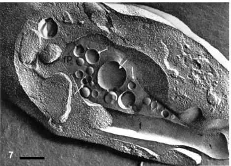
Fig. 7 – Freeze-fracture view of the anterior region of a promastigote forms ofHerpetomonas. A large number of vesicles (arrows) are seen within theflagellar pocket (FP). F,flagellum. Small arrows point to an aggregation of intramembranous particles, which form theflagellar-cell body adhesion structure. Bar, 0.12µm. After De Souza et al. 1979.
Fig. 8 – Immunocytochemical localization of cysteine proteinase in epimastigotes ofTrypanosoma cruzi. This protein is synthesized in the ER and concentrated in structures which are part of the endocytic pathway of this protozoan (asterisks) and in the plasma membrane (arrows). Bar, 0.5µm.
After Souto-Padrón et al. 1990.
with the fusion of the ESV with the cell surface. It is important to point out that the cyst wall pro-teins are not glycosylated but they have potential N- and O-glycosylation sites (Lujan et al. 1995b, Mowatt et al. 1995). It has been considered that the cyst wall, which is formed by interconnectedfi
Figs. 9-11 – Immunocytochemical localization of cysteine proteinase in amastigotes ofLeishmania amazonensis. This protein is stored in the megasomes (M) and secreted into theflagellar pocket. Labeling of the cell surface (arrow infigure 11) and of the cisternae of the endoplasmic reticulum is observed. FP,flagellar pocket; K, kinetoplast; L, lipidic inclusion; M, megasome. Bar, 0.25µm.
Micrographs from T Ueda-Nakamura and W de Souza.
2003). More recently it was suggested that a typical exocytosis does not occur. During membrane fusion some membrane segments appeared to be disrupted and released into the extracellular medium where could be resealed forming empty vesicles (Benchi-mol 2004).
During the period of encystation G. lamblia maintains a constitutive pathway for the synthesis of the variant surface proteins. These proteins are not mixed with the ESV proteins. Therefore, the protozoan may have sorting mechanisms to distin-guish these two export pathways.
CELL SECRETION IN ENTAMOEBA
a known marker of the ER, has also been identi-fied inE. histolytica(Ghosh et al. 1999). Proteins present on the surface ofE. histolytica, as the Ser-rich protein and the Gal or GalNAc lectin are in-serted into the plasma membrane via fusion of secre-tory vesicles to the membrane. During the process of encystation, which has been studied mainly in E. invadens, proteins such as chitinase, localized using an immunocytochemical approach, were seen in many secretory vesicles (Ghosh et al. 1999). Dur-ing encystation ofE. histolyticatrophozoites large vacuoles with a densely packed filamentous content were observed. They contained chitin since were labeled when cells were incubated in the presence of calcofluor (Chávez-Munguía et al. 2004). It was suggested that these vacuoles are equivalent to the encystation vesicles described during encysta-tion ofG. lamblia.
ACKNOWLEDGMENTS
The work carried out in the author’s laboratory has been supported by Conselho Nacional de Desen-volvimento Científico e Tecnológico (CNPq), Fun-dação Carlos Chagas Filho de Amparo à Pesquisa do Estado do Rio de Janeiro (FAPERJ) and Pro-grama de Núcleos de Excelência (PRONEX).
RESUMO
Processos de secreção celular desempenham papel rele-vante na biologia e no ciclo de vida de protozoários pato-gênicos. A presente revisão analisa, sob uma perspec-tiva de biologia celular, o processo de secreção em (a) micronemas, roptrias e grânulos densos encontrados em membros do grupo Apicomplexa, onde essas estruturas participam da penetração do protozoário no interior da célula hospedeira, na sua sobrevivência intravacuolar e no posterior egresso da célula hospedeira, (b) a fenda de Maurer, encontrada emPlasmodium, uma estrutura en-volvida na secreção de proteínas sintetizadas pelo proto-zoário intravacuolar e transportada, através de vesículas, para a superfície do eritrócito, (c) a secreção de macro-moléculas na bolsaflagelar de tripanosomatídeos, e (d) a secreção de proteínas que fazem parte da parede cística de
GiardiaeEntamoebae que se concentram nas vesículas de encistamento.
Palavras-chave: protozoários parasitas, secreção
celu-lar, apicomplexa, tripanosomatídeos, vesículas de encis-tamento.
REFERENCES
ACHBAROUA, MERCEREAU-PUIJALONO, AUTHER -MAN JM, FORTIER B, CAMUS D AND DUBRE -METZJF. 1991. Characterization of microneme pro-teins ofToxoplasma gondii. Mol Biochem Parasitol 47: 223–234.
ADISAA, RUGM, FOLEYMANDTILLEYL. 2002. Characterization of a delta-COP homologue in the malaria parasite,Plasmodium falciparum. Mol Bio-chem Parasitol 123: 11–21.
ADJOGBLE KDZ, MERCIER C, DUBREMETZ JF, HUCKE C, MACKENZIE CR, CESBRON-DELAW MFANDDAUBENERW. 2004. GRA 9, a new Toxo-plasma gondiidense granule protein associated with the intravacuolar network of tubular membranes. Int J Parasitol 34: 1255–1264.
ARRIZABALAGAGANDBOOTHROYDJC. 2004. Role of calcium duringToxoplasma gondiiinvasion and egress. Int J Parasitol 34: 361–368.
ASAIT, O’SULLIVAN WJANDTATIBANA M. 1983. A potent nucleoside triphosphate hydrolase from the parasitic protozoanToxoplasma gondii. J Biol Chem 258: 6816–6822.
ASAIT, MIURAS, SIBLEYLD, OKABAYASHIHAND TAKEUCHI T. 1995. Biochemical and molecular characterization of nucleosidase triphosphate hydro-lase isoenzymes from the parasitic protozoan Toxo-plasma gondii. J Biol Chem 270: 11391–11397. BARRAGANA, BROSSIERF ANDSIBLEY LD. 2005.
Transepithelial migration of Toxoplasma gondii involves an interaction of intercellular adhesin molecule 1 (ICAM-1) with the parasite adhesin MIC2. Cell Microbiol 7: 561–568.
BECKERBANDMELKONIANM. 1996. The secretory pathway of protists: spatial and functional organiza-tion and evoluorganiza-tion. Microbiol Rev 60: 697–721. BECKERS CJM, DUBREMETZ JF, MERCEREAU
the parasitophorous vacuole membrane, surrounding the intracellular parasite, and is exposed to the host cell cytoplasm. J Cell Biol 127: 947–961.
BECKERS CJM, WAKEWFIELD T AND JOINER KA. 1997. The expression of Toxoplasmaproteins in Neosporum caninumand the identification of a gene encoding a novel rhoptry protein. Mol Biochem Par-asitol 89: 209–223.
BENCHIMOLM. 2004. The release of secretory vesicle in encystationGiardia lamblia. FEMS Microbiol Lett 235: 81–87.
BLACKMAN MJANDBANNISTERLH. 2001. Apical organelles of Apicomplexa: biology and isolation by subcellular fractionation. Mol Biochem Parasitol 117: 11–25.
BLISNICKT, VINCENSINIL, BARALE JC, NAMANE AANDBRETONCB. 2005. LANCL1, an erythro-cyte protein recruited to the Maurer’s clefts during Plasmodium falciparum development. Mol Bio-chem Parasitol 141: 39–47.
BORST P, BITTER W, BLUNDELL PA, CHAVES I, CROSS M, GERRITS H, VAN LEEUWEN F, MC -CULLOCHR, TAYLORMANDRUDENKOG. 1998. Control of VSG gene expression sites in Trypano-soma brucei. Mol Biochem Parasitol 91: 67–76. BOUCHOTA, ZIEROLDK, BONHOMMEA, KILIANL,
BELLONIA, BALOSSIERG, PINONJMANDBON -HOMMEP. 1999 Tachyzoite calcium changes during cell invasion byToxoplasma gondii. Parasitol Res 85: 809–818.
BRADLEY PJ ANDBOOTHROYDJC. 2001. The pro region ofToxoplasmaROP1 is a rhoptry-targeting signal. Int J Parasitol 31: 1177–1186.
BRADLEY PJ ET AL. 2006. Proteomic analysis of rhoptry organelles reveals many novel constituents for host-parasite interaction inToxoplasma gondii. J Biol Chem 280: 34245–34258.
BRECHTS, CARRUTHERSVB, FERGUSONDJ, GID -DINGS OK, WANGG, JACKLE U, HARPER JM, SIBLEY LD AND SOLDATI D. 2001. The toxo-plasma micronemal protein MIC4 is an adhesin com-posed of six conserved apple domains. J Biol Chem 276: 4119–4127.
BROSSIER F, JEWETT TJ, LOVETT JL AND SIBLEY LD. 2003. C-terminal processing of theToxoplasma
protein MIC2 is essential for invasion into host cells. J Biol Chem 278: 6229–6234.
BROSSIERF, JEWETT TJ, SIBLEY LD ANDURBAN S. 2005. A spatially localized rhomboid protease cleaves cell surface adhesions essential for invasion byToxoplasma. Proc Natl Acad Sci USA 102: 4146– 4151.
BRYDGES SD, SHERMAN GD, NOCKEMANN S, LOYENS A, DAUBNER W, DUBREMETZJF AND CARRUTHERSVB. 2000. Molecular characteriza-tion of TgMIC5, a proteolitically processed antigen secreted from the micronemes ofToxoplasma gondii. Mol Biochem Parasitol 111: 51–66.
CARRUTHERSVB AND SIBLEY LD. 1997. Sequen-tial protein secretion from three distinct organelles ofToxoplasma gondiiaccompanies invasion of hu-man fibroblasts. J Cell Biol 73: 114–123.
CARRUTHERSVBANDSIBLEYLD. 1999. Mobiliza-tion of intracellular calcium stimulates microneme discharge inToxoplasma gondii. Mol Microbiol 31: 421–428.
CARRUTHERSVB, GIDDINGSOKANDSIBLEY LD. 1999a. Secretion of micronemal proteins is associ-ated with toxoplasma invasion of host cells. Cell Microbiol 1: 225–235.
CARRUTHERS VB, MORENOSNJ ANDSIBLEY LD. 1999b. Ethanol and acetaldehyde elevate intra-cellular Ca2+and stimulate microneme discharge in Toxoplasma gondii. Biochem J 342: 379–386. CARRUTHERSVB, SHERMANGDANDSIBLEY LD.
2000. The Toxoplasma adhesive proteins MIC2 is proteolitically processed at multiple sites by two parasite-derived proteases. J Biol Chem 275: 14346– 14353.
CARVALHO TU, SOUTO-PADRÓN T AND DESOUZA W. 1991. Localization of lectin-binding sites and sugar-binding proteins in tachyzoites ofToxoplasma gondii. J Parasitol 77: 156–161.
CÉRÈDE O, DUBREMETZ JF, SOÊTE M, DESLÉED, VIAL H, BOUT DAND LEBRUNM. 2005. Syn-ergistic role of micronemal proteins inToxoplasma gondiivirulence. J Exp Med 201: 453–463. CESBRON-DELAUWMFET AL. 1989. Molecular
CHÁVEZ- MUNGUÍA B, HERNÁNDEZ- RAMÍREZV, ÁNGELA, RÍOSA, TALAMÁS-ROHANAP, GON -ZÁLEZ-ROBLES A, GONZÁLEZ-LÁZARO M AND MARÍNEZ-PALOMOA. 2004. Entamoeba histoly-tica: ultrastructure of trophozoites recovered from experimental liver lesions. Exp Parasitol 107(1-2): 39–46.
COPPENS I, ANDRIES M, LIU JL AND CESBRON -DELAUW MF. 1999. Intracellular trafficking of dense granule proteins in Toxoplasma gondii and experimental evidences for a regulated exocytosis. Eur J Cell Biol 78: 463–472.
DESOUZAW. 1984. Cell Biology ofTrypanosoma cruzi. Int Rev Cytol 86: 197–283.
DESOUZAW. 2002. Special organelles of some patho-genic protozoa. Parasitol Res 88: 1013–1025.
DESOUZAWANDSOUTO-PADRÓN T. 1978. Ultra-structural localization of basic proteins on the conoid, rhoptries and micronemes ofToxoplasma gondii. Z Parasitenkd 56: 123–127.
DESOUZA W, CHÁVEZB ANDMARTINEZ-PALOMO A. 1979. Freeze-fracture study of the cell mem-brane ofHerpetomonas samuelpessoai. J Parasitol 65: 109–116.
DI CRISTINAM, SPACCAPELO R, SOLDATID, BIS -TONI BANDCRISANTI A. 2000. Two conserved amino acid motifs mediate protein targeting to the mi-cronemes of the apicomplexan parasiteToxoplasma gondii. Mol Cell Biol 20: 7332–7341.
DINIZ JA, SILVA EO,DE SOUZA WAND LAINSON R. 2002. Some observations on the fine structure of trophozoites of the haemogregarineCyrilia lignieresi (Adeleina: Haemogregarinidae) in erythrocytes of the fishSynbranchus marmoratus(Synbranchidae). Parasitol Res 88: 593–597.
DOWSETJ, PASCALLJC, BROWNKDANDSOLDATI D. 2005. Apicomplexan rhomboids have a potential role in microneme protein cleavage during host cell invasion. Int J Parasitol 35: 747–756.
DUBOISESM, VANNIER-SANTOSMA, COSTA-PINTO D, RIVAS L, PAN AA, TRAUB-CSEKO Y, DE SOUZAWANDMCMAHON-PRATTD. 1994. The biosynthesis, processing and immunolocalization of Leishmania pifanoiamastigote cysteine proteinases. Mol Biochem Parasitol 68: 119–132.
ERLANDSENSL, MACECHKOPT, KEULENHVAND JARROLEL. 1996. Formation of theGiardiacyst wall: studies on extracellular assembly using im-munogold labeling and high resolution field emission SEM. J Euk Microbiol 43: 416–429.
ETZION Z, MURRAY MCAND PERKINS ME. 1991. Isolation and characterization of rhoptries of Plas-modium falciparum. Mol Biochem Parasitol 47: 51– 62.
FOURMAUX MN, ACHBAROU A, MERCEREAU -PUIJALONO, BIDERREC, BRICHEI, LOYENSA, ODBERG-FERRAGUTC, CAMUSDANDDUBRE -METZ JF. 1996. The MIC1 microneme protein of Toxoplasma gondii contains a duplicated receptor-like domain and binds to host cell surface. Mol Biochem Parasitol 83: 201–210.
FOTHB, PIANIA, CURTISJM, ILGT, MCCONVILLE M AND HANDMAN E. 2002. Leishmania major proteophosphoglycans exist as membrane-bound and soluble forms and localize to the cell membrane, the flagellar pocket and the lysosome. Int J Parasitol 32: 1701–1708.
FOUSSARDF, LERICIMAANDDUBREMETZJF. 1991. Characterization of the lipid content ofToxoplasma gondiirhoptries. Parasitology 102: 367–370. GHOSH SK, FIELD J, FRISARDI M, ROSENTHAL B,
MAI Z, ROGERS R AND SAMUELSON J. 1999. Chitinase secretion by encysting Entamoeba inva-densand transfectedEntamoeba histolytica tropho-zoites: localization of secretory vesicles, endoplas-mic reticulum and Golgi apparatus. Infect Immu 67: 3073–3081.
GILLINFD, REINERDSANDMCCAFFERYM. 1991. Organelles of protein transport inGiardia lamblia. Parasitol Today 7: 113–116.
GILLINFD, REINERDSANDMCCAFFERYM. 1996. Cell biology of the primitive eukaryoteGiardia lam-blia. Ann Rev Microbiol 50: 679–705.
HARPER JM, ZHOU XW, PSZENNY V, KAFSACK BFCANDCARRUTHERSVB. 2004. The novel coc-cidian micronemal protein MIC 11 undergoes proteolytic maturation by sequential cleavage to re-move an internal propeptide. Int J Parasitol 34: 1047–1058.
SAMUEL BU AND ROBERTS CW. 2005. Toxo-plasma gondiidense granule protein 3 (GRA3) is a type I transmembrane protein that possesses a cy-toplasmic dilysine (KKXX) endoplasmic reticulum (ER) retrieval motif. Parasitology 131: 169–179. HOFF EF, COOK SH, SHERMAN GD, HARPER JM,
FERGUSON DJP, DUBREMETZ JF AND CARRU -THERSVB. 2001. Toxoplasma gondii: molecular cloning and characterization of a novel 18-KDa secretory antigen, TgMIC10. Exp Parasitol 97(2): 77–88.
HUYNH MH, BARENAN KE, HARPER JM, BEATTY WL, SIBLEY LD AND CARRUTHERS VB. 2003. Rapid invasion of host cells byToxoplasma gondii requires secretion of the MIC2-M2AP adhesive pro-tein complex. EMBO J 22: 2082–2090.
ISHINOT, CHINZEI YANDYUDAM. 2005. A Plas-modium sporozoite protein with a membrane attack complex domain is required for breaching the liver sinusoidal cell layer prior to hepatocyte infec-tion. Cell Microbiol 7: 199–208.
JEWETTTJANDSIBLEY LD. 2004. The toxoplasma proteins MIC2 and M2AP form a hexameric complex necessary for intracellular survival. J Biol Chem 279: 9362–9369.
JOINER KA AND ROOS DS. 2002. Secretory traffic in the eukaryote parasiteToxoplasma gondii: less is more. J Cell Biol 157: 557–563.
KAISERK, CAMARGON, COPPENSI, MORRISEYJM, VAIDYAABANDKAPPESHI. 2004. A member of a conservedPlasmodiumprotein family with mem-brane-attack complex / perforin (MACPF)-like do-mains localizes to the micronemes of sporozoites. Mol Biochem Parasitol 133: 15–26.
KARASOVAO, BOOTHROYDJCANDARRIZABALAGA G. 2005. Identification and disruption of a rhoptry-localized homologue of sodium hydrogen exchangers inToxoplasma gondii. Int J Parasitol 35: 285–291. KIMKANDBOOTHROYDJC. 1993. Gene replacement
inToxoplasma gondiiwith chloramphenicol acetyl-transferase as selectable marker. Science 262: 911– 914.
LANDFEARSMANDIGNATUSHCHENKOM. 2001. The
flagellum and flagellar pocket in trypanosomatids. Mol Biochem Parasitol 115: 1–17.
LANFREDI-RANGELA, ATTIAS M, CARVALHO TU, KATTENBACH WAND DESOUZAW. 1998. The peripheral vesicles of trophozoites of the primitive protozoonGiardia lambliamay correspond to early and late endosomes and to lysosomes. J Struct Biol 123: 225–235.
LANFREDI-RANGELA, KATTENBACHW, DINIZJRJA AND DESOUZAW. 1999. Trophozoites ofGiardia lambliamay have a Golgi-like structure. FEMS Mi-crobiol Lett 181: 245–251.
LANFREDI-RANGELA, ATTIASM, REINERDS, GIL -LINFDAND DESOUZAW. 2003. Fine structure of the biogenesis ofGiardia lambliaencystation secre-tory vesicles. J Struct Biol 143: 153–163.
LANGRETHSG, JENSENJB, REESERTANDTRAGER W. 1978. Fine structure of human malaria in vitro. J Protozool 25: 443–452.
LANZERM, WICKERTH, KHRONEG, VINCENSINIL ANDBRETON CB. 2006. Maurer’s clefts: a novel multi-functional organelle in the cytoplasm of Plas-modium falciparum-infected erythrocytes. Int J Par-asitol 36: 23–36.
LERICHEMAANDDUBREMETZJF. 1991. Characteri-zation of the proteins content of rhoptries and dense granule ofToxoplasma gondiitachyzoites by subcel-lular fractionation and monoclonal antibodies. Mol Biochem Parasitol 45: 249–260.
LOURENÇO EV, PEREIRA SR, FAÇAVM, COELHO -CASTELOAAM, MINEOJR, ROQUE-BARREIRA MC, GREEN LJ AND PANUNTO- CASTELO A. 2001.Toxoplasma gondiimicronemal protein MIC1 is a lactose-binding lectin. Glicobiology 11: 541– 547.
LOVETTJLANDSIBLEYLD. 2003. Intracellular cal-cium stores inToxoplasma gondii govern invasion of host cells. J Cell Sci 116: 3009–3016.
LOVETT JL, MARCHESINI N, MORENO SNJ AND SIBLEY LD 2002. Toxoplasma gondiimicronemal secretion involves intracellular Ca2+ release from inositol 1,4,5-triphosphate (IP3)ryanodine-sensitive
stores. J Biol Chem 29: 25870–25876.
LUJANHD, MOWATTMR, CONRADJT, BOWERS B ANDNASH TE. 1995b. Identification of a novel Giardia lambliacyst wall protein with leucine-rich repeats. Implications for secretory granule formation and protein assembly into the cyst wall. J Biol Chem 270: 29307–29313.
LYCKE E, NORRBY R AND REMINGTON J. 1968. Penetration-enhancing factor extracted from Toxo-plasma gondii, which increases its virulence for mice. J Bacteriol 96: 785–788.
MANNINGP, ERLANDSENSLANDJARROLEL. 1992. Carbohydrate and aminoacid analyses of Giardia muriscysts. J Protozool 39: 290–296.
MARTIMANDHEHLAB. 2003. Encystation-specific vesicles in Giardia: a primordial Golgi or just another secretory compartment? Trends Parasitol 19: 440–446.
MARTIM, LIY, SCHRANEREM, WILDP, KOHLERP ANDHEHLAB. 2003. The secretory apparatus of an ancient eukaryote: protein sorting to separate export pathways occurs before formation of transient Golgi-like compartments. Mol Biol Cell 14: 1433–1447. MARTIM, BAUMJ, RUGM, TILLEYLANDCOWMAN
AF. 2005. Signal-mediated export of proteins from the malaria parasite to the host erythrocyte. J Cell Biol 171: 587–592.
MATTHIESENSH, SHENOYSM, KIMK, SINGERRH ANDSATIRBH. 2001a. A parafusin-related Toxo-plasma protein in Ca2+-regulated secretory orga-nelles. Eur J Cell Biol 80: 775–783.
MATTHIESENSH, SHENOYSM, KIMK, SINGERRH ANDSATIRBH. 2001b. Role of the parafusin or-thologue, PRP1, in microneme exocytosis and cell invasion in Toxoplasma gondii. Cell Microbiol 5: 613–624.
MAZZUCO A, BENCHIMOL M AND DE SOUZA W. 1997. Endoplasmic reticulum and Golgi-like ele-ments inEntamoeba. Micron 28: 241–247. MCCAFFERY JM, FAUBERT GM AND GILLIN FD.
1994. Giardia lamblia: traffic of a trophozoite vari-ant surface protein and a major cyst wall epitope dur-ing growth, encystations, and antigenic switchdur-ing. Exp Parasitol 79: 236–249.
MEISSNER M, REISS N, VIEBIG N, CARRUTHERS VB, TROUSELC, TOMAVO S, AJIOKAJW AND
SODATI D. 2002. A family of transmembrane microneme proteins ofToxoplasma gondii contain EGF-like domains and function as escorters. J Cell Sci 115: 563–574.
MERCIERC, LECORDIER L, DARCY F, DESLÉED, MURRAYA, TOURVIEILLEB, MAESSP, CAPRON AANDCESBRON-DELAUWMF. 1993. Molecular characterization of a dense granule antigen (GRA2), associated with the network of the parasitophorous vacuole inToxoplasma gondii. Mol Biochem Para-sitol 58: 71–82.
MERCIER C, CESBRON-DELAUW MF AND SIBLEY LD. 1998a. The amphiphatic and alpha helices of the Toxoplasma protein GRA2 mediate post-secretory membrane association. J Cell Sci 111: 2171–2180.
MERCIERC, HOWE DK, MORDUE D, LINGNAUM ANDSIBLEYLD. 1998b. Targeted disruption of the GRA2 locus inToxoplasma gondiidecreases the vir-ulence in mice. Infect Immun 66: 4176–4182. METSISA, PETTSERSEN EANDPETERSENE. 1995.
Toxoplasma gondii: characterization of a mono-clonal antibody recognizing antigens of 36 and 38 kDa with acid phosphatase activity located in dense granules and rhoptries. Exp Parasitol 81: 472–479. MORENOSNJANDZHONGL. 1996. Acidocalcisomes
inToxoplasma gondiitachyzoites. Biochem J 813: 655–659.
MORRIS MT, COPPIN A, TOMAVO S AND CARRU -THERS VB. 2002. Functional analysis of Toxo-plasma gondiiprotease inhibitor 1. J Biol Chem 277: 45259–45266.
MOWATTMR, LUJANHD, COTTONDB, BOWERSB, YEEJ, NASHTEANDSTIBBSHH. 1995. Devel-opmentally regulated expression ofGiardia lamblia cyst wall protein gene. Mol Microbiol 15: 955–963. NEUDECKA, STACHELHAUSS, NISCHIK N, STRIE -PENB, REICHMANNGANDFISCHERHG. 2002. Expression variance, biochemical and immunolog-ical properties of Toxplasma gondiidense granule protein GRA 7. Microbes Infect 4: 581–590. NGÔHM, HOPPEHCANDJOINERK. 2000.
rhop-tries in Apicomplexan parasites secretory granules or secretory lysosomal granules? Mol Microbiol 52: 1531–1541.
OSSORIOPN, SCHWARTZMANJDANDBOOTHROYD JC. 1992.Toxoplasma gondiirhoptry protein associ-ated with host cell penetration has an unusual charge asymmetry. Mol Biochem Parasitol 50: 1–16. OSSORIOPN, DUBREMETZJFANDJOINERKA. 1994.
A soluble secretory protein of the intracellular par-asiteToxoplasma gondii associated with the para-sitophorous vacuole membrane through hydrophobic interactions. J Biol Chem 269: 15350–15357. PERIZ J, GILL AC, KNOTT V, HANDFORD PAAND
TOMLEYFM. 2005. Calcium binding activity of the epidermal growth factor-like domains of the apicom-plexan microneme protein EtMIC4. Mol Biochem Parasitol 143: 192–199.
PEZZELAN, BOUCHOTA, BONHOMMEA, PINGRET L, KLEIN C, BURLET H, BALOSSIER G, BON -HOMMEP ANDPINONJM. 1997. Involvement of calcium and calmodulin inToxoplasma gondii tachy-zoite invasion. Eur J Cell Biol 74: 92–101. PRZYBORSKI JM AND LANZER M. 2005. Protein
transport and trafficking inPlasmodium falciparum -infected erythrocytes. Parasitology 130: 373–388. PRZYBORSKI JM, WICKERT H, KROHNE G AND
LANZERM. 2003. Maurer’s cleft-a novel secretory organelle? Mol Biochem Parasitol 132: 17–26. PSZENNYV, LEDESMABE, MATRAJTM, DUSCHAK
VG, BONTEMPIEJ, DUBREMETZJFANDANGEL SO. 2002. Subcellular localization and post-secre-tory targeting of TgP1, a serine proteinase inhibitor from Toxoplasma gondii. Mol Biochem Parasitol 121: 283–286.
RABENAUKE, SOHRABIA, TRIPATHY A, REITTER C, AJIOKAJW, TOMLEYFMANDCARRUTHERS VB. 2001. TgM2AP participates in Toxoplasma gondiiinvasion of host cells and is tightly associated with the adhesive protein TgMIC2. Mol Microbiol 41: 537–547.
REISS M, VIEBIG N, BRECHT S, FORMAUX MN, SOETE M, DICRISTINA M, DUBREMETZJ AND SOLDATID. 2001. Identification and characteriza-tion of an escorter for two secretory adhesions in Tox-oplasma gondii. J Cell Biol 152: 563–578.
SAFFERLD, MERCEREAU-PUIJALONO, DUBREMETZ JFANDSCHWARTZMANJD. 1992. Localization of Toxoplasma gondiirhoptry protein by immunoelec-tron microscopy during and after cell penetration. J Protozool 39: 526–530.
SAM-YELLOWETY. 1999. Rhoptry organelles of the Apicomplexa: their role in host cell invasion and in-tracellular survival. Parasitol Today 12: 308–316. SAM-YELLOWETY, SHIOHANDPERKINSME. 1988.
Secretion ofPlasmodium falciparumrhoptry protein into the plasma membrane of the host erythrocytes. J Cell Biol 106: 1507–1513.
SANCHEZ-LOPEZR, GAMA-CASTROS, RAMOSMA, MERINOE, LIZARDIPMANDALAGONA. 1998. Cloning and expression of theEntamoeba histolytica ERD2 gene. Mol Biochem Parasitol 92: 355–359. SAOUROS S ET AL. 2005. A novel galectin-like
do-main fromToxoplasma gondiimicronemal protein 1 assists the folding, assembley, and transport of a cell adhesion complex. J Biol Chem 280: 38583–38591. SCHWAB JC, BECJERSCJMANDJOINERKA. 1994. The parasitophorous vacuole membrane surround-ing intracellularToxoplasma gondii functions as a molecular sieve. Proc Natl Acad Sci 91: 509–513. SCHWARTZMANJD. 1986. Inhibition of a
penetration-enhancing factor of Toxoplasma gondii by mono-clonal antibodies specific for rhoptries. Infect Immun 51: 760–764.
SCHWARZJA, FOUTS AE, CUMMINGSCA, FERGU -SON DJP AND BOOTHROYD JC. 2005. A novel rhoptry protein in Toxoplasma gondii bradyzoites and merozoites. Mol Biochem Parasitol 144: 159– 166.
SHAWMK, ROOSDSANDTILNEYLG. 1998. Acidic compartments and rhoptry formation inToxoplasma gondii. Parasitology 117: 308–316.
SIBLEY LD, NEISMANJR, ASAIT ANDTAKEUCHI T. 1994. Toxoplasma gondii: Secretion of a po-tent nucleoside triphosphate hydrolase into the para-sitophorous vacuole. Exp Parasitol 79: 301–311. SIBLEYLD, NEISMANJR, PARMLEYSFANDCES
SILVERMAN JA, QI H, RIEHL A, BECKERS C, NAKAARVANDJOINERKA. 1998. Induced acti-vation of theToxoplasma gondiinucleoside triphos-phatase hydrolase leads to depletion of host cell ATP levels and rapid exit of intracellular parasites from infected cells. J Biol Chem 273: 12352–12359. SIM BKL. 1995. EBA-175: An erythrocyte-binding
ligand ofPlasmodium falciparum. Parasitol Today 11: 213–217.
SINAI APAND JOINER KA. 2001. TheToxoplasma gondiiprotein ROP2 mediates host organelle associ-ation with the parasitophorous vacuole. J Cell Biol 154: 95–108.
SINAIAP, WEBSTER PANDJOINER KA. 1997. As-sociation of the host cell endoplasmic reticulum and mitochondria with theToxoplasma gondii parasito-phorous vacuole membrane: a high affinity interac-tion. J Cell Sci 110: 2117–2128.
SOLDATID, DUBREMETZJFANDLEBRUNM. 2001. Microneme proteins: structural and functional re-quirements to promote adhesion and invasion by the apicomplexan parasiteToxoplasma gondii. Int J Par-asitol 31: 1293–1302.
SOUTO-PADRÓN T, CAMPETELLA OE, CAZZULOJJ AND DESOUZAW. 1990. Cysteine proteinase in
Trypanosoma cruzi: Immunocytochemical localiza-tion and involvement in parasite-host cell invasion. J Cell Sci 96: 485–490.
STIERHOFY-D, ILGT, RUSSELDG, HOHENBERGH ANDOVERATHP. 1994. Characterization of poly-mer release from theflagellar pocket ofLeishmania mexicanapromastigotes. J Cell Biol 125: 321–331. STOMMEL EW, ELY KH, SCHWARTZMAN JD AND KASPER LH. 1997. Toxoplasma gondii: dithiol-induced Ca2+ flux causes egress of parasites from the parasitophorous vacuole. Exp Parasitol 87: 88– 97.
STRIEPEN B, SOLDATI D, GARCIA-REGUET N, DUBREMETZJFANDROOSDS. 2001. Targeting of soluble proteins to the rhoptries and micronemes inToxoplasma gondii. Mol Biochem Parasitol 92: 325–338.
SULTANAA, THATHYV, FREVERTU, ROBSONKJH, CRISANTI A, NUSSENZWEIG V, NUSSENZWEIG RSANDMENARDR. 1997. TRAP is necessary for gliding motility and infectivity of plasmodium sporo-zoites. Cell 90: 511–522.
TOBIEJEANDCOATNEYGR. 1961. Fluorescent anti-body staining of human malarial parasites. Exp Par-asitol 11: 128–132.
TOUZMC, NORESMJ, SLAVINI, CARMONAC, CON -RADJT, MOWATTMR, NASHTE, CORONELCE ANDLÚJANHD. 2002. The activity of a develop-mentally regulated cysteine proteinase is required for cyst wall formation in the primitive eukaryote Giardia lamblia. J Biol Chem 277: 8474–8481. TRAGERW, RUDZINSKAMA AND BRADBURY PC.
1966. Thefine structure ofPlasmodium falciparum and its host erythrocyte in natural malarial infections in man. Bull Wld Hlth Org 35: 883–885.
TRIGGSVPANDBANGSJD. 2003. Glycosylphospha-tidylinosito-dependent protein trafficking in blood-stream stageTrypanosoma brucei. Euk Cell 2: 76– 83.
VIEIRAMCFANDMORENOSNJ. 2000. Mobilization of intracellular calcium upon attachment of Toxo-plasma gondii tachyzoites to human fibroblasts is required for invasion. Mol Biochem Parasitol 106: 157–162.
WAN KL, CARRUTHERS VB, SIBLEY LD AND AJIOKAJW. 1997. Molecular characterization of an expressed sequence tag locus of Toxoplasma gondiiencoding the micronemal protein MIC2. Mol Biochem Parasitol 84: 203–214.
WICKERT H, WISSING F, ANDREWSKT, STICHA, KROHNEGANDLANZER M. 2003. Evidence for trafficking of PfEMP1 to the surface ofP. falciparum -infected erythrocytes via a complex membrane net-work. Eur J Cell Biol 82: 271–284.