online | memorias.ioc.fiocruz.br
Circulating natural killer and
γδ
T cells decrease soon after infection
of rhesus macaques with lymphocytic choriomeningitis virus
Juan D Rodas1, Cristiana Cairo2,Mahmoud Djavani2,Juan Carlos Zapata2,Tracy Ruckwardt3, Joseph Bryant2, C David Pauza2,Igor S Lukashevich2, Maria S Salvato2/+
1Grupo de Investigaciones en Ciencias Veterinarias Centauro, Facultad de Ciencias Agrarias, Universidad de Antioquia,
Medellín, Colombia 2Institute of Human Virology, University of Maryland, School of Medicine, 725 West Lombard St., Baltimore,
MD 21201, USA 3Vaccine Research Center, National Institutes of Health, Bethesda, MD, USA
Rhesus macaques infected with the WE strain of lymphocytic choriomeningitis virus (LCMV-WE) serve as a model for human infection with Lassa fever virus. To identify the earliest events of acute infection, rhesus macaques were monitored immediately after lethal infection for changes in peripheral blood mononuclear cells (PBMCs). Changes in CD3, CD4, CD8 and CD20 subsets did not vary outside the normal fluctuations of these blood cell populations;
however, natural killer (NK) and γδ T cells increased slightly on day 1 and then decreased significantly after two
days. The NK subsets responsible for the decrease were primarily CD3-CD8+ or CD3-CD16+ and not the NKT (pri-marily CD3+CD56+) subset. Macaques infected with a non-virulent arenavirus, LCMV-Armstrong, showed a similar
drop in circulating NK and γδ T cells, indicating that this is not a pathogenic event. Vγ9 T cells, representing the majority of circulating γδ T cells in rhesus macaques, displayed significant apoptosis when incubated with LCMV
in cell culture; however, the low amount of cell death for virus-co-cultured NK cells was insufficient to account for the observed disappearance of this subset. Our observations in primates are similar to those seen in LCMV-infected mice, where decreased circulating NK cells were attributed to margination and cell death. Thus, the disappearance of these cells during acute hemorrhagic fever in rhesus macaques may be a cytokine-induced lymphopenia common to many virus infections.
Keywords: NK cells - γδ T - rhesus macaque - LCMV - hemorrhagic fever
Financial support: National Institutes of Health (AI5252367 to ISL, RR138980 to ISL, AI53620 to MSS, AI53619 to MSS)
+ Corresponding author: [email protected] Received 4 November 2008
Accepted 15 May 2009
Arenaviruses are rodent-borne pathogens that have occasionally been known to cause lethal diseases in hu
-man beings (Oldstone 2002, Salvato & Rodas 2005). Every year, Lassa fever and the South American hem
-orrhagic fever viruses account for almost half a mil
-lion cases worldwide, with approximately 16% mortal
-ity (Jahrling et al. 1985, McCormick et al. 1986, 1987,
Fisher-Hoch et al. 2000). Although rodents serve as
a reservoir for arenaviruses, studies of hemorrhagic fever have relied on guinea pig, hamster and primate models rather than mouse models because the disease mechanism is fundamentally different in non-reservoir species (Peters et al. 1987).
Numerous murine studies of viral persistence and cell-mediated immunity have involved the proto
-type arenavirus, lymphocytic choriomeningitis virus (LCMV) (Zinkernagel & Doherty 1974, Ahmed et al. 1984, Salvato et al. 1991). The WE strain of LCMV is hepatotropic in mice, guinea pigs and primates (Ri-viere et al. 1985, Zinkernagel et al. 1986, Lukashevich et al. 2002, 2003), but in contrast to murine LCMV
infec-tions, LCMV-WE infection of rhesus macaques can re
-semble Lassa hemorrhagic fever in human beings (Ja-hrling et al. 1980, Peters et al. 1987, Lukashevich et al. 2002, 2003). The Armstrong (ARM) strain of LCMV does not cause overt disease in monkeys, even after in
-travenous inoculation (Danes et al. 1963, Peters et al. 1987, Lukashevich et al. 2002, 2004).
Several immunological studies have shown the importance of CD8 T lymphocytes in both protection and in induction of immuno-pathogenesis in the mouse model (Doherty & Zinkernagel 1974, Zinkernagel & Doherty 1974, Bonilla et al. 2002). More recently, we demonstrated the importance of cell-mediated immuni
-ty in survival after a lethal intravenous challenge in the primate model (Rodas et al. 2004). However, no studies in primates have yet described the earliest changes in peripheral blood subsets after LCMV infection. Signifi
2003). This study describes a 20-70% decrease in cir
-culating NK and γδ cells and explores some possible explanations for these observations.
MATERIALS AND METHODS
Virus stocks and cell culture- Stocks of serum-free
LCMV were produced in Vero E6 cells and stored at 107 to108plaque-forming units (pfu)/mL for use in all mon -key inoculations. The LCMV-WE and LCMV-ARM strains have been well characterized (Salvato & Shimo
-maye 1989, Djavani et al. 1998) and their use in monkeys has been previously described by our group. A dose of 103 pfu LCMV-WE inoculated intravenously in unvac
-cinated macaques is uniformly lethal (Lukashevich et al. 2002, 2003, 2004, Rodas et al. 2004).
Rhesus macaque inoculations and challenges- Rhe
-sus monkeys were housed in a BSL-2/3 facility. The 12 animals used in this study were all 2-5-year-old females with normal weights and activity levels. All monkeys were first anesthetized (ketamine, 20 mg/kg) and then intravenously inoculated (via the left saphenous vein) with a lethal dose (103pfu) of LCMV-WE in 0.5 mL of PBS. Animals were observed and sacrificed at the rate of one or two per day during the first week after infec
-tion. Two additional animals, Rh-iv3 (ARM) and Rh-ig8 (ARM), were infected with LCMV-ARM as previously described (Rodas et al. 2004). All experimental pro
-cedures and protocols were reviewed and approved by the Institutional Animal Care and Use Committee. The methods were consistent with the recommendations of Panel on Euthanasia of the American Veterinary Medi
-cal Association. Necropsy was conducted in a negative-pressure, Hepa-filtered room and protective gear in
-cluded respirators (Hepa 12 Air-supply units; Lab Safety Supply #0E-67642, Janesville, WI) and barrier clothing (i.e., disposable Tyvek jump suits, boots, gloves and face shields; Fisher Scientific, Pittsburgh, PA).
Blood collection - Two blood samples were taken per
animal, one before infection and one just before eutha
-nasia. Samples were submitted to the clinical laboratory for complete blood counts. Peripheral blood mononu
-clear cells (PBMCs) were obtained from anticoagulant-treated blood and centrifuged over Ficoll-Paque (Amer
-sham Biosciences) as described elsewhere (Pauza et al. 1997). Other human and monkey samples were obtained from healthy donors at the Institute of Human Virology.
Analysis of lymphocyte subsets and apoptosis of in-fected rhesus macaques - Flow cytometry was used to
monitor changes in white blood cell subsets after infection. PBMCs were paraformaldehyde-fixed and stained with anti-human monoclonal antibodies known to cross-react with rhesus CD3+, CD4+, CD8+, CD16+, CD20+, CD56+ and Vγ9 T-cell receptor (TCR) lymphocyte subsets. NK cells are considered CD3-CD8+ because 99% of these cells are CD16+ or CD56+ (classical NK markers) (Dykhuizen et al. 2000). FITC-conjugated monoclonal antibodies against CD3, CD20 (Becton-Dickinson, Mountain View, CA) and Vγ9 TCR (Beckman Coulter, Miami, FL), a PerCP-conjugated monoclonal antibody against CD4, an
APC-conjugated monoclonal antibody against CD8 and PE-conjugated monoclonal antibodies against CD16 and CD56 (Becton-Dickinson) were used with relevant iso
-type controls as described previously (Pauza et al. 1997, Lukashevich et al. 2002). Samples were analyzed on a FACScan flow cytometer (Becton-Dickinson) and data were processed using FlowJo 2.7.8 software (Three star, 1997-1998). Absolute counts were determined by multi
-plying the percentage for each subset by the absolute lym
-phocyte count obtained from clinical hematology data. Apoptosis induced by viral infection was detected by staining with 7-amino actinomycin D (7-AAD) as de
-scribed previously (Lecoeur et al. 1997). Briefly, 2.5 x 10⁵PBMCs were labelled for surface markers and then incubated for 20 min at 4ºC in PBS that contained 20 µg/ mL of 7-AAD (Sigma, St. Louis, MO). Samples were washed in PBS + 2% FBS containing 20 µg/mL of non-fluorescent actinomycin D (AD, Sigma) and fixed in the same buffer containing 1% paraformaldehyde. Samples were analyzed 15 min later (10,000 events/sample) using the FL-3 channel to detect 7-AAD staining.
Analysis of apoptosis and lymphocyte subsets after culturing PBMCs with or without virus - Blood samples
were taken from healthy monkey and human donors and cultured in the presence or absence of LCMV and/or IL-2, to determine the level of cell survival after 24, 48 or 72 h. Cells were monitored by trypan blue and 7-AAD staining. The cells from the human samples were counted at every time point to determine the absolute counts for each cell subset. In all of the cases where error bars are depicted, samples were run in triplicate to estimate the average and standard deviation for each time point and treatment.
Detection of infectious virus in plasma -LCMV-WE
was detected in plasma by plaque assay as described (Lu
-kashevich et al. 2002). In brief, 10-fold dilutions of plasma samples were added to monolayers of Vero cells (about 2.5 x 10⁵ cells/well) in 6-well plates and incubated for 1 h at 37ºC. After incubation, wells were overlaid with 1% agarose in MEM + 2% FBS and incubated again at 37ºC. After five days, the wells were treated with formaldehyde for 30 min. The agarose overlay was then removed and monolayers were stained with crystal violet, washed with tap water and dried and visible plaques were counted to estimate the pfu/mL of virus in plasma.
RT-PCR for detection of viral RNA - RNA was ex
-tracted from PBMCs or tissue samples using the Trizol reagent (Invitrogen, Carlsbad, CA). Isolated RNA was treated with DNAse (Promega, Madison, WI) 1 unit/µg, at 37°C for 30 min and purified again with the RNAeasy Mini Kit (Qiagen, Valencia, CA). RNA was converted to cDNA with the avian myeloblastosis virus reverse tran
-scriptase (5 units, RT, Promega) using random hexamers (Invitrogen) for 1 h at 42°C. The cDNA was subsequent
-ly amplified using standard PCR conditions as described (Lukashevich et al. 2002).
RESULTS
Viremia was detected 4-6 days after inoculation with
LCMV-WE - The viral infection of rhesus macaques in
RT-PCR using specific primers for the envelope glyco
-protein as described (Lukashevich et al. 2002). Since the infection data for these animals has been published (Lu
-kashevich et al. 2004), we will summarize here that viral sequences were detected in plasma as early as four days after inoculation and virus was detected by plaque assay of plasma at 4-6 days after the inoculation. As our group and others have previously described, the only early evi
-dence of infection in the rhesus model, besides circulat
-ing virus, is a dramatic increase of liver enzymes (Luka
-shevich et al. 2003). Other signs of the viral disease are barely detectable before 8-9 days after inoculation.
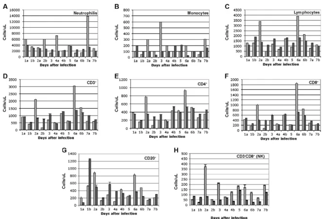
Circulating NK cells decrease soon after LCMV infec-tion - According to flow cytometric analysis and manual
differential cell counts, there were no dramatic or con
-sistent changes in the major circulating blood cell popu
-lations (Fig. 1A-C). Absolute numbers of CD3+CD4+, CD3+CD8+ and CD20+ cells decreased in only two of the monkeys (2nd and 6th), while the values of these subsets did not change significantly for the other monkeys (Fig. 1D-G). On the other hand, the number of cells that were
CD3-CD8+, also recognized as the phenotype for NK cells (Dykhuizen et al. 1998, 2000), increased slightly during the first day after infection, though the absolute numbers then decreased consistently in all the monkeys tested > 2 days after inoculation (Fig. 1H).
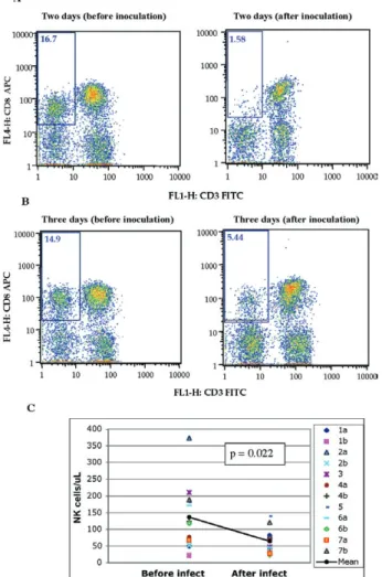
Specific examples of decreasing NK cells (CD3 -CD8+) are seen by flow cytometry (Fig 2A, B). The ab -solute numbers and average of NK cells are also shown for two groups of samples: those taken before inocula
-tion and those taken on the day of euthanasia (between days 2 and 7) (Fig. 2C). The t-test establishes a statis
-tically significant probability (p = 0.022) that the two groups of samples are different.
As a control for the analysis of lymphocyte subsets in LCMV-WE-infected monkeys, we also examined two LCMV-ARM-infected monkeys that never experienced disease (i.e., no rise in liver enzymes or body tempera
-ture) (Rodas et al. 2004). These animals were analysed before intravenous infection with 103 pfu (Arm iv3) or intragastric infection with 108 pfu(Arm ig8) of LCMV-ARM strain, two weeks following infection, before and two weeks after a lethal challenge with 103 pfu of
Fig. 1: fluctuations in circulating blood cell subsets were normal, except for a 20-70% drop in natural killer (NK) cells from day 2 after infec
-tion. Light bars represent cells before infection (pool of 3 timepoints) and dark bars represent cells from one animal after infection (1 timepoint indicated on the abscissa). 1a, b: to samples from two different monkeys (a and b), both taken on day 1. All data in this figure are from monkeys infected with the virulent LCMV-WE (A-C). Absolute counts for neutrophils, monocytes and lymphocytes are normalized according to the manual differential counts provided by the clinical laboratory. D-H: absolute numbers for the CD3+ subset are normalized to the percentage of
CD3+ cells out of the gated lymphocytes in agreement with the absolute number of lymphocytes from the manual differential. Absolute number
of CD4+ and CD8+ were obtained from the percentage of the absolute numbers of CD3 that are CD4+ or CD8+. CD20+ is a subpopulation of
lymphocytes directly stained for CD20 and CD3-CD8+ corresponds to the phenotype of non-T lymphocytes considered NK cells in primates
LCMV-WE i.v. given eight weeks after the first inocula
-tion and then, after surviving the LCMV-WE challenge unscathed, both were sampled before a second challenge with LCMV-WE. A week after the second challenge circulating NK cell numbers increased rather than de
-creased (absolute and percentage). Similar to the LCMV-WE-infected animals, they displayed a 16-30% decrease in circulating NK cells after infection (Fig. 3). This was less than the mean decrease seen with the LCMV-WE-infected monkeys, but the difference was not statistical
-ly significant because so few animals were examined. By three weeks after infection, circulating NK cells had been restored to the numbers seen before infection.
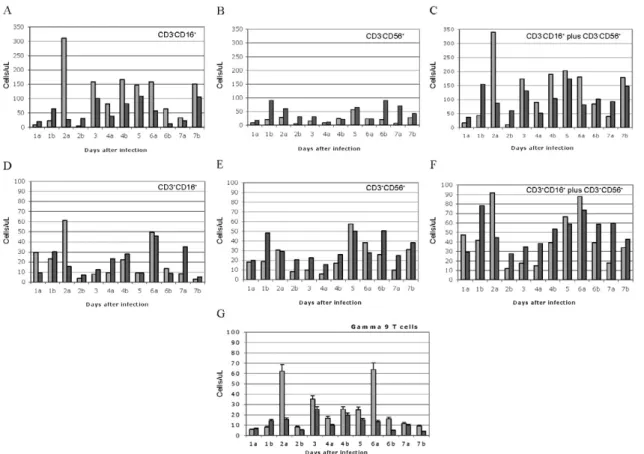
NK and NKT cell subsets - To better characterise the
NK cells, several markers previously reported for this
cell subtype were employed. Using monoclonal antibod
-ies for CD16 and CD56, it appeared that the numbers of CD3-CD16+ cells (i.e., the majority of the CD3-CD8+ NK cells) disappeared from the circulation during the course of infection. The CD3-CD56+ cells are a barely detect -able sub-population of the CD3-CD8+ NK cells (Fig. 4A-C). Absolute numbers of cells are presented in Fig. 4, but a similar picture is obtained by looking at percentages of circulating PBMCs (not shown). In the overall picture for NK cells, whether we consider the CD3-CD8+ cells or the majority of these that are CD3-CD16+, we see that they represented 4-15% of PBMC before infection and they became 2-7% of PBMC after two days of infection.
NKT cells were also observed in the LCMV-infect
-ed monkeys. The numbers of circulating NKT cells (CD3+CD16+ and CD3+CD56+) were even lower than the numbers of NK cells (note the different scales of CD3+ and CD3- cells: 100 compared to 350 on the “y” axis on Fig. 4D-F versus Fig. 4A-C), representing only 1-2% of PBMCs. According to Judy Thomas, who has been working with rhesus NK cells, the proportion of circu
-lating NKT cells is lower than 0.23 ± -0.12 (personal communication). We saw a slight increase in the NKT population (both percentage and absolute number) dur
-ing the course of infection, but this was not statistically significant (Fig. 4D-F).
Since CD3+CD16+ cells often include γδ T cells (Eberl et al. 2005), we also examined that population using flow cytometry. Vγ9 T cells, which represent approximately 80% of the γδ T cells in primate peripheral blood, exhib
-ited a similar pattern of reduction in the percentage and absolute numbers as the NK cells (CD3-CD8+) after the second day of infection (Fig. 4G).
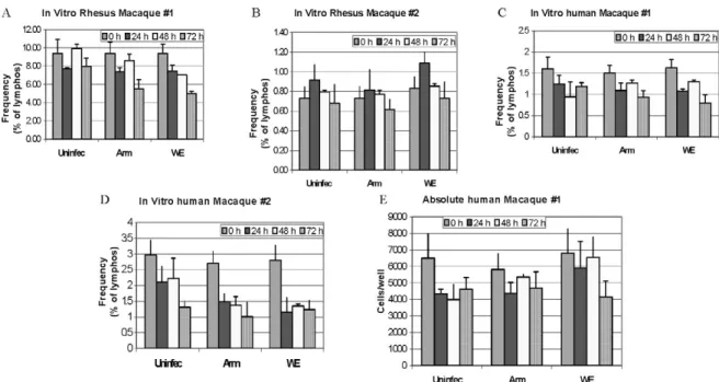
Some of the NK cell decrease may be attributable to cell death - To determine whether the decrease in circu
-lating NK cells could be attributable to virus-mediated
Fig. 2: examples of the decreased populations of natural killer (NK) (CD3-CD8+) cells two and three days after infection. A, B: flow cy
-tometry of peripheral blood mononuclear cells from monkeys 2a and 3; C: the probability that the two populations of NK cells before infec
-tion and NK cells after infec-tion are the same is lower than 0.05 (p < 0.05), meaning that the differences between the two means are statis
-tically significant. Statistical significance using the Student t-test can be shown for the drop in NK cells when data from multiple animals and timepoints are combined: these data are from the pre-infection data points (n = 12) and post-infection data points (day 1-7; n = 12).
Fig. 3: flow cytometry of monkey peripheral blood mononuclear cells (PBMC) infected with lymphocytic choriomeningitis virus (LCMV)-Armstrong (ARM) shows natural killer (NK) decrease. PBMC taken just before infection with either 103 plaque-forming units (Pfu) intra
-venously (Arm iv3), or 108 Pfuintragastrically (Arm ig8) of
LCMV-ARM strain and monitored two weeks later, showed a 16-30% drop in circulating NK cells. A similar drop occurred two weeks after these animals received a lethal challenge (103 Pfu of LCMV-WE iv)
eight weeks after the first inoculation. No drop occurred a week after these monkeys were given a second challenge (103 Pfu of LCMV-WE
iv), 12 weeks after the initial inoculation. The pre-infection (Preinf), pre-challenge (Prech), and pre-boost (Preb) samples were all samples taken minutes before infection, lethal challenge or second lethal chal
cell death, we incubated monkey or human PBMCs with virus (LCMV-ARM or LCMV-WE) at a multiplicity of one pfu/cell and looked for apoptosis using 7-AAD staining. The reduction in absolute numbers of NK cells in the human PBMC samples (Fig. 5E) is similar to the reduction of percentages seen in the monkey and human samples (Fig. 5A-D). Absolute cell counts were deter
-mined by counting cells in triplicate cultures and then using the percentage of particular markers determined by flow cytometry.
The apoptosis identified via 7-AAD staining was negligible (< 2%) in both monkey and human samples (not shown). Although the number of cells undergoing apoptosis was negligible, absolute NK cell numbers were reduced almost 30% over 72 h of cell culture, in
-dicating significant cell death due to culture alone (Fig. 5E) (trypan blue staining). The WE-infected cultures experienced approximately 7% more cell death than the uninfected and ARM-infected cultures, raising the pos
-sibility that virus-mediated cell death could be contrib
-uting (less than 10%) to reductions in NK cells.
The reduction in circulating NK cells is not likely to be due to cell death, but the reduction in Vγ9 TCR-posi
-tive cells may be partially explained by apoptosis. Apop
-tosis was not a significant cause of the NK cell decrease, but it did seem to affect the γδ T cell population. Two of the monkeys depicted in Fig. 1, which showed typical reductions in NK cells (CD3-CD8+) and γδ T cells, were also monitored for apoptosis of circulating NK cells. Apoptosis was measured via 7-AAD staining and was insignificant (< 5%) in the circulating NK cell popula
-tion during the course of infec-tion (Fig. 6). The reduc
-tion of Vγ9 T cells, on the other hand, was accompanied by 10-50% 7-AAD staining, indicating rather high levels of apoptotic cell death.
DISCUSSION
The virulence of a pathogen is often determined by innate responses of the host soon after infection. To understand the events leading to disease, we described changes in circulating blood cells occurring soon after infection. In 10 rhesus macaques, we observed a mean 55% reduction in circulating NK cells from 2-7 days
Fig. 4: peripheral natural killer (NK) cells (CD3-CD16+) and γδ (Vγ9δ+) T cells are reduced but NKT cells (CD3+CD56+) are not reduced. Light
bars represent cells collected minutes before infection and dark bars represent cells after infection (1 timepoint indicated on the abscissa). A-C: NK cells; D-F: NKT cells; G: γδ T cells. Most CD3-CD8+ cells (NK cells) are also CD3-CD16+ lymphocytes, and in panel 4A they are reduced
by day 2 after infection (p = 0.041). The major contributor for the CD3- NK cells seems to be CD16+ over CD56+ (Dykhuizen et al. 2000). Note
the different scales of CD3+ and CD3- cells (100 compared to 350); G: γδ T cells in peripheral blood are also reduced. The Vγ9δ subtype of T
after lethal LCMV-WE infection. The reduction in NK cells, which ranged from 20-70%, encompassed the NK cell drop of 16-30% observed in two monkeys infected with the non-pathogenic strain, LCMV-ARM. Whereas the monkeys infected with the benign virus (ARM) re
-covered their circulating NK cells within two weeks, le
-thally infected monkeys (WE) generally succumbed by day 12 and showed no recovery of circulating NK cells.
Another 20-30% drop in NK cells was observed when the LCMV-ARM-infected animals were given a lethal challenge with LCMV-WE, but when they were given a second challenge, NK numbers were not reduced. Thus, either the second challenge failed to elicit an inductive signal or the NK cells had become unresponsive to the inductive signal. Although our study failed to show a significant difference between the benign and virulent infections, it is possible that a study with more time points could detect kinetic differences in NK loss dif
-ferentiating these two infections. Since ARM-infection immunises monkeys from lethal LCMV-WE challenge (Danes et al. 1963, Peters et al. 1987, Rodas et al. 2004), it is notable that a drop in NK cells is seen during a WE challenge infection in the absence of pathogenesis.
Murine infections with LCMV have provided a large body of information on innate immune responses after viral inoculation. Interferon type I (or IFN α/β) is pro
-duced by infected cells soon after LCMV infection of the mouse and it then activates NK cells to proliferate and carry out their cytotoxic role (Biron et al. 1996, Nguyen et al. 2002). Type I IFN inhibits the expression of IL-12 (Biron et al. 1996, Cousens et al. 1999, Pien et al. 2000) and in the absence of IL-12, type II interferon
Fig. 6: absolute numbers of natural killer (NK) and γδ T cells are re
-duced by lymphocytic choriomeningitis virus-WE infection in vivo without an increase in staining by 7-AAD. The shaded bars are a measure of cells/µL for NK cells (A) and γδ T cells (B). Error bars represent the variation of three blood draws per monkey. A: NK CD3
-CD8+ and CD3-CD16+ subsets similarly are decreased after infection
but no increase in 7-AAD; B: γδ T cells show no increase in percent apoptotic cells as a result of infection.
(IFN-γ) increases in peripheral organs without inhibi
-tion (Cousens et al. 1997, 1999, Pien et al. 2000). Again, in the mouse model during the acute phase of LCMV infection, NK cells increase in the peripheral blood by 10% over three days and NK cell margination results in a > 30% increase in the liver (McIntyre & Welsh 1986, Biron et al. 2002). Murine NK cells have been pheno typically characterised as NK 1.1+TCR-, CD3-, CD8+, asialo+ and CD16+; they are known to stimulate the induction of T cell-mediated immunity by secreting IFN-γ and they also cause some cell death in the liver (Liu et al. 2000). In our monkey study, where NK cells are primarily CD3 -CD16+, we did not investigate the migration of NK cells to peripheral organs, but in other published studies, we have demonstrated the presence of unidentified cellular infiltrates in the liver and lymphoid tissues (Lukashevich et al. 2002, 2003, 2004). Here, we demonstrated in cell culture that virus-mediated cell death could not satisfac
-torily explain the reduction in circulating NK cells. Although NK cells account for approximately 5% of circulating PBMCs, the high interest in NKT cells caused us to monitor changes in that population as well, even though they account for less than 1% of circulating PBMCs in primates (Koyasu 1994). NKT is a subpopu
-lation of T cells with common markers for NK cells, but higher heterogeneity. NKT cells have been identified in the mouse as either double negative for CD4 and CD8 or as positive for CD4 with a very limited diversity of α/β TCR, most commonly Vα14 Vβ281 (Emoto & Kauf
-mann 2003). In human beings, the NKT TCR is Vα24, which cross-reacts with the rhesus monkey cell deter
-minant for NKT (Gansuvd et al. 2003). In rhesus ma
-caques, 80% of Vα24TCR+ cells from the spleen also ex -press CD56 (Gansuvd et al. 2003). Although it has been accepted that rhesus macaque NKTs are CD56+, there is some controversy about the presence of CD16 on their surface (Gansuvd et al. 2003), so we used both the CD16 and CD56 as markers. Unlike the CD3- NK cells, the NKTs in our monkey studies seemed unaffected or even a little increased by day 7 of the study. A rise in NKTs, concomitant with a drop in NK cells has been reported in murine studies (Eberl & MacDonald 2000, Hobbs et al. 2001) and has been interpreted to indicate comple
-mentary functions for the two cell types. Specifically, murine infection with both ARM and LCMV-WE resulted in a 30% increase in NK cells in the liver
by day 3 (Pien et al. 2000), whereas LCMV-ARM in
-fection resulted in an apoptotic reduction of NKT cells in the liver, where this population is commonly most abundant (Hobbs et al. 2001).
Blood cells before and after infection were examined for alterations in apoptosis using 7-AAD, a fluorescent DNA-binding agent that preferentially labels cells with reduced plasma membrane integrity (Lecoeur et al. 1997). No significant increases in the levels of apoptosis were observed in NK cells derived from fresh PBMCs or from cells cultured in the presence of virus, although apoptotic cell death of about 30% (of infected or non-infected cells equally) was observed in these cultures. In the murine system, apoptosis was only seen in NKT cells that were already residing in the periphery (Hobbs
et al. 2001). Since the NKT cells were not susceptible to LCMV infection, they were most likely eliminated by a bystander effect such as that described during HIV in
-fection (Finkel et al. 1995, Abbate et al. 2000, Yang et al. 2003). One likely mechanism is that abundant IFNα/β in peripheral organs increases double-strand RNA-de
-pendent protein kinase in uninfected cells that induces apoptosis (Biron 1999, Biron et al. 2002).
Ebola infection of non-human primates resulted in apoptosis of NK cells, as shown by TUNEL assays of NK cells in PBMCs, liver, spleen and lymph nodes (Gei
-sbert et al. 2000, 2003a, b, Reed et al. 2004). Although we saw little evidence of apoptosis in LCMV-infected monkey PBMCs, we could not rule out an almost 10% non-apoptotic reduction in NK cell numbers cultured in the presence of LCMV-WE. Since virus does not infect NK cells, the virus-mediated loss of NK was most likely due to indirect effects or to cell-surface signalling by vi
-ral particles. Therefore, the reduction in circulating NK cells in the primate model can most likely be attributed to margination of NK cells to peripheral organs with a lesser proportion due to virus-mediated cell death.
Evidence that NK cells have little influence on vi
-rus loads in murine LCMV infections first came from the Welsh laboratory. They have reported that (i) virus titers were unperturbed during the early stages of infec
-tion in mice depleted of NK cells by cyclophosphamide (Biron et al. 1996); (ii) Beige mice (NK deficient) syn
-thesized normal levels of virus (Roder & Duwe 1979); (iii) adoptive transfer of NK cells into 5-day-old mice with low NK cell activity did not confer LCMV resis
-tance even though it conferred resis-tance to murine CMV (Bukowski et al. 1985) and (iv) depletion of NK cells (using antibody to asialo-GM1) had no effect on the synthesis of LCMV-WE or LCMV-ARM in acute or persistent infections (Bukowski et al. 1983, Welsh et al. 1984). Although NK cells have little impact on LCMV titers, they are purported to play a role in con
-trolling viral dissemination (Welsh et al. 1984) and in modulating T cell responses during infection (Su et al. 2001). NK cells produce IFNγ, which induces the ex
-pression of major histocompatibility complex class I on antigen-presenting cells, thereby stimulating CD8+ lymphocytes that play an important role in controlling LCMV infection (Oldstone 2002). If NK cells do impact virus dissemination, one might predict that intravenous infections that are systemically disseminated and rap
-idly established in peripheral organs might escape the effects of NK cells, whereas more slowly disseminated infections, such as mucosal or subcutaneous infections, might be attenuated by NK cells.
Similar to NK cells, γδ T cells have little effect on murine LCMV infection and CTL-mediated clearance (Pien et al. 2002, Eberl et al. 2005). γδ T cells have been implicated in immunosurveillance of tumours and infec
-tions via cytokine secretion and cytolysis (Poccia et al. 2002, Jacobs et al. 2005). In contrast with the absence of virus-mediated cell death in cultured NK cells, γδ T cells display a much higher sensitivity to virus exposure in cul
the virus (Cairo et al. 2005). Thus, the decrease in circu
-lating γδ T cells may constitute a viral strategy for elimi
-nating a potential suppressor of viral infection.Recently, Baize et al. (2009) have reported a NK cell decrease early in Lassa virus-infected cynomolgous macaques like we described in LCMV-infected rhesus macaques.
ACKNOWLEDGEMENTS
To excellent animal care facilities and pathology support from Harry Davis.
REFERENCES
Abbate IF, Dianzani MR, Capobianch I 2000. Activation of sig
-nal transduction and apoptosis in healthy lymphomonocytes exposed to bystander HIV-1-infected cells. Clin Exp Immunol
122: 374-380.
Ahmed RA, Salmi LD, Butler JM, Chiller MB, Oldston E 1984. Se
-lection of genetic variants of lymphocytic choriomeningitis virus in spleens of persistently infected micE role in suppression of cy
-totoxic T lymphocyte response and viral persistence. J Exp Med
160: 521-540.
Baize S, Marianneau P, Loth P, Reynard S, Journeaux A, Chevallier M, Tordo N, Deubel V, Contamin H 2009. Early and strong im
-mune responses are associated with control of viral replication and recovery in Lassa virus-infected cynomolgus monkeys. J Virol 83: 5890-5903.
Biron CA 1999. Initial and innate responses to viral infections-pattern setting in immunity or disease. Curr Opin Microbiol 2: 374-381. Biron CA, Nguyen KB, Pien GC 2002. Innate immune responses to
LCMV infections: natural killer cells and cytokines. Curr Top
Microbiol Immunol 263: 7-27.
Biron CA, Su HC, Orange JS 1996. Function and regulation of natural killer (NK) cells during viral infections: characterization of re
-sponses in vivo. Methods 9: 379-393.
Bonilla WV, Pinschewer DD, Klenerman P, Rousson V, Gaboli M, Pandolfi PP, Zinkernagel R M, Salvato MS, Hengartner H 2002. Effects of promyelocytic leukemia protein on virus-host balance.
J Virol 76: 3810-3818.
Bukowski JF, Biron CA, Welsh RM 1983. Elevated natural killer cell-mediated cytotoxicity, plasma interferon and tumor cell rejection in mice persistently infected with lymphocytic choriomeningitis virus.J Immunol 131: 991-996.
Bukowski JF, Warner JF, Dennert G, Welsh RM 1985. Adoptive trans- 1985. Adoptive trans
-fer studies demonstrating the antiviral effect of natural killer cells in vivo. J Exp Med 161: 40-52.
Cairo C, Propp N, Hebbeler AM, Colizzi V, Pauza CD 2005. The Vgamma2/Vdelta2 T-cell repertoire in Macaca fascicularis: func
-tional responses to phosphoantigen stimulation by the Vgamma2/ Jgamma1.2 subset. Immunology 115: 197-205.
Cousens LP, Orange JS, Su HC, Biron CA 1997. Interferon-alpha/beta inhibition of interleukin 12 and interferon-gamma production in vitro and endogenously during viral infection. Proc Natl Acad Sci
USA 94: 634-639.
Cousens LP, Peterson R, Hsu S, A Dorner, Altman JD, Ahmed R, Biron CA 1999. Two roads diverged: interferon alpha/beta- and interleukin 12-mediated pathways in promoting T cell in
-terferon gamma responses during viral infection. J Exp Med
189: 1315-1328.
Danes L, Benda R, Fuchsova M 1963. Experimental inhalation infec-Experimental inhalation infec
-tion of monkeys of the Macacus cynomolgus and Macacus rhesus species with the virus of lymphocytic choriomeningitis (WE). Bratisl Lek Listy 2: 71-79.
Djavani M, Lukashevich IS, Salvato MS 1998. Sequence comparison of the large genomic RNA segments of two strains of lympho
-cytic choriomeningitis virus differing in pathogenic potential for guinea pigs. Virus Genes 17: 151-155.
Doherty PC, ZinkernageL RM 1974. T-cell-mediated immunopathol
-ogy in viral infections. Transplant Rev 19: 89-120.
Dykhuizen M, Ceman J, Mitchen J, Zayas M, MacDougall A, Helge
-land J, Rakasz E, Pauza CD 2000. Importance of the CD3 marker for evaluating changes in rhesus macaque CD4/CD8 T-cell ratios. Cytometry 40: 69-75.
Dykhuizen M, Mitchen JL, Montefiori DC, Thomson J, Acker L, Lardy H, Pauza CD 1998. Determinants of disease in the simian immunodeficiency virus-infected rhesus macaque: characteriz
-ing animals with low antibody responses and rapid progression.
J Gen Virol 79 (Pt 10): 2461-2467.
Eberl G, MacDonald HR 2000. Selective induction of NK cell prolif
-eration and cytotoxicity by activated NKT cells. Eur J Immunol 30: 985-992.
Eberl M, Engel R, Aberle S, Fisch P, Jomaa H, Pircher H 2005. Human Vgamma9/Vdelta2 effector memory T cells express the killer cell lectin-like receptor G1 (KLRG1). J Leukoc Biol 77: 67-70. Emoto M, Kaufmann SH 2003. Liver NKT cells: an account of hetero
-geneity. Trends Immunol 24: 364-369.
Finkel TH, Tudor-Williams G, Banda NK, Cotton MF, Curiel T, Monks C, Baba TW, Ruprecht RM, Kupfer A 1995. Apoptosis occurs predominantly in bystander cells and not in productively infected cells of HIV and SIV-infected lymph nodes. Nat Med 1:
129-134.
Fisher-Hoch SP, Hutwagner L, Brown B, McCormicK JB 2000. Ef
-fective vaccine for lassa fever. J Virol 74: 6777-6783.
Gansuvd B, Hubbard WJ, Hutchings A, Thomas FT, Goodwin J, Wil
-son SB, Exley MA, Thomas JM 2003. Phenotypic and functional characterization of long-term cultured rhesus macaque spleen-derived NKT cells. J Immunol 171: 2904-2911.
Geisbert TW, Hensley LE, Gibb TR, Steele KE, Jaax NK, Jahrling PB 2000. Apoptosis induced in vitro and in vivo during infection by Ebola and Marburg viruses. Lab Invest 80: 171-186.
Geisbert TW, Hensley LE, Larsen T, Young HA, Reed DS, Geisbert JB, Scott DP, Kagan E, Jahrling PB, DaviS KJ 2003a. A patho
-genesis of Ebola hemorrhagic fever in cynomolgus macaques: evidence that dendritic cells are early and sustained targets of infection. Am J Pathol 163: 2347-2370.
Geisbert TW, Young HA, Jahrling PB, Davis KJ, Larsen T, Kagan E, HensleY LE 2003b. Pathogenesis of Ebola hemorrhagic fever in primate models: evidence that hemorrhage is not a direct effect of virus-induced cytolysis of endothelial cells. Am J Pathol 163:
2371-2382.
Hobbs JA, Cho S, Roberts TJ, Sriram V, Zhang J, Xu M, Brutkiewicz RR 2001. Selective loss of natural killer T cells by apoptosis fol
-lowing infection with lymphocytic choriomeningitis virus. J Vi-rol 75: 10746-10754.
Jacobs R, Heiken H, Schmidt RE 2005. Mutual interference of HIV and natural killer cell-mediated immune response. Mol Immunol 42: 239-249.
Jahrling PB, Frame JD, Smith SB, Monson MH 1985. Endemic Lassa fever in Liberia. III: Characterization of Lassa virus isolates.
Trans R Soc Trop Med Hyg 79: 374-379.
Koyasu S 1994. CD3+CD16+NK1.1+B220+ large granular lymphocytes
arise from both alpha-beta TCR+CD4-CD8- and gamma-delta
TCR+CD4-CD8- cells. J Exp Med 179: 1957-1972.
Lecoeur H, Ledru E, Prevost MC, Gougeon ML 1997. Strategies for phenotyping apoptotic peripheral human lymphocytes comparing ISNT, annexin-V and 7-AAD cytofluorometric staining methods.
J Immunol Methods 209: 111-123.
Liu ZX, Govindarajan S, Okamoto S, Dennert G 2000. NK cells cause liver injury and facilitate the induction of T cell-mediated immu
-nity to a viral liver infection. J Immunol 164: 6480-6486. Lukashevich IS, Djavani M, Rodas JD, Zapata JC, Usborne A, Em
-erson C, Mitchen J, Jahrling PB, Salvato MS 2002. Hemorrhagic fever occurs after intravenous, but not after intragastric, inocula
-tion of rhesus macaques with lymphocytic choriomeningitis vi
-rus. J Med Virol 67: 171-186.
Lukashevich IS, Rodas JD, Tikhonov II, Zapata JC, Yang Y, Djavani M, Salvato MS 2004. LCMV-mediated hepatitis in rhesus ma
-caques: WE but not ARM strain activates hepatocytes and in
-duces liver regeneration. Arch Virol 149: 2319-2336.
Lukashevich IS, Tikhonov I, Rodas JD, Zapata JC, Yang Y, Djavani M, Salvato MS 2003. Arenavirus-mediated liver pathology: acute lymphocytic choriomeningitis virus infection of rhesus macaques is characterized by high-level interleukin-6 expression and hepa
-tocyte proliferation. J Virol 77: 1727-1737.
McCormick JB, Walker DH, King IJ, Webb PA, Elliott LH, Whitfield SG, JohnsoN KM 1986. Lassa virus hepatitis: a study of fatal Lassa fever in humans. Am J Trop Med Hyg 35: 401-407. McCormick JB, Webb PA, Krebs JW, Johnson KM, Smith ES 1987.
A prospective study of the epidemiology and ecology of Lassa fever. J Infect Dis 155: 437-444.
McIntyre KW, Welsh RM 1986. Accumulation of natural killer and cytotoxic T large granular lymphocytes in the liver during virus infection. J Exp Med 164: 1667-1681.
McNally JM, Zarozinski CC, Lin MY, Brehm MA, Chen HD, Welsh RM 2001. Attrition of bystander CD8 T cells during virus-in
-duced T-cell and interferon responses. J Virol 75: 5965-5976. Nguyen KB, Salazar-Mather TP, Dalod MY, Van Deusen JB, Wei XQ,
Liew FY, Caligiuri MA, Durbin JE, Biron CA 2002. Coordinated and distinct roles for IFN-alpha beta, IL-12, and IL-15 regulation of NK cell responses to viral infection. J Immunol 169: 4279-4287. Oldstone MB 2002. Biology and pathogenesis of lymphocytic cho
-riomeningitis virus infection. Curr Top Microbiol Immunol
263: 83-117.
Pauza CD, Hinds PW, Yin C, McKechnie TS, Hinds SB, Salvato MS 1997. The lymphocytosis-promoting agent pertussis toxin affects virus burden and lymphocyte distribution in the SIV-infected rhesus macaque. AIDS Res Hum Retroviruses 13: 87-95. Peacock CD, Kim SK, Welsh RM 2003. Attrition of virus-specific
memory CD8+ T cells during reconstitution of lymphopenic envi
-ronments. J Immunol 171: 655-663.
Peters CJ, Jahrling PB, Liu CT, Kenyon RH, McKee KT JR, Barrera Oro JG 1987. Experimental studies of arenaviral hemorrhagic fe
-vers. Curr Top Microbiol Immunol 134: 5-68.
Pien GC, Nguyen KB, Malmgaard L, Satoskar AR, Biron CA 2002. A unique mechanism for innate cytokine promotion of T cell re
-sponses to viral infections. J Immunol 169: 5827-5837.
Pien GC, Satoska ARR, Takeda K, Akira S, Biron CA 2000. Cutting edge: selective IL-18 requirements for induction of compart
-mental IFN-gamma responses during viral infection. J Immunol
165: 4787-4791.
Poccia F, Gougeon ML, Agrati C, Montesano C, Martini F, Pauza CD, Fisch P, Wallace M, Malkovsky M 2002. Innate T-cell immunity in HIV infection: the role of Vgamma9Vdelta2 T lymphocytes. Curr Mol Med 2: 769-781.
Reed DS, Hensley LE, Geisbert JB, Jahrling PB, Geisbert TW 2004. Depletion of peripheral blood T lymphocytes and NK cells dur
-ing the course of ebola hemorrhagic fever in cynomolgus ma
-caques. Viral Immunol 17: 390-400.
Riviere Y, Ahmed R, Southern PJ, Buchmeier MJ, Oldstone MB 1985. Genetic mapping of lymphocytic choriomeningitis virus patho
-genicity: virulence in guinea pigs is associated with the L RNA segment. J Virol 55: 704-709.
Rodas JD, Lukashevich IS, Zapata JC, Cairo C, Tikhonov I, Djavani M, Pauza CD, Salvato MS 2004. Mucosal arenavirus infection of primates can protect them from lethal hemorrhagic fever. J Med Virol 72: 424-435.
Roder J, Duwe A 1979. The beige mutation in the mouse selectively impairs natural killer cell function. Nature 278: 451-453. Salvato M, Borrow P, Shimomaye E, Oldstone MB 1991. Molecular
basis of viral persistence: a single amino acid change in the glyco
-protein of lymphocytic choriomeningitis virus is associated with suppression of the antiviral cytotoxic T-lymphocyte response and establishment of persistence. J Virol 65: 1863-1869.
Salvato MS, Rodas JD 2005. The arenaviruses. In L Collier, B Mahy (eds.). Topley and Wilson’s principles of bacteriology, virology and immunity, Edward Arnold Ltd, London, p. 1059-1084.
Salvato MS, Shimomayr EM 1989. The completed sequence of lym
-phocytic choriomeningitis virus reveals a unique RNA structure and a gene for a zinc finger protein. Virology 173: 1-10.
Su HC, Nguyen KB, Salazar-Mather TP, Ruzek MC, Dalod MY, Biron CA 2001. NK cell functions restrain T cell responses during viral infections. Eur J Immunol 31: 3048-3055.
Welsh RM, Biron CA, Bukowski JF, McIntyre KW, Yang H 1984. Role of natural killer cells in virus infections of mice. Surv Synth Pathol Res 3: 409-431.
Yang Y, Tikhonov I, Ruckwardt TJ, Djavani M, Zapata JC, Pauza CD, Salvato MS 2003. Monocytes treated with human immunodefi
-ciency virus Tat kill uninfected CD4(+) cells by a tumor necrosis factor-related apoptosis-induced ligand-mediated mechanism. J Virol 77: 6700-6708.
Zinkernagel RM, Doherty PC 1974. Restriction of in vitro T cell-mediated cytotoxicity in lymphocytic choriomeningitis within a syngeneic or semiallogeneic system. Nature 248: 701-702. Zinkernagel RM, Haenseler E, Leist T, Cerny A, Hengartner H,
Althage A 1986. T cell-mediated hepatitis in mice infected with lymphocytic choriomeningitis virus. Liver cell destruc
-tion by H-2 class I-restricted virus-specific cytotoxic T cells as a physiological correlate of the 51Cr-release assay? J Exp Med