Silencing of the integrin-linked kinase gene suppresses the proliferation,
migration and invasion of pancreatic cancer cells (Panc-1)
Xiang-Yu Zhu, Ning Liu, Wei Liu, Shao-Wei Song
*and Ke-Jian Guo
*Department of Pancreatic and Gastrointestinal Surgery,
The First Affiliated Hospital of China Medical University, Shenyang, Liaoning Province, China.
Abstract
Integrin-linked kinase (ILK) is an ankyrin repeat-containing serine-threonine protein kinase that is involved in the reg-ulation of integrin-mediated processes such as cancer cell proliferation, migration and invasion. In this study, we ex-amined the effect of a lentivirus-mediated knockdown of ILK on the proliferation, migration and invasion of pancreatic cancer (Panc-1) cells. Immunohistochemical staining showed that ILK expression was enhanced in pancreatic can-cer tissue. The silencing of ILK in human Panc-1 cells led to cell cycle arrest in the G0/G1 phase and delayed cell pro-liferation, in addition to down-regulating cell migration and invasion. The latter effects were mediated by up-regulating the expression of E-cadherin, a key protein in cell adhesion. These findings indicate that ILK may be a new diagnostic marker for pancreatic cancer and that silencing ILK could be a potentially useful therapeutic ap-proach for treating pancreatic cancer.
Key words:E-cadherin, epithelial-mesenchymal transition (EMT), integrin-linked kinase (ILK), Panc-1 cell line, RNA interference (RNAi).
Received: December 20, 2011; Accepted: March 16, 2012.
Introduction
Pancreatic cancer is one of the most lethal cancers, with 5-year survival rates averaging less than 5% (Shaibet al., 2006). Although several chemotherapeutic agents are active against pancreatic cancer, there is still no satisfactory chemical treatment for this disease (Yoganathan et al., 2000; Chenet al., 2010). Consequently, surgery is still the most effective treatment for pancreatic cancer. In most cases, however, surgery is not possible because the cancer cells have already invaded other tissues by the time of diag-nosis. Less than 20% of the patients undergo surgical treat-ment, with the remainder receiving chemotherapy and/or radiotherapy, both of which are associated with serious side effects (Gunaratnamet al., 2001). The development of new treatments with fewer side effects is therefore an important area of pancreatic cancer research.
Integrin-mediated cell adhesion regulates gene ex-pression through the activation of transcription factors. These activations are mediated through integrin-linked kinase (ILK), an intracellular serine/threonine kinase that interacts with the cytoplasmic domains of integrinb1 and
b3 subunits (Giancotti and Ruoslahti, 1999). Elevated ILK
expression has been observed in ovarian and lung cancer (Ahmedet al., 2004; Hanniganet al., 2005). ILK activity is stimulated by the binding of extracellular matrix compo-nents and growth factors. In addition, the association of ILK with tumor invasion is significantly correlated with all of the epithelial-mesenchymal transition (EMT) markers examined so far, including the loss of E-cadherin and Snail expression (Kanget al., 2004; Barneset al., 2010). Conse-quently, the inhibition of ILK may represent a novel ap-proach for treating pancreatic cancer. The aim of this study was to assess the expression of ILK in pancreatic cancer and examine the effects of siRNA-mediated knockdown of ILK in pancreatic cancer cells.
Material and Methods
Immunohistochemistry
Immunohistochemistry was done using formalin-fixed, paraffin-embedded tissue sections, as previously de-scribed (Ramos-Vara, 2005). Sixty-one pairs of resected pancreatic cancer tissue, 26 samples of corresponding adja-cent tissue and four samples of normal tissue were fixed in 10% formalin solution and embedded in paraffin. Histo-logical sections 3.5mm thick were deparaffinized in xylene following dehydration with ethanol. Endogenous peroxi-dase was blocked with 0.3% H2O2in methanol for 20 min at
room temperature (RT). After antigen retrieval, the sections
Send correspondence to Ke-Jian Guo. Department of Pancreatic and Gastrointestinal Surgery, The First Affiliated Hospital of China Medical University, 110001 Shenyang, Liaoning Province, China. E-mail: cmuzxy_81@126.com.
*These two authors contributed equally to this work.
were blocked with 5% bovine serum albumin for 20 min at RT and then probed with rabbit anti-ILK antibody (diluted 1:500; sc-20019, Santa Cruz Biotechnology Inc., CA, USA) at 4 °C overnight. After washing, the sections were incubated with biotinylated goat anti-rabbit immunoglo-bulins at RT for 1 h and immunoreactivity was detected using peroxidase-conjugated streptavidin and diaminoben-zidine followed by counterstaining with Mayer’s hema-toxylin. The stained sections were examined and scored by a pathologist blinded to all clinical data. The tissues were scored positive when more than 10% of the cells reacted with the anti-ILK antibody and showed cytoplasmic stain-ing.
Plasmids and reagents
A viral packaging system (pHelper 1.0 vector and pHelper 2.0 vector) and the vector pGCSIL/ILK-A were purchased from Genechem Co. Ltd (Shanghai, China).
AgeI,EcoRI and SYBR Green Master Mix kits were pur-chased from TaKaRa (Dalian, China). An RNeasy Midi kit was obtained from Qiagen (Valencia, CA, USA). Dulbec-co’s modified Eagle’s medium (DMEM) and fetal bovine serum (FBS) were obtained from Gibco (Cambrex, MD, USA). Lipofectamine 2000, TRIzol and Super ScriptII re-verse transcriptase were purchased from Invitrogen (Carlsbad, CA, USA). All other chemicals were obtained from Sigma (St. Louis, MO, USA). The antibodies were obtained from Santa Cruz Biotechnology and included polyclonal anti-ILK (sc-20019, diluted 1:500), anti-E-ca-dherin (sc-33743, diluted 1:500) and anti-GAPDH (SC-32233, diluted 1:500).
Cell culture
Pancreatic cancer cells (Panc-1) and human embry-onic kidney HEK293T cells were purchased from the Ame-rican Type Culture Collection (ATCC). Both cell lines were cultured in DMEM supplemented with 10% FBS, 2 mM glutamine and penicillin/streptomycin/amphotericin (100mg each/mL) at 37 °C in a 5% CO2atmosphere.
Construction of ILK-specific shRNA lentivirus
The oligonucleotides encoding a negative control (NC) small interfering RNA (siRNA) (5’-TTCTCCGAACGTGTCACGT-3’), ILK siRNA (5’-CGAAGCTCAACGAGAATCA-3’), and stem-loop-stem oligos (short-hairpin RNAs, shRNAs) were synthe-sized, annealed and ligated into the vector AgeI/EcoRI-linearized pGCSIL-GFP. Expression of the lentiviral shRNA was confirmed by DNA sequencing. The plasmids generated were referred to as pGCSIL-shILK or -shNC. HEK293T cells (1 x 10⁷) were seeded in 10 cm dishes and cultured for 24 h to 70%-80% confluence. Two hours be-fore transfection, the medium was replaced with serum-free DMEM. Plasmids along with 20mg of pGCSIL-shILK or -shNC, 15mg of packaging vector pHelper 1.0 and 10mg of
VSVG expression plasmid pHelper 2.0 were added to 200mL of Opti-MEM and 15mL of Lipofectamine 2000. Lentiviruses were harvested in serum-free medium two days after transfection, filtered and concentrated in primed Centricon Plus-20 filter devices (Millipore). Subsequently, Panc-1 cells were grown to 75% confluence and infected with ILK-shRNA lentivirus or control lentivirus at a MOI (Multiplicity of infection) of 30. The number of green fluo-rescent protein (GFP)-positive cells was determined micro-scopically four days post-transduction.
RT-PCR and real-time PCR analysis
RNA was extracted from infected cells using an RNeasy Midi kit according to the manufacturer’s protocol. cDNA was synthesized using SuperScriptII reverse tran-scriptase. In brief, a mixture containing 1mg of total RNA, 0.5mg of oligo-dT primer (Shanghai Sangon) and nucle-ase-free water in a total volume of 15 mL was heated at 70 °C for 5 min and then cooled on ice for another 5 min. The mixture was supplemented with 2mL of 10 buffer (supplied with the kit) and 200 U of SuperScriptII reverse transcriptase to a final volume of 20mL followed by incu-bation at 42 °C for 60 min.
Real-time quantitative PCR was done using a SYBR Green Master Mix kit in a DNA Engine Opticon system (MJ Research, Waltham, MA, USA). Each PCR mixture containing 10mL of 2 SYBR Green Master Mix, 1mL of sense and antisense primers (5 mmol/mL) and 1 mL of cDNA (10 ng) in a total volume of 20mL was run for 45 cy-cles that included denaturation at 95 °C for 15 s, annealing at 60 °C for 30 s and extension at 72 °C for 30 s. For relative quantification, 2-DDCTwas calculated and used as an indica-tion of the relative expression levels. The primer sequences used to amplify the desired cDNA were: ILK forward and reverse primers: 5’-TCCACCTGCTCCTCA TCC-3’ and 5’-CCTCATCAATCATTACACTACGG-3’ and GAPDH forward and reverse primers: 5’-TGACTTCAACAGCGACACCCA-3’ and 5’-CACCC TGTTGCTGTAGCCAAA-3’. The PCR products were electrophoresed on 1.5% agarose gels, visualized by ethi-dium bromide staining and quantified using AlphaEase® gel image-analysis software (Alpha Innotech, San Leandro, CA, USA).
Western blotting
Western blotting was done using standard procedures (Towbinet al., 1979). Cells were collected 96 h after infec-tion and washed with ice-cold PBS. Whole-cell extracts were prepared using cell lysis buffer (20 mM Tris, pH 7.5, 0.1% Triton X-100, 0.5% deoxycholate, 1 mM phenyl-methylsulfonyl fluoride, 10mg of aprotinin/mL and 10mg of leupeptin/mL) and cleared by centrifugation at 12,000g
elec-trophoresis, proteins were transferred to polyvinylidene difluoride (PVDF) membrane (Millipore). Following the blockade of non-specific sites, anti-ILK (1:500), anti-E-cadherin (1:500) and anti-GAPDH (1:500) antibodies were used to detect the respective proteins. Enhanced chemi-luminescence detection was done in accordance with the manufacturer’s instructions. Blots were visualized using an image analyzer and protein expression was quantified with an ImageQuant densitometric scanner (Molecular Dynam-ics).
Cell proliferation assay
Antiproliferative activity was assayed using MTT (3-(4,5-dimethyl-2-yl)-2,5-diphenyl-tetrazolium bromide). For this, infected and non-infected Panc-1 cells were cul-tured in 96-well plates at an initial density of 2 x 10⁴ cells/well. After 1, 2, 3, 4 and 5 days of infection, the cells were washed twice with phosphate buffered saline (PBS) and 100mL of MTT solution (5 mg/mL) was added to each well. After incubation for 4 h the MTT solution in each well was removed by suction and 100mL of dimethyl sulfoxide (DMSO) was added to solubilize the formazan salt. The op-tical density was measured at 490 nm with a UV microplate reader (Tecan Austria GmbH, Groedig, Austria). The rela-tive cell viability was calculated by comparison with the NC cells.
Fluorescence-activated cell sorting
The cell cycle distribution was analyzed by fluores-cence-activated cell sorting (FACS; Becton-Dickinson, Franklin Lakes, NJ, USA). The cells (1 x 10⁶) were seeded onto six-well plates and allowed to attach overnight after which they were collected, washed in PBS and fixed in 70% cold ethanol. The cells were then treated with DNase-free RNase (100mg/mL) and incubated for 30 min at 37 °C. Propidium iodide (50mg/mL; Sigma) was added to the cell suspension and 10,000 fixed cells were analyzed by FACs.
Assay for apoptosis
Apoptosis was assessed using ApoScreen Annexin V Apoptosis kits (Southern Biotech Inc, Birmingham, AL). Cells infected with lentivirus carrying shILK and shRNA negative controls (NC) were grown to 75% confluence. The cells were then trypsinized, centrifuged, washed in 1x bind-ing buffer and resuspended in 1 mL of Annexin V bindbind-ing buffer. Subsequently, 5 x 10⁵cells (100mL of cell suspen-sion) were stained with 5mL of Annexin V-APC followed by 5mL of PI. For each experiment, 20,000 cells were ana-lyzed using a FACSCalibur flow cytometer (BD Biosci-ences).
Cell migration and invasion
Cell migration and invasion was assayedin vitro us-ing a 24-well transwell unit (8mm pore size) containing
polyvinylpyrrolidone-free polycarbonate filters that were (invasion assay) or were not (migration assay) coated with 500mg/mL of BD Matrigel Basement Membrane Matrix (BD, Franklin Lakes, US). The cells were placed in the up-per compartment of the migration chamber and allowed to attach for 8 h prior to incubation in FBS-free medium for 24 h at 37 °C in a 5% CO2atmosphere. The lower
compart-ment of the migration chamber contained DMEM with 10% FBS. After incubation, the filter inserts were removed from the wells and the cells on the upper side of the filter were removed using cotton swabs. The cells that migrated to the lower surface of the membrane were fixed with meth-anol and stained with 0.5% crystal violet for 10 min. The phenotypes of the migrating cells were determined by counting the cells that migrated to the lower side of the fil-ter using a Leica DM6000B microscope at a magnification of 100x (Leica Microsystems Wetzlar GmbH, Germany). Three migration chambers were used per condition. The number of migrating cells was calculated by averaging the number of cells in at least three bright fields for each of three filters.
Statistical analysis
The results are presented as the mean±SD of at least three independent experiments done on separate days using freshly prepared reagents. Differences between the means of the individual groups were assessed by one-way ANOVA with Duncan’s multiple range tests. A value of p < 0.05 indicated significance. The statistical software package, SPSS v.16 (SPSS Inc., Chicago, Illinois, USA), was used for the analysis.
Results
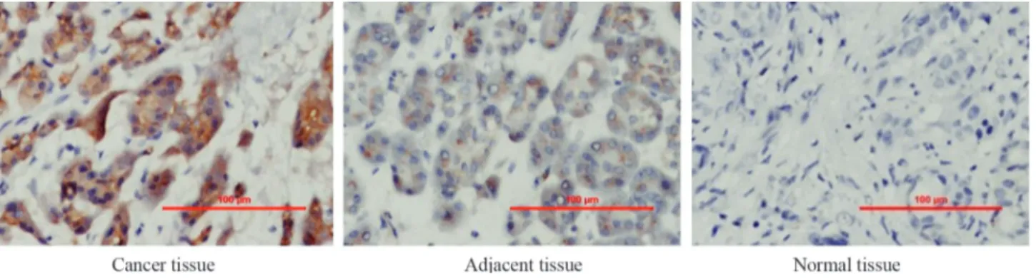
Table 1 and Figure 1 summarize the expression of ILK in pancreatic cancer tissue, adjacent tissue and normal pancreatic tissue based on immunohistochemical analysis. No ILK expression was detected in 7 (11.5%) of the 61 pan-creatic cancer tissue samples whereas 21 samples (34.4%) were hadro-positive and 19 samples (31.1%) were positive; the remaining samples were weakly positive. In contrast to pancreatic tissue, 88.5% and 100% of the adjacent and nor-mal tissue samples, respectively, showed no ILK expres-sion. These findings indicate that ILK expression was enhanced in pancreatic cancer tissue compared to normal and adjacent pancreatic tissues. This enhanced ILK expres-sion may be related to the pathogenesis and progresexpres-sion of pancreatic cancer.
siRNA also markedly attenuated ILK protein expression (Figure 2C). These findings indicate that siRNA directed
towards ILK was effective in specifically knocking down the ILK gene in Panc-1cells.
Table 1- Expression of ILK in pancreatic cancer tissue and normal tissue.
Tissue type Number of cases ILK expression
Negative (-) Weakly positive (+) Positive (++) Hadro-positive (+++)
Cancer 61 ¯7 (11.5%) 14 (23%) 19 (31.1%) 21 (34.4%)
Adjacent 26 20 (77%) 3 (11.5%) 3 (11.5%) 0 (0%)
Normal 4 4 (100%)
¯, decreased ILK expression in cancer tissue compared to adjacent and normal tissues. , enhanced ILK expression in cancer tissue compared to adjacent and normal tissues.
Figure 1- Immunohistochemical analysis of ILK expression in prostate cancer tissue, adjacent tissue and normal tissue. Scale bars are in micrometers.
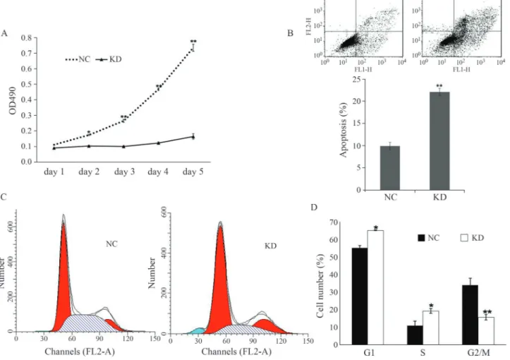
The MTT-based cell proliferation assay revealed markedly less proliferation in pGCSIL-shILK infected Panc-1 cells compared to pGCSIL-shNC infected cells at 3, 4 and 5 days post-infection (Figure 3A). The optical density corresponding to the number of cells was four-fold lower in shILK-infected cells compared to shNC-infected cells. These findings indicate that ILK is necessary for Panc-1 cell proliferation.
The deletion of ILK resulted in a significant accumu-lation of Panc-1 cells in the G1phase and a decrease in the
number of cells in the S and G2/m phases (Figure 3C).
Sig-nificantly more pGCSIL-shILK-infected cells were in the G1 phase compared to pGCSIL-shNC-infected cells
(65.2%vs.55.1%; p < 0.05). These results indicated that ILK knockdown caused cellular arrest in the G1phase of
the cell cycle (Figure 3D). In Panc-1 cells infected with shILK the level of apoptosis reached 22.2%, as shown by FACS of annexin V-stained cells (Figure 3B). This finding suggested that ILK suppressed cell proliferation possibly by inducing apoptosis.
Cancer metastasis requires invasiveness by cancer cells. We used crystal purple staining and the transwell as-say to examine the effect of ILK knockdown on the migra-tion and invasion of Panc-1 cancer cells. As shown in Figure 4A,B, the migration of shILK-infected pancreatic cancer cells was significantly reduced (p < 0.01) compared to the negative control cells. The migration rate was ap-proximately six-fold lower after ILK knockdown. Interest-ingly, the invasion rate of shILK-infected cells was also significantly lower (p < 0.05) than in shNC-infected cells.
E-cadherin plays an essential role in normal physio-logical processes and in pathophysio-logical states such as epithe-lial-mesenchymal transition (EMT). Down-regulation of E-cadherin usually leads to tumor dedifferentiation, infil-tration and the metastasis of cancer cells. We therefore used western blotting to examine the effect of shILK trans-fection on E-cadherin expression. Deletion of ILK led to enhanced E-cadherin expression (Figure 4C) and this may have contributed to the reduction in the rates of invasion and migration.
Discussion
Pancreatic cancer is the fourth most common cause of cancer-related deaths worldwide (Hariharanet al., 2008). Pancreatic cancer often has a poor prognosis, with the 1-and 5-year relative survival rates are 25% 1-and 6%, respec-tively, for all stages combined. The mechanisms involved in the invasion and metastasis of pancreatic cancer are be-ing intensively studied.
ILK is an intracellular protein that interacts with the cytoplasmic domains of the integrinb1 andb3 subunits. Previous work has established a central role for ILK in con-necting integrins to the actin cytoskeleton (Brakebusch and Fassler, 2003). Increased levels of ILK expression have been observed in a variety of human tumors such as malig-nant melanoma, prostate cancer, colon cancer, thyroid can-cer, lung cancan-cer, Ewing’s sarcoma and primitive ectoder-mal tumors (David and Parham, 2001). ILK expression is elevated in virtually all of these cases, indicating the ILK could be a useful diagnostic marker for these types of can-cer.
The adhesion and metastasis of cancer cells in the extracellular matrix are also regulated by ILK. EMT has long been known to play a significant role in tumor pro-gression, dedifferentiation, infiltration and metastasis. There is increasing evidence that ILK is a critical media-tor of the induction of EMT. E-cadherin, a member of the cadherin superfamily of calcium-dependent, trans-membrane glycoproteins, plays an essential role in nor-mal physiological processes and in pathological states such as EMT. ILK may regulate E-cadherin expression indirectly by modulating the transcription ofsnailthat in turn represses E-cadherin expression (Kang and Mas-sague,2004; Barneset al., 2010). The down-regulation of E-cadherin leads to reduced cell-cell adhesion. Under confluent conditions, these cells detach, grow in suspen-sion and undergo EMT that is characterized by reduced expression of E-cadherin and increased expression of Snail. The over-expression of ILK increases the invasive potential of cancer cells by stimulating the expression of invasion-related genes such as MMP-9 (Matsui et al., 2012). Based on studies in vivo, Yau et al. (2005) re-ported that ILK plays a prominent role in oncogenic phosphatidylinositol 3-kinase/PKB signaling and sug-gested that ILK inhibitors might be useful for treating pancreatic cancer patients. Knockdown of the ILK gene may inhibit IL-1a-induced activation of the MAPK/AP-1 signaling pathway by regulating GSK-3 phosphorylation (Kumaret al., 2004). This regulation of AP-1, a major transcription factor that regulates MMP-9 expressions, could have contributed to the lower inva-siveness of ILK-repressed pancreatic cancer cells (Troussardet al., 2000).
In conclusion, the results of this study show that ILK up-regulation plays a major role in the survival and prolif-eration of pancreatic cancer (Panc-1) cells and that silenc-ing the ILK gene suppresses the proliferation, migration and invasion of these cells. The reduced migration and in-vasion of Panc-1 cells may be related to the enhanced ex-pression of E-cadherin, a key protein involved in cell-cell adhesion. Together, these findings suggest that ILK gene suppression could be a potentially useful approach for trea-ting pancreatic cancer.
Acknowledgments
This research was supported by the Science and Tech-nology Research Foundation of the Department of Educa-tion of Liaoning province, China (grant No. 2009A783) and the Science and Technology Program Foundation of Liaoning province of China (grant No. 2011225019).
References
Ahmed N, Oliva K, Rice GE and Quinn MA (2004) Cell-free 59 kDa immunoreactive integrin-linked kinase: A novel narker for ovarian carcinoma. Clin Cancer Res 10:2415-2420.
Barnes EA, Kenerson HL, Jiang XY and Yeung RS (2010) Tu-berin regulates e-cadherin localization: Implications in epi-thelial-mesenchymal transition. Am J Pathol 177:1765-1778.
Brakebusch C and Fassler R (2003) The integrin-actin connection, an eternal love affair. EMBO J 22:2324-2333.
Chen H, Tu H, Meng ZQ, Chen Z, Wang P and Liu LM (2010) K-ras mutational status predicts poor prognosis in unre-sectable pancreatic cancer. Eur J Surg Onc 36:657-662.
David M and Parham MD (2001) Neuroectodermal and neu-roendocrine tumors principally seen in children. Am J Clin Pathol 115:S113-S128.
Giancotti FG and Ruoslahti E (1999) Integrin signaling. Science 13:1028-1032.
Gunaratnam NT, Sarma AV, Norton MID and Wiersema MJ (2001) A prospective study of EUS-guided celiac plexus neurolysis for pancreatic cancer pain. Gastrointest Endosc 54:316-324.
Hannigan G, Troussard AA and Dedhar S (2005) Integrin-linked kinase: A cancer therapeutic target unique among its ILK. Nat Rev Cancer 5:51-63.
Hariharan D, Saied A and Kocher HM (2008) Analysis of mortal-ity rates for pancreatic cancer across the world. HPB (Ox-ford) 10:58-62.
Kang YB and Massague J (2004) Epithelial-mesenchymal transi-tions: Twist in development and metastasis. Cell 118:277-279.
Kumar AS, Naruszewicz I, Wang P, Leung-Hagesteijin CL and Hannigan GE (2004) ILKAP regulates ILK signaling and in-hibits anchorage-independent growth. Oncogene 23:3454-3461.
Matsui Y, Assi K, Ogawa O, Raven PA, Dedhar S, Gleave ME, Salh B and So AI (2012) The importance of integrin-linked kinase in the regulation of bladder cancer invasion. Int J Cancer 130:521-531.
Ramos-Vara JA (2005) Technical aspects of immunohistoche-mistry. Vet Pathol 42:405-426.
Shaib YH, Davila JA and El-serag HB (2006) The epidemiology of pancreatic cancer in the United States: Changes below the surface. Aliment Pharmacol Ther 24:87-94.
Towbin H, Staehelint T and Gordon J (1979) Electrophoretic transfer of proteins from polyacrylamide gels to nitro-cellulose sheets: Procedure and some applications. Proc Natl Acad Sci USA 76:4350-4354.
Troussard AA, Costello P, Yoganathan TN, Kumagai S, Ros-kelley CD and Dedhar S (2000) The integrin linked kinase (ILK) induces an invasive phenotype via AP-1 transcription factor-dependent upregulation of matrix metalloproteinase 9 (MMP-9). Oncogene 19:5444-5452.
Yau CY, Wheeler JJ, Sutton KL and Hedley DW (2005) Inhibi-tion of integrin-linked kinase by a selective small molecule inhibitor, QLT0254, inhibits the PI3K/PKB/mTOR, Stat3, and FKHR pathways and tumor growth, and enhances gem-citabine-induced apoptosis in human orthotopic primary pancreatic cancer xenografts. Cancer Res 65:1497-1504. Yoganathan TN, Costello P, Chen XY, Jabali M, Yan J, Zhang
ZH, Yee A, Dedhar S and Sanghera J (2000) Integrin/linked kinase (ILK): A “hot” therapeutic target. Biochem Phar-macol 60:1115-1119.
Associate Editor: Jeremy A. Squire