Iranian Journal of Basic Medical Sciences
ijbms.mums.ac.ir
Alteration in cardiac uncoupling proteins and eNOS gene
expression following high-intensity interval training in favor
of increasing mechanical efficiency
Ali Asghar Fallahi
1, Shahnaz Shekarfroush
3*, Mostafa Rahimi
4, Amirhossain Jalali
5, Ali
Khoshbaten
21 Faculty of Education and Psychology,Shiraz University, Shiraz, Iran
2 Sport Physiology Research Center, Baghiyatallah University of Medical Sciences, Tehran, Iran 3 Department of Physiology, Arsanjan Branch, Islamic Azad University, Fars, Iran
4 Department of Physical Education, University of Kashan, Iran
5 Institute of Biotechnology and Bioengineering, Isfahan University of Technology, Isfahan, Iran A R T I C L E I N F O A B S T R A C T
Article type: Original article
Objective(s): High-intensity interval training (HIIT) increases energy expenditure and mechanical
energy efficiency. Although both uncoupling proteins (UCPs) and endothelial nitric oxide synthase (eNOS) affect the mechanical efficiency and antioxidant capacity, their effects are inverse. The aim of this study was to determine whether the alterations of cardiac UCP2, UCP3, and eNOS mRNA expression following HIIT are in favor of increased mechanical efficiency or decreased oxidative stress. Materials and Methods: Wistar rats were divided into five groups: control group (n=12), HIIT for an acute bout (AT1), short term HIIT for 3 and 5 sessions (ST3 and ST5), long-term training for 8 weeks (LT) (6 in each group). The rats of the training groups were made to run on a treadmill for 60 min in three stages: 6 min running for warm-up, 7 intervals of 7 min running on treadmill with a slope of 5° to 20° (4 min with an intensity of 80-110% VO2max and 3 min at 50-60% VO2max), and 5-min running for cool-down. The control group did not participate in any exercise program. Rats were sacrificed and the hearts were extracted to analyze the levels of UCP2, UCP3 and eNOS mRNA by RT-PCR.
Results:UCP3 expression was increased significantly following an acute training bout. Repeated HIIT
for 8 weeks resulted in a significant decrease in UCPs mRNA and a significant increase in eNOS expression in cardiac muscle.
Conclusion:This study indicates that Long term HIIT through decreasing UCPs mRNA and increasing
eNOS mRNA expression may enhance energy efficiency and physical performance. Article history:
Received: Jul 22, 2015 Accepted: Oct 8, 2015
Keywords: eNOS
High-intensity interval training
UCP2 UCP3
►
Please cite this article as:Fallahi AA, Shekarfroush Sh, Rahimi M, Jalali AH, Khoshbaten A. Alteration in cardiac uncoupling proteins and eNOS gene expression following high-intensity interval training in favor of increasing mechanical efficiency. Iran J Basic Med Sci 2016; 19:258-264.
Introduction
High-intensity interval training (HIIT) rapidly stimulates metabolic adaptations in skeletal muscle, improves aerobic ability and results in concomitant changes in mitochondrial-associated mRNA and protein levels (1). The increase in exercise-induced energy expenditure is twice the training load (2). The increase in mechanical energy efficiency suggests that the energy needs for daily activities decrease in trained subjects (3). The mechanisms by which exercise could influence energy consumption and mechanical energy efficiency are largely unknown.
Uncoupling proteins (UCPs) are mitochondrial inner membrane proteins, which can disintegrate the proton gradient across the inner mitochondrial membrane and thereby reduce reactive oxygen species (ROS) generation and oxidative stress. Therefore, UCPs may participate in antioxidant defense, but reduced
intracellular ATP production due to uncoupling of the mitochondrial oxidative phosphorylation is associated with less efficient metabolism (4). There is very strong evidence that UCP2 and UCP3 are negatively related to mechanical energy efficiency, suggesting that decreased UCPs with training increases mechanical energy efficiency (3). The different expression patterns of UCPs have been reported previously in organs with different metabolic outlines, such as muscle and heart. In contrast to UCP1, which is expressed exclusively in brown adipose tissue, UCP2 is distributed in a variety of tissues including the heart and UCP3 is highly expressed in skeletal muscle and to a lesser extent, the heart. UCP2 and UCP3, due to their close genetic mapping, have a role in metabolic regulation and calcium homeostasis (5). Both of them are involved in the aging heart dysfunction and upregulated under pathophysiological conditions, such as heart failure (6).
Endothelial nitric oxide synthase (eNOS) is accompanied by a number of physiological processes involved in the regulation of metabolism. eNOS expressed in cardiomyocytes in part mediates the length-dependent increase in cardiac contraction force (7). There is evidence that ablation of eNOS reduces mechanical efficiency and exercise capacity (8). However, the myocardium eNOS uncoupling following exercise inhibits ample NO synthesis and limits the reaction between NO and O₂- to form peroxynitrite, which is cytotoxic (9).
No data concerning the expression of the UCPs and eNOS mRNA in the cardiac muscle following HIIT has been published so far. Due to the adverse effects of these factors on the mechanical efficiency and antioxidant capacity, the present research was designed to characterize whether changes in the expression of the UCP2, UCP3, and eNOS mRNA following HIIT in the cardiac muscle of rats are in favor of increasing mechanical efficiency or decreasing oxidative stress.
Materials and Methods
AnimalsA total of 36 male Wistar rats (250±20 g initial weight, 6 rats/group) were acquired from Pasteur Institute of Iran and kept under controlled conditions (12 hr light/12 hr dark cycle; 22±3 °C). The rats had free access to tap water and standard rat food. The ethical codes of treating laboratory animals, set by the Iranian Society for Supporting Laboratory Animals Used for Scientific Purposes, were strictly followed in the present study. The University of Tehran approved the treating and handling method, the training program, and the sampling type of animals conducted in the present study. The animals were randomly divided into control (cont), HIIT for an acute bout (AT1), short term HIIT (ST), and long-term HIIT (LT).
Exercise training program
The training program was developed based on our previous study (10). The rats were habituated to the treadmill (Iranian model, Tehran, Iran) over the first week. Acute HIIT consisted of one exercise session. Animals in the LT group were exercised for 8 weeks, 5 days/wk, and in the ST group were made to run on the treadmill for 3 or 5 sessions (ST3 and ST5). The control group, comprising 6 rats for acute and short-term training and 6 rats for long term training, were placed on a treadmill without running for 60 min/day.
Every session consisted of one hour exercise training performed in three stages:
Warm-up: running for 6 min at 50-60% VO2max
Main training: 7 intervals of 7-min running at 5°–20° slope consisting of 4-min running at 80-110% VO and 3-min running at 50-60% VO
Cool-The intensity of the training program in terms of VO2max was obtained based on the relation of VO2max to speed and treadmill slope (11). Since the best time for rats is during the dark cycle to do exercise training, the training program was conducted in the afternoon during the dark cycle (between 6 pm and 12 am) under red light (12).Using this protocol for 8 weeks was shown to induce mild heart hypertrophy and protect cardiac muscle against ischemia-reperfusion injury in rats (10).
Sampling procedure
The animals in AT1 and other groups were anesthetized with ketamine and pentobarbital (13) immediately or 24 hr after the last exercise session, respectively. The ventricles were excised, dropped into liquid nitrogen, and stored at -80 °C.
RNA extraction and cDNA synthesis
Total RNAs from the heart tissue samples were extracted using the miRCURY™ RNA )solation Kit Exiqon, Denmark according to the manufacturer’s instructions. The quality and concentration of total RNA were determined at A 260 nm and impurities were assessed by the A 260 nm:A280 nm ratio using Picodrop P200 system (Alpha Biotech Ltd. UK). RNase-free DNase I (Fermentase, Germany) was used to remove any genomic DNA contamination. First-strand cDNA was synthesized from μg of RNA using Thermo Scientific Maxima First Strand cDNA Synthesis Kit (GmbH, Germany).
Real-time PCR
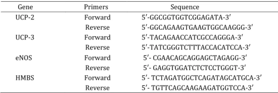
Real-time PCR was performed using the StepOne™ Real-Time PCR System (Life technologies, USA) and SYBR® Green I PCR Master Mix kit (Life Technologies, USA). As an internal control, HMBS (Hydroxymethylbilane synthase) gene was used for
UCP2, UCP3, and eNOS gene expression
normalization (14). Primers for the specific genes are listed in Table 1. Each 5 μl reaction volume contained 400–8 nM primers, .5 μl SYBR®
Green ) PCR Master Mix and .5 μl of sample cDNA.
After a primary denaturation step at 95 °C for 10 min, amplification was performed with 40 cycles
of denaturation at 95 °C for 15 sec and annealing at 60 °C for 1 min with collection of fluorescent data. Finally, the specificity of the amplified product was confirmed using a melt curve analysis for each pair of primer at a temperature between 60 °C and 99 °C. The presence and sizes of all PCR products were verified by inspection of the dissociation curve and by gel electrophoresis. The normalized level of expression of genes in each sample was calculated using the Pfaffl method (15). Values were expressed as fold of the control.
Table1. Primer sequences used in the Real-time PCR for uncoupling proteins, eNOS, and HMBS genes
Gene Primers Sequence
UCP-2 Forward 5′-GGCGGTGGTCGGAGATA- ′
Reverse 5′-GGCAGAAGTGAAGTGGCAAGGG- ′
UCP-3 Forward 5′-TACAGAACCATCGCCAGGGA- ′
Reverse 5′-TATCGGGTCTTTACCACATCCA- ′
eNOS Forward 5′- CGAACAGCAGGAGCTAGAGG- ′
Reverse 5′- GAGGTGGATCTCTCCTGGGT- ′
HMBS Forward 5′- TCTAGATGGCTCAGATAGCATGCA- ′
Reverse 5′- TGTTCAGCAAGAAGATGGTCCA- ′
presented as the mean ± SE. Statistically significant differences between groups were calculated by the one-way ANOVA (Tucky posthoc comparison) or the independent t-test. A value of P<0.05 was considered significant.
Results
The effects of HIIT on mRNA expression of eNOS, UCP2, and UCP3 in the cardiac muscle were studied. Animals in acute training were killed immediately and the other rats 24 hr after the last exercise bout. There was not a significant exercise effect on HMBS gene expression (P>0.05). The effects of HIIT on mRNA expression of eNOS, UCP2, and UCP3 in the cardiac muscle are presented in Figures 1, 2 and 3.
The effect of HIIT on eNOS mRNA expression As shown in Figure 1A, an acute bout of HIIT induced a small increase in eNOS mRNA level, but
there was no significant difference compared to the control group (P=0.19). eNOS mRNA were significantly up-regulated, 2.9 and 3.5 fold, in the short term HIIT for 3 and 5 bouts, respectively, compared to the control group (Figure 1B) and by 2.5 fold in the long term HIIT for 8 weeks (Figure 1C).
The effect of HIIT on UCPs mRNA expression The expression level of UCP2 mRNA did not change following an acute training and short-term training. Cardiac UCP2 mRNA expression was significantly decreased by 44% as compared to that of the control rats after 8 weeks (Figure 2).
UCP3 mRNA was elevated by ∼ 4-fold following an acute bout of training (P<0.01). UCP3 mRNA did not change after short-term training while it was decreased by 48% in long term training compared to that of the control group (Figure 3).
Figure 1. Relative abundance of endothelial nitric oxide (eNOS) mRNA in rat cardiac muscle in response to an acute bout of high-intensity interval training (HIIT, AT1), short term HIIT for 3 and 5 bouts (ST3 and ST5), and long term HIIT for 8 weeks (LT). It normalized with HMBS (using the corresponding 18S rRNA values). The results are expressed as mean±SEM, * P<0.05, **P<0.01 vs. control rats
0 0.5 1 1.5 2
Cont AT1
eN
O
S
mR
N
A
e
xp
re
ss
ion
A
0 1 2 3 4
Cont ST3 ST5
e
N
O
S
m
R
N
A
e
x
p
r
e
ss
io
n *
** B
0 0.5 1 1.5 2 2.5 3
Cont LT
eN
O
S
mR
N
A
e
xp
re
ss
ion
Figure 2. Relative abundance of uncoupling protein 2 (UCP2) mRNA in rat cardiac muscle in response to an acute bout of high-intensity interval training (HIIT, AT1), short term HIIT for 3 and 5 bouts (ST3 and ST5), and long term HIIT for 8 weeks (LT). It normalized with HMBS (using the corresponding 18S rRNA values). The results are expressed as mean±SEM. *P<0.05 vs. control rats
Figure 3. Relative abundance of uncoupling protein 3 (UCP3) mRNA in rat cardiac muscle in response to an acute bout of high-intensity interval training (HIIT, AT1), short term HIIT for 3 and 5 bouts (ST3 and ST5), and long term HIIT for 8 weeks (LT). It normalized with HMBS (using the corresponding 18S rRNA values). The results are expressed as mean±SEM. *P<0.05 vs. control rats
0 0.5 1 1.5
Cont AT1
U
C
P
2
m
R
N
A
e
x
p
r
e
ss
io
n A
0 0.5 1
Cont ST3 ST5
U
C
P
2
m
R
N
A
E
x
p
r
e
ss
io
n B
0 0.5 1 1.5
Cont LT
U
C
P
2
m
R
N
A
e
x
p
re
ss
io
n
* C
0 1 2 3 4
Cont AT1
U
C
P
3
m
R
N
A
e
x
p
r
e
ss
io
n
** A
0 0.5 1 1.5
Cont ST3 ST5
U
C
P
3
m
R
N
A
E
x
p
r
e
ss
io
n
B
0 0.5 1 1.5
Cont LT
U
C
P
3
m
R
N
A
e
x
p
r
e
ss
io
n
*
Discussion
This study describes the changes in the cardiac UCP2, UCP3, and eNOS gene expression following different periods of HIIT. The current data provides evidence that an acute bout of HIIT induced an increase in UCP3 and eNOS mRNA expression, while long-term HIIT for 8 weeks decreased UCPs mRNA expression and increased eNOS mRNA expression.
HIIT represents a time efficient strategy alternative to classic endurance training (ET) that enhances rapid adaptations in skeletal muscle and exercise performance. Gibala et al demonstrated 2.5 hr of HIIT produced molecular and cellular adaptations in human skeletal muscles similar to 10.5 hr of endurance training (16). However, Holloway et al reported that ET and HIIT induce divergent signals in the heart. So that ET for 4 weeks increased eNOS content, while HIIT did not. They found that in the presence of hypertension, HIIT and ET have opposing effects on cardiac remodeling (17). Swimming training for 10 weeks led to increased expression of eNOS of skeletal muscle paralleled with decreases in blood pressure and heart rate in spontaneously hypertensive rats (18). These results suggest that different types of exercise training in different situations have diverse effects on biomarkers expression. In addition, the alterations in eNOS are tissue-specific. Our results showed that eNOS mRNA was increased after HIIT
in the cardiac muscle. Previous studies
demonstrated that downregulation of eNOS exerts following effects on ATP levels and oxidative phosphorylation complexes in skeletal muscles of rodents (8, 19). On the other hand, nitric oxide production through eNOS plays an important role in the regulation of cardiac hypertrophy (20) and protection against myocardial ischemia-reperfusion injury (21). Taken together, eNOS upregulation following HIIT has a role in increased energy efficiency and physical performance and enhances cardioprotection.
Our data revealed that UCP3 gene expression is up-regulated in response to an acute bout of HIIT in rat cardiac tissue. An increase in UCP3 gene expression is thermodynamically unfavorable because it decreases the efficiency of oxidative phosphorylation (22). Acute exercise can result in oxidative stress more than trained exercise. In rat heart, an increase in antioxidant enzyme activities was more in acute exercise as compared to those in chronic exercise, possibly the heart copes with the enhanced production of ROS during exhaustive exercise (23). Previous studies have shown that UCP3 expression can be rapidly upregulated in response to an acute bout of exercise or contractile activity in mammalian skeletal muscles, possibly owing to increased ROS generation (22). UCPs overexpression appears to be a physiological
response to elevated temperature and oxidative stress of exercise in the cardiac muscle (24). Thus, increased UCP3 serves as an early response to antioxidant protection. It is suggested that this
adaptation precedes MnSOD upregulation
temporally and it could be an important molecular
mechanism to maintain the integrity of
mitochondria at the expense of ATP production and oxidative phosphorylation efficiency under potential oxidative stress (22). On the other hand, increased UCP3 mRNA after an acute exercise seems to be related to changes in fatty acid metabolism. It was postulated that the primary function of UCP3 would be in the handling of those fatty acids that cannot be oxidized (25). After acute exercise in the fasted state more fatty acids are released from the adipose tissue than can be oxidized, thus explaining the upregulation of UCP3 (3).
Although during an acute bout of exercise, the first strategy of mitochondria to reduce oxidative
stress is decreasing the production of O2- by overexpressing UCP2/UCP3, endurance training
reduces acute stress-induced UCP2/UCP3
somewhat conflicting results. In obese mice, swim training for 6 weeks increased the levels of UCPs mRNA and proteins in adipose tissue and skeletal muscles (32). Treadmill exercise at low and moderate intensities for 8 weeks increased UCP2 protein expression of brown adipose tissue in Zucker rats compared to the control group (30). These discrepancies between the reports may be due to variations in the type and duration of training. Additionally, the findings suggest that acute exercise and repeated training with different intensities might have opposite effects on UCPs mRNA expression.
Conclusion
The present data suggested that an acute bout of HIIT induced a significant increase in eNOS and UCP3 mRNA level, whereas long-term HIIT resulted in a significant decrease in UCPs and a significant increase in eNOS mRNA levels compared to the control group. After considering all the results, long term HIIT may induce an adaptive physiological response improving metabolic efficiency and physical performance and enhance cardioprotection by downregulation of UCPs expression and upregulation of eNOS in the cardiac muscle. Future studies are needed to compare the alterations of antioxidant enzymes between acute and chronic HIIT.
Acknowledgment
This work was supported by a grant from the Iran National Science Foundation, Sports Physiology Research Center, Baghiyatallah University of Medical Sciences, Tehran, Iran.
Conflict of interest
No conflict of interest to disclose.
References
1. Vincent G, Lamon S, Gant N, Vincent PJ, MacDonald JR, Markworth JF, et al. Changes in mitochondrial function and mitochondria associated protein expression in response to 2-weeks of high intensity interval training. Front Physiol 2015; 6:51.
2. Westerterp KR. Alterations in energy balance with exercise. Am J Clin Nutr 1998; 8:970S-94S.
3. Schrauwen P, Hesselink M. Uncoupling protein 3 and physical activity: the role of uncoupling protein 3 in energy metabolism revisited. Proc Nutr Soc 2003; 62:635-643.
4. Safari F, Bayat G, Shekarforoush S, Hekmatimoghaddam S, Anvari Z, Moghadam MF, et al. Expressional profile of cardiac uncoupling protein-2 following myocardial ischemia reperfusion in losartan- and ramiprilat-treated rats. J Renin Angiotensin Aldosterone Syst 2014; 15:209-217. 5. Trenker M, Malli R, Fertschai I, Levak-Frank S, Graier WF. Uncoupling proteins 2 and 3 are fundamental for mitochondrial Ca2+ uniport. Nat Cell
6. Rupprecht A, Brauer AU, Smorodchenko A, Goyn J, Hilse KE, Shabalina IG, et al. Quantification of uncoupling protein 2 reveals its main expression in immune cells and selective up-regulation during T-cell proliferation. PLoS One 2012; 7:e41406.
7. Balligand JL, Feron O, Dessy C. eNOS activation by physical forces: from short-term regulation of contraction to chronic remodeling of cardiovascular tissues. Physiol Rev 2009; 89:481-534.
8. Lee-Young RS, Ayala JE, Hunley CF, James FD, Bracy DP, Kang L, et al. Endothelial nitric oxide synthase is central to skeletal muscle metabolic regulation and enzymatic signaling during exercise in vivo. Am J Physiol Regul Integr Comp Physiol 2010; 298:R1399-1408.
9. Farah C, Kleindienst A, Bolea G, Meyer G, Gayrard S, Geny B, et al. Exercise-induced cardioprotection: a role for eNOS uncoupling and NO metabolites. Basic Res Cardiol 2013; 108:389.
10. Fallahi AA, Gaeini A, shekarfroush S, khoshbaten A. Cardioprotective effect of preconditioning with high intensity interval training (HIIT), nitric oxide Metabolites (NO2-, NO3-) mechanisms. Iran J Public Health 2015; 44:In Press.
11. Hoydal MA, Wisloff U, Kemi OJ, Ellingsen O. Running speed and maximal oxygen uptake in rats and mice: practical implications for exercise training. Eur J Cardiovasc Prev Rehabil 2007; 14:753-760. 12. Farrell PA, Fedele MJ, Hernandez J, Fluckey JD, Miller JL, 3rd Lang CH, et al. Hypertrophy of skeletal muscle in diabetic rats in response to chronic resistance exercise. J Appl Physiol (1985) 1999; 87:1075-1082.
13.Shekarforoush S, Fatahi Z, Safari F. The effects of pentobarbital,ketamine–pentobarbitaland Ketamine-xylazine anesthesia in a rat myocardial ischemic reperfusion injury model. Lab Anim 2015; In press. 14.Vesentini N, Barsanti C, Martino A, Kusmic C, Ripoli A, Rossi A, et al. Selection of reference genes in different myocardial regions of an in vivo ischemia/reperfusion rat model for normalization of antioxidant gene expression. BMC Res Notes 2012; 5:124.
15. Pfaffl MW. A new mathematical model for relative quantification in real-time RT-PCR. Nucletic Acids Res 2001; 29:e45.
16. Gibala MJ, Little JP, van Essen M, Wilkin GP, Burgomaster KA, Safdar A, et al. Short-term sprint interval versus traditional endurance training: similar initial adaptations in human skeletal muscle and exercise performance. J Physiol 2006; 575:901-911. 17. Holloway TM, Bloemberg D, da Silva ML, Simpson JA, Quadrilatero J, Spriet LL. High intensity interval and endurance training have opposing effects on markers of heart failure and cardiac remodeling in hypertensive rats. PLoS One 2015; 10:e0121138. 18. Ma ZY, Zhao YC. [Effects of aerobic exercise training on antihypertension and expressions of VEGF, eNOS of skeletal muscle in spontaneous hypertensive rats]. Zhongguo Ying Yong Sheng Li Xue Za Zhi 2014; 30:320-324.
20. Yang L, Jia Z, Zhu M, Zhang J, Liu J, Wu P, et al. Exercise protects against chronic beta-adrenergic remodeling of the heart by activation of endothelial nitric oxide synthase. PLoS One 2014; 9:e96892. 21. Calvert JW, Condit ME, Aragon JP, Nicholson CK,
Moody BF, Hood RL, et al. Exercise protects against myocardial ischemia-reperfusion injury via
stimulation of beta(3)-adrenergic receptors and increased nitric oxide signaling: role of nitrite and nitrosothiols. Circ Res 2011; 108:1448-1458.
22. Jiang N, Zhang G, Bo H, Qu J, Ma G, Cao D, et al. Upregulation of uncoupling protein-3 in skeletal muscle during exercise: a potential antioxidant function. Free Radic Biol Med 2009; 46:138-145. 23. Somani SM, Frank S, Rybak LP. Responses of antioxidant system to acute and trained exercise in rat heart subcellular fractions. Pharmacol Biochem Behav 1995; 51:627-634.
24. Bo H, Jiang N, Ma G, Qu J, Zhang G, Cao D, et al. Regulation of mitochondrial uncoupling respiration during exercise in rat heart: role of reactive oxygen species (ROS) and uncoupling protein 2. Free Radic Biol Med 2008; 44:1373-1381.
25. Schrauwen P, Saris WH, Hesselink MK. An alternative function for human uncoupling protein 3: protection of mitochondria against accumulation of nonesterified fatty acids inside the mitochondrial matrix. FASEB J 2001; 15:2497-2502.
26. Bo H, Jiang N, Ji LL, Zhang Y. Mitochondrial redox metabolism in aging: Effect of exercise interventions.
J Sport Health Sci 2013; 2:67-74.
27. Noland RC, Hickner RC, Jimenez-Linan M, Vidal-Puig A, Zheng D, Dohm GL, et al. Acute endurance exercise increases skeletal muscle uncoupling protein-3 gene expression in untrained but not trained humans. Metabolism 2003; 52:152-158.
28. Boss O, Samec S, Desplanches D, Mayet MH, Seydoux J, Muzzin P, et al. Effect of endurance training on mRNA expression of uncoupling proteins 1, 2, and 3 in the rat. FASEB J 1998; 12:335-339. 29. Bayat G, Hajizadeh S, Javan M, Forouzandeh Moghaddam M, Safari F, Azizi H, et al. Decreased Uncoupling Protein 2 and 3 (UCP2 and UCP3) mRNA expression by endurance exercise training with and without chronic administration of nandrolone in rat heart. Physiol Pharmacol 2011; 15:330-340.
30. Kim DH, Kim SH, Kim WH, Moon CR. The effects of treadmill exercise on expression of UCP-2 of brown adipose tissue and TNF-alpha of soleus muscle in obese Zucker rats. J Exerc Nutrition Biochem 2013; 17:199-207.
31. Schrauwen P, Troost FJ, Xia J, Ravussin E, Saris WH. Skeletal muscle UCP2 and UCP3 expression in trained and untrained male subjects. Int J Obes Relat Metab Disord 1999; 23:966-972.