JSCS–4614 548.7:543.42–74:547.415
Original scientific paper
Structural, spectral and thermal properties of
2-(2-pyridylamino)pyridinium trihydrogen pyromellitate
DEJAN POLETI1*#, JELENA ROGAN1#, LIDIJA RADOVANOVIĆ2# and MARKO RODIĆ3#
1Department of General and Inorganic Chemistry, Faculty of Technology and Metallurgy,
University of Belgrade, Karnegijeva 4, 11000 Belgrade, Serbia, 2Innovation Center, Faculty
of Technology and Metallurgy, University of Belgrade, Karnegijeva 4, 11000 Belgrade, Serbia and 3Faculty of Sciences, University of Novi Sad, Trg Dositeja Obradovića 3,
21000 Novi Sad, Serbia
(Received 7 June, revised 24 October, accepted 25 October 2013) Abstract: The title compound, (Hdipya)(H3pyr), where Hdipya is protonated 2,2′-dipyridilamine and H3pyr is the monoanion of pyromellitic acid (H4pyr), was obtained from a reaction mixture containing Zn(II) ions, dipya, Na4pyr (in the mole ratio 2:2:1) and HNO3. The products (micro- and single-crystalline) were characterized by X-ray structure determination, FT-IR spectroscopy and TG/DSC analysis. The most striking structural feature of (Hdipya)(H3pyr) are short inter- and extremely short intramolecular hydrogen bonds. These bonds mutually connect cations and anions making thin layers parallel to the crys-tallographic (223) plane. In addition, quite unusual, linear proton-bound poly-meric {H(H2pyr)-}
n were identified in the structure. The results of crystal
struc-ture determination are compared with FT-IR and TG/DSC data. The low posi-tion of the νas(COO) vibration at 1660 cm-1 is in accordance with strong hyd-rogen bonding. This value could be used as a measure of the C=O bond order, which was calculated to be 1.81.
Keywords: pyromellitic acid; 2,2′-dipyridylamine; crystal structure; FT-IR spectra; TG/DSC analysis.
INTRODUCTION
Pyromellitic or 1,2,4,5-benzenetetracarboxylic acid (H4pyr), one of 12
ben-zenepolycarboxylic acids, is used in the production of polyesters and polyamides. Anions of H4pyr, in particular pyr4–, are well-known as very prospective ligands
in coordination chemistry due to the presence of eight O atoms as potential ligating sites that can coordinate up to ten metal centers.1 The crystal structure of
* Corresponding author. E-mail: [email protected] # Serbian Chemical Society member.
H4pyr was published in 1971,2 and earliest transition metal (TM) complexes with
the tetra-anion of H4pyr were published just after that.3 The first structurally
characterized complexes were described at the beginning of the 1990s.4 Later,
during structural studies of TM complexes, it was shown that the tetra-anion pyr4– usually acts as a centrosymmetric bridging ligand and can build discrete,5
chain,6 layered,7 or framework8 structures. A survey of the Cambridge Structural
Database, CSD,9 showed that the partially deprotonated forms: Hpyr3–, H2pyr2–
and H3pyr– are, as a rule, not coordinated.
Binary and ternary TM complexes with anions of aromatic polycarboxylic acids have been studied for a long time.10 In line with this subject, attempts were
made to prepare mixed ligand Mn(II) and Zn(II) complexes with pyr4– and 2,2′-
-dipyridylamine (dipya) as an N,N′ ligand. Surprisingly, the title compound
con-taining only potential ligands was obtained instead. Such compounds can be clas-sified into two major groups, as co-crystals (adducts) or organic salts (proton-transfer complexes),11 but between them there are so-called “disordered solid
forms”,12,13 where hydrogen atoms are not undoubtedly located or they are
dis-ordered. Although such compounds are much less frequent in comparison with single-component organics or the corresponding complexes, the number of stu-died examples permanently increases since their possible importance for pharma-ceutical chemistry was recognized.14,15 Even ferroelectric properties were found
in some compounds.16 Therefore, it seemed worthwhile to study the obtained
compound in detail.
In the CSD9 there is a very limited number of structurally characterized
compounds concerning molecule of H4pyr or its partially deprotonated ions.
There are, however, several similar organic salts: (Hpy)(H3pyr), where py is
pyridine,13 (NMe4)(H3pyr),17 a series of H2pyr2– salts with different pyridinium
derivatives,18 (cytosinium)2(H2pyr)·H2O,19 and (Hbipy)2(H2pyr)(H4pyr), where
bipy is 2,2′-bipyridine, which was characterized structurally20 and by FT-IR and
NMR spectroscopy.21
On the other hand, dipya is well known as a chelating ligand. A search of the CSD revealed some 170 compounds containing dipya.9 Most of them were TM
complexes, and there were no examples of uncoordinated dipya in its neutral form. At the same time, there were only eight compounds containing protonated dipya, which can be present as monoprotonated 2-(2-pyridylamino)pyridinium, Hdipya, or diprotonated 2,2′-iminodipyridinium(2+), H2dipya. Such protonated
forms are not coordinated, and in five out of eight cases tetrahalidometalates, [MX4]2– (M = Co, Cu or Hg; X = Cl or Br), were present as counter ions.9
Herein, crystal structure of the title compound, (Hdipya)(H3pyr), together
EXPERIMENTAL
The starting reagents pyromellitic acid (> 95 %), 2,2′-dipyridylamine (98 %), Mn(II) nitrate solution, 45–50 % in dilute nitric acid (all Sigma–Aldrich) and Zn(NO)3·6H2O (p.a. Kemika, Zagreb) were used without any purifications.
Preparation of the microcrystalline product. Into 100 cm3 of an aqueous solution con-taining 0.74 g (2.5 mmol) of Zn(NO3)2·6H2O, first 5 cm3 of HNO3 solution (c = 1 mol dm-3), then 0.43 g of dipya (2.5 mmol) dissolved in 10 cm3 of an EtOH/H
2O mixture (3:1 volume ratio) was added. The pH value of this mixture was 2. Then, 50 cm3 of an aqueous solution of Na4pyr (0.32 g; 1.25 mmol) was slowly added under stirring. The final pH value was 4. After standing for 24 h, the obtained precipitate was filtered and rinsed with H2O, EtOH and Et2O. Yield: 84.6 %; Anal. Calcd. for C20H15N3O8: C, 56.48; H, 3.55; N, 9.88 %. Found: C, 56.27, H, 3.62, N, 9.91 %; FT-IR (KBr, cm-1): 3470 (bw), 3117 (m), 3097 (m), 3061 (m), 3024 (m), 2993 (m), 2920 (w), 1715 (m), 1660 (s), 1604 (s), 1562 (s), 1451 (s), 1311 (w), 1292 (w), 1270 (w), 1250 (w), 1170 (m), 1151 (m), 1005 (m), 955 (m), 901 (m), 771 (s), 748 (s), 594 (m), 559 (w), 532 (m). The compound was soluble in DMSO but insoluble in H2O and EtOH.
A similar procedure using Mn(II) instead of Zn(II) ions resulted in a white microcrys-talline powder in a comparable yield (78.9 %). As revealed by elemental analysis and FT-IR spectroscopy, this product was identical to the product obtained in the presence of Zn(II) ions.
Preparation of single-crystals. Into 50 cm3 of an aqueous suspension containing 0.25 g (1 mmol) of H4pyr was added 0.17 g of dipya (1 mmol) dissolved in 5 cm3 of EtOH. Then, 50 cm3 of an aqueous solution containing 0.30 g (1 mmol) of Zn(NO
3)2·6H2O was added drop-wise under continuous stirring. The mixture was gently heated at 50 °C for 3 h, filtered to remove a small quantity of unreacted H4pyr, and left standing under ambient conditions. The first crystals appeared after 24 h, but single-crystals of appropriate size were collected after 10 days.
Characterization. The FT-IR spectra were recorded on a Bomem MB-100 (Hartmann Braun) spectrophotometer (4000–600 cm-1 region) using KBr pellets. The thermal properties of the compound were examined from room temperature up to 380 °C using an SDT Q600 TGA/DSC instrument (TA Instruments) in a dry nitrogen atmosphere (flow rate: 100 cm3 min-1) at a heating rate of 20 °C min-1. The sample mass was 8.14 mg.
Fig. 1. Molecular structure of (Hdipya)(H3pyr) with atomic numbering scheme. Displacement ellipsoids are drawn at the 50 % probability level. Disordered Hdipya ions are marked by
different shades of gray. Symmetry codes: (i) 1 – x, 1 – y, 1 – z; (ii) 1 – x, 1 – y, –z; (iii) 1 + x, 1 + y, –z.
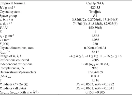
TABLE I. Crystal data and structure refinement for (Hdipya)(H3pyr)
Empirical formula C20H15N3O8
M / g mol-1 425.35
Crystal system Triclinic
Space group P1
a, b, c / Å 3.8268(2), 9.2726(6), 13.3494(8)
α, , / º 74.761(6), 81.845(5), 82.935(6)
V / Å3 450.59(5)
Z 1
ρc / g cm-3 1.568
/ mm-1 1.056
F(000) 220
Crystal dimensions, mm 0.09×0.10×0.31
θmax / ° 72.12
Range of h, k, l –4 ≤ h ≤ 3, –11 ≤ k ≤ 11, –16 ≤ l ≤ 16
Reflections collected 7605
Independent reflections 1770 (Rint = 0.0361)
Completeness, % 99.6
Data/restraints/parameters 1770/6/169
(Δ/σ)max 0.001
S 1.116
RESULTS AND DISCUSSION
Synthesis
As described in the Experimental, the title compound was obtained during attempts to synthesize ternary Zn(II)–dipya-pyr complexes or their Mn(II) anal-ogues. This demonstrates the difficulties in the preparation of such complexes, in spite of being known.9 A similar experience was already published by Ha,25
when (Hdipya)2[MnCl4] was obtained instead of the expected Mn(II)–dipya
complex, and it was not reported previously for Zn(II) complexes. Such behavior can be partly explained by an increase on the thermodynamic stability of the protonated dipya in respect to the neutral molecule,26 but since dipya complexes
of many TMs are known, the specific role of Zn(II)/Mn(II) ions and the pH of the reaction mixture should not be neglected.
Crystal structure
In the structure of (Hdipya)(H3pyr), both the Hdipya and H3pyr ions are
positioned around the symmetry centre, and only half of each species belongs to the asymmetric unit (Fig. 1). Besides, Hdipya is disordered over a centre of sym-metry with equal allocation of both entities. A comparable disorder was found in some organostannates,26 and in the analogous (Hdipya)(Hpht), where Hpht is the
hydrogen phthalate ion.27 One of pyridyl N atoms has to be protonated, but it
was not possible to locate the corresponding H atom. Both N atoms from the pyridyl rings could be protonated because N1A···O1 and N1B···O1 distances are 3.018(7) and 3.121(5) Å, respectively, i.e., they could form hydrogen bonds.
Therefore, the H atom is very likely spread over four possible positions.
The whole Hdipya is practically perfectly planar with a negligible dihedral angle between the mean-planes of the two pyridyl rings. In three known dipya polymorphs and five crystallographically different dipya molecules, this angle varied between about 4 and 40°.28 A very similar variety of angles was also
found in some TMs–dipya complexes.10 The planarity of Hdipya might be
explained by symmetry constraints or by extended delocalization of the π elec-trons. However, the influence of intramolecular (intra-H) and intermolecular (inter-H) hydrogen bonds could also be important. According to Haddad et al.,29
weak intra-H bonding is also possible between the protonated and unprotonated N atom from adjacent pyridyl rings (Fig. 1, atoms N1A and N1B). As usual, pyri-dyl rings are less regular than benzene rings.
At first sight, the H3pyr ion (Fig. 1) is fully protonated, i.e., it should be
described as a neutral H4pyr molecule, not as a monoanion. However, the H2
atom is in special position (site symmetry:
1
) and with a 0.5 site occupancy factor. The geometry data for the H3pyr anion are listed in Table II. While theC–C bond distances are as expected, the angles, in particular C1–C2–C3 and C2–C3–C5, strongly deviate from the values found in H4pyr·2H2O (max.
intra-H bond O2–H1…O3 and the formation of seven-membered, S(7), pseudo- -rings. In respect to the aromatic ring, the C–COO groups are inclined in the opposite sides, with the corresponding angles of 13.3° for the C2C1O1O2 group and 73.6° for the C3C5O3O4 group. This intra-H bond is extremely short and clearly asymmetrical (Table III, Fig. 1). Very similar geometries were already found in H2pyr and H3pyr anions.13,18–20 and in some Hpht complexes.31,32
TABLE II. Bond distances and angles in H3pyr ion (For O–H bond distances, see Table III) Bond distance, Å
O1–C1 1.202(4) O2–C1 1.297(3)
O3–C5 1.225(3) O4–C5 1.251(3)
C1–C2 1.527(3) C2–C3 1.406(3)
C2–C4 1.391(3) C3–C4 1.391(3)
C3–C5 1.513(3)
Bond angle, °
O1–C1–O2 121.2(3) O1–C1–C2 118.9(2)
O2–C1–C2 119.9(2) C1–C2–C3 129.2(2)
C1–C2–C4 113.0(2) C3–C2–C4 117.8(2)
C2–C3–C4 118.0(2) C2–C3–C5 126.8(2)
C4–C3–C5 115.2(2) C2–C4–C3 124.2(2)
C2–C4–H4 117.9(2) C3–C4–H4 117.9(2)
O3–C5–O4 122.2(3) O3–C5–C3 121.9(2)
O4–C5–C3 115.8(2)
TABLE III. Geometry of hydrogen bonds in (Hdipya)(H3pyr); Symmetry codes: (iv) –x, –y + 2, –z + 1; (v) x – 1, y, z
D–H···A d(D–H) / Å d(H···A) / Å Angle (DHA) / ° d(D···A) / Å
O2–H1···O3 1.11(4) 1.34(4) 167(3) 2.435(3)
O4–H2···O4iv 1.24a 1.24a 180a 2.477(2)
N2–H2A···O1v 0.86 1.87 165 2.706(4)
aAtom H2 is in a special position and its coordinates were fixed during refinement
Another, inter-H bond O4–H2…O4iv ((iv) –x, –y + 2, –z + 1) is symmetrical
and also very short, but a little longer than the intra-H bond (Table III). In com-parison to typical O…O distances found in related Hpht compounds,27 the intra-H
bond is slightly longer, whereas inter-H bond is shorter than expected. This shows how strong the inter-H bond is in this case. Being that the inter-H bond is sym-metrical, H3pyr could also be viewed as a polymeric proton-bound trihydrogen
pyromellitate anion, {H(H2pyr)–}n (Fig. 2). Again, there is great similarity with
Hpht compounds, in which proton-bound anions are also found.33–35 The main
difference is that {H(H2pyr)–}n is polymeric, while [H(Hpht)2]– ions are only
dime-ric. This is because H4pyr could be considered as developed from H2pht by
mirroring two COOH groups in the ortho-position to another pair of COOH
while Hpht– is restricted to dimeric forms.36 To the best of our knowledge, a
similar polyanion was found only in (NMe4)(H3pyr), described by
Rodriguez-Cuamatzi et al.17 However, those chains were of a zig–zag type, while here the
presented chains are linear (Fig. 2) and so could be regarded as unprecedented.
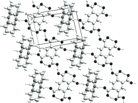
Fig. 2. Motif of hydrogen bonds (thin dark lines) in the (223) crystallographic plane. Color codes for atoms: H – white, C – light gray, N – dark gray and O – black.
In the structure, there is another hydrogen bond between the amine N2 atom and O1 (Fig. 1, Table III). Since all hydrogen bonds are concentrated in one plane, thin layers approximately parallel to the crystallographic (223) plane are formed (Fig. 2). The {H(H2pyr)–}n and Hdipya ions are assembled in alternating rows
making a non-rectangular grid. The layers are stacked together by short parallel- -displaced π–π interactions. For example, all distances between the plane of one benzene ring and benzene C atoms from the nearest layer are 3.490(5) Å. Anal-ogous layers also exist in (cytosinium)2(H2pyr)·H2O,19 and in (NMe4)(H3pyr).17
Spectral and thermal properties
A sequence of weak and sometimes broad IR bands between 3500 and 3200 cm–1 is indicative for numerous hydrogen bonds. According to the literature,21
even weak and very broad bands centered at 2900, 2430 and 1900 cm–1 could also
be ascribed to strong hydrogen bonding. In some cases, they could be extremely strong and broad.37 Bands between about 3120 and 2990 cm–1 are characteristic
It seems that the stretching N–H band was shifted from 3244 cm–1 in dipya to
3117 or 3097 cm–1 in (Hdipya)(H3pyr), suggesting stronger hydrogen bonding.
In the “fingerprint” region, there are bands of different origin typical for substituted benzene rings and secondary amines; they agree with the bands in the individual components. Especially characteristic for dipya is a prominent doublet due to N–H out of plane bending at 771 and 748 cm–1. Both vibrations were
slightly shifted with respect to pure dipya (768 and 735 cm–1) and very likely
overlap with the corresponding C–H vibrations.38
The most interesting part of the FT-IR spectrum is the 1720–1250 cm–1
region (Fig. 3), where many strong bands are expected: asymmetrical and sym-metrical stretching vibrations of COOH and the corresponding COO groups, C–C and C–N stretching vibrations of aromatic rings and N–H bending in a secondary aromatic amine.39 (Asymmetrical νas(COOH) vibrations, characteristic for
orga-nic acids, are often labelled ν(C=O) and called carbonyl, due to the presence of double C=O bond). Consistent to the literature,37 νas(COOH) and νs(COOH) frequencies for H4pyr are at 1710 and 1266 (as doublet at 1278 and 1255) cm–1,
respectively, and the present experimental data agree within ±5 cm–1 with these
values. These bands are broad and usually the most intense in the spectrum.38
Surprisingly, the corresponding bands in the FT-IR spectrum of (Hdipya)(H3pyr)
are medium to weak, but one additional very prominent band at 1660 cm–1
appeared. At first sight, this band could be attributed to the secondary amine N–H bending group frequency, which is located at 1604 cm–1 in dipya. However,
such high wavenumbers are observed only in the case of coordinated dipya and the band is typically very sharp.10 Therefore, the band at 1660 cm–1 should be
ascribed to the νas(COOH) or carbonyl C=O vibrations. Such a low position of νas(COOH) is not expected,38 and, together with no unambiguous identification of νs(COOH) vibrations, has to be correlated to the extremely short and strong hydrogen bonds involving all O atoms from H3pyr. In (Hbipy)2(H2pyr)(H4pyr),20,21
just the opposite behavior was observed, νas(COOH) is shifted to the higher wavenumbers (1726 cm–1), which was explained by the absence of hydrogen bonds.
In the tetrathiafulvalene–p-chloranil (TTF–CA) complex, which is a widely
studied charge-transfer organic compound,40 the position of C=O vibrations was
used to determine the degree of charge transfer from TTF to CA. In other words, νas(COOH)/ν(C=O) could be related to the C=O bond order. For H4pyr νas(COOH) was at 1710 cm–1,39 and this corresponds to a bond order of 2. In alkali metal
salts, the COOH groups are ionized and, due to delocalization of the electrons in COO groups, the bond order should be 1.5. The available data for the two alkali metal salts K4pyr39 and Na4pyr (this study) give an identical value, 1580 cm–1
for νas(COO). From these values, it is easy to calculate the C=O bond order in (Hdipya)(H3pyr), which was 1.81. By analogy to the “degree of charge transfer”
this quantity could be used to measure the “degree of proton transfer”,41 and
However, the observed geometry of H3pyr ions (Table II) does not enable a
simple explanation of the calculated value. For O1–C1 and O2–C1 pair, bond distances can be judged as normal when compared to the H4pyr and common
COOH values.2 Nevertheless, O3–C5 bond is longer than expected and O4–C5
bond distance corresponds to the typical values found in ionized COO groups.9
This could be a possible cause for the low value of νas(COOH), although an extensive coupling of vibrational modes should also be kept in mind.
Fig. 3. FT-IR spectrum (KBr pellets) of (Hdipya)(H3pyr).
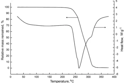
As shown in Fig. 4, the decomposition of (Hdipya)(H3pyr) began at 237 °C
(onset temperature) and ended at about 325 °C with a total mass loss. No melting
was observed and the decomposition started around the boiling point of dipya (222 °C), suggesting that this molecule escaped first. Although the TG curve decreased rapidly, the shape of the DSC curve indicated that the disintegration occurred in at least three, probably four, highly overlapped steps, which could not be resolved. The total molar decomposition enthalpy was relatively high,
o dec mH
∆ =461.2 kJ mol–1. In comparison to TM complexes with dipya and some polycarboxylate ligands,10 the initial decomposition temperature of
(Hdipya)(H3pyr) was lower, very likely owing to the absence of coordinative
bonds. In analogous systems (py)2(H2tpht), where H2tpht is terephthalic acid,
and (py)3(H3tms), where H3tms is trimesic acid, the total degradation was found
to be at 339 and 366 °C, respectively.13 There, however, loss of py molecules
started just above room temperature, although the boiling point of py is 115 °C. CONCLUSIONS
A new organic salt (Hdipya)(H3pyr) was prepared unpredictably during
efforts to obtain ternary Zn(II) or Mn(II) complexes with dipya and pyr ligands. Besides the higher stability of Hdipya with respect to dipya,26 the presence of
Zn2+ or Mn2+ ions, as well as a starting mixture pH value of 4 most likely
affected the reaction mechanism and the formation of (Hdipya)(H3pyr). The
compound consisted of disordered Hdipya cations and H3pyr anions connected
by hydrogen bonds. There were short inter- and extremely short intra-H bonds. In this way, thin layers parallel to the (223) plane were formed. Due to the very strong and symmetrical inter-H bond, the H3pyr anions could furthermore be
regarded as polymeric {H(H2pyr)–}n anions. The position and shape of the
cha-racteristic bands revealed by FT-IR spectroscopy undoubtedly confirmed the structural findings in (Hdipya)(H3pyr), and the value of νas(COO) vibration was
used for measuring the “degree of proton transfer”, which was calculated to be 1.81. The TG/DSC analysis proved that (Hdipya)(H3pyr) decomposed in several
overlapping steps and a total molar decomposition enthalpy of 461.2 kJ mol–1
was calculated.
SUPPORTING INFORMATION
Crystallographic data can be obtained free of charge at http://www.ccdc.cam.ac.uk/cgi- -bin/catreq.cgi.
И З В О Д
СТРУКТУРНА, СПЕКТРАЛНАИТЕРМИЧКАСВОЈСТВА
2-(2-ПИРИДИЛАМИНО)ПИРИДИНИJУМ-ТРИХИДРОГЕНПИРОМЕЛИТАТА
ДЕЈАНПОЛЕТИ1
, ЈЕЛЕНАРОГАН1
, ЛИДИЈАРАДОВАНОВИЋ2
иМАРКОРОДИЋ3
1Катедразаопштуинеорганскухемију, Технолошко–металуршкифакултет,Универзитету
Београду, Карнегијева 4, 11000 Београд, 2ИновационицентарТехнолошко–металуршкогфакултета,
УниверзитетуБеограду, Карнегијева 4, 11000 Београди3Природно–математичкифакултет,
УниверзитетуНовомСаду, ТргДоситејаОбрадовића 3, 21000 НовиСад
Описано једињење, (Hdipya)(H3pyr), где је Hdipya протоновани 2,2′-дипиридил
-амин, а H3pyr моноанјон пиромелитне киселине (H4pyr), добијено је из реакционе смешекојајесадржала Zn(II) јоне, dipya, Na4pyr (молскиоднос 2:2:1) и HNO3. Произ
-води (микро- имонокристални) окарактерисанисурендгенскомструктурноманализом, FT-IR спектроскопијом и TG/DSC анализом. Врло необично структурно својство
(Hdipya)(H3pyr) јестеприсуствократкихинтер- иекстремнократкихинтрамолекулских водоничнихвеза, којемеђусобноповезујукатјонеианјонеградећитанкеслојевепара
-лелнекристалографскојравни (223). Такође, уструктурисунађенидосадавеомаретки,
линеарни полимерни анјони {H(H2pyr)-}nнасталиповезивањем прекопротона. Резул -тати рендгенскеструктурне анализеупоређенисусаподацима добијеним FT-IR спек
-троскопијомиТG/DSC анализом. Нискафреквенцијаν
as(COO) вибрацијена 1660 cm-1у
сагласностијесапостојањемјакихводоничнихвеза. Овавредностискоришћенајезаодре
-ђивањереда C=O везе (“степенапреносапротона”) иизрачунатоједаонизноси 1,81.
(Примљено 7. јуна, ревидирано 24. октобра, прихваћено 25. октобра 2013)
REFERENCES
1. Y.-G. Li, N. Hao, E.-B. Wang, Y. Lu, Ch.-W. Hu, L. Xu, Eur. J. Inorg. Chem. (2003) 2567
2. F. Takusagawa, K. Hirotsu, A. Shimada, Bull. Chem. Soc. Jpn. 44 (1971) 1274
3. a) T. Ota, H. Imai, Technol. Rep. Kansai Univ. 13 (1972) 25; b) T. Ota, H. Imai, Technol. Rep. Kansai Univ. 15 (1974) 61
4. a) B. T. Usubaliev, A. N. Shnulin, H. S. Mamedov, Koord. Khim. 8 (1982) 1532; b) D. L. Ward, D. C. Luehrs, Acta Crystallogr., Sect. C: Cryst. Struct. Commun. 39 (1983) 1370 5. a) D. Poleti, B. Prelesnik, R. Herak, Đ. Stojaković, Acta Crystallogr., C 44 (1988) 242; b)
A. M. Atria, G. Corsini, L. Gonzalez, M. T. Garland, R. Baggio, Acta Crystallogr., C 65 (2009) m250
6. a) D. Poleti, Lj. Karanović, Acta Crystallogr., C 45 (1989) 1716; b) A. Majumder, V. Gramlich, G. M. Rosair, S. R. Batten, J. D. Masuda, M. S. El Fallah, J. Ribas, J.-P. Sutter, C. Desplanches, S. Mitra, Cryst. Growth Des. 6 (2006) 2355
7. a) J. C. Geng, L. W. Liu, S. L. Xiao, G. H. Cui, Transition Met. Chem. 38 (2013) 143; b) X.-Y. Yu, J. Lu, J.-H. Yu, X. Zhang, J.-Q. Xu, T.-G. Wang, Z. Anorg. Allg. Chem. 633 (2007) 490
8. a) Sh.-Y. Yang, L.-Sh. Long, R.-B. Huang, L. S. Zheng, S. W. Ng, Acta Crystallogr., E 59 (2003); m731; b) Sh.-Y. Yang, L.-Sh. Long, R.-B. Huang, L. S. Zheng, S. W. Ng, Acta Crystallogr., E 59 (2003) m921; c) D. Poleti, Lj. Karanović, J. Serb. Chem. Soc. 70 (2005) 1441
10. a) J. Rogan, D. Poleti, Thermochim. Acta 413 (2004) 227; b) J. Rogan, D. Poleti, Lj. Karanović, Z. Anorg. Allg. Chem. 632 (2006) 133; c) J. Rogan, D. Poleti, Lј. Karanović, Z. Jagličić, J. Mol. Struct. 985 (2011) 371
11. A. D. Bond, CrystEngComm 9 (2007) 833
12. S. Mohamed, D. A. Tocher, M. Vickers, P. G. Karamertzanis, S. L. Price, Cryst. Growth Des. 9 (2009) 2881
13. S. H. Dale, M. Elsegood, M. Hemmings, A. L. Wilkinson, CrystEngComm 6 (2004) 207 14. P. Vishweshwar, J. A. McMahon, J. A. Bis, M. J. Zaworotko, J. Pharm. Sci. 95 (2006) 499 15. A. Lemmerer, J. Bernstein, V. Kahlenberg, J. Chem. Crystallogr. 41 (2011) 991 16. S. Horiuchi, Y. Tokura, Nat. Mater. 7 (2008) 357
17. P. Rodriguez-Cuamatzi, O. I. Arillo-Flores, M. I. Bernal-Uruchurtu, H. Hopfl, Supramol. Chem. 19 (2007) 559
18. K. Biradha, M. J. Zaworotko, Cryst. Eng. 1 (1998) 67 19. R. Thomas, G. U. Kulkarni, J. Mol. Struct. 873 (2008) 160
20. D. Mrvoš-Sermek, Z. Popović, D. Matković-Čalogović, Acta Crystallogr., C 52 (1996) 2538
21. P. Novak, S. Sekušak, D. Vikić-Topić,Z. Popović, J. Chem. Soc., Faraday Trans. 94 (1998) 1051
22. A. Altomare, M. C. Burla, M. Camalli, G. L. Cascarano, C. Giacovazzo, A. Guagliardi, A. G. G. Moliterni, G. Polidori, R. Spagna, J. Appl. Crystallogr. 32 (1999) 115
23. G. M. Sheldrick, Acta Crystallogr., Sect. A: Found. Crystallogr. 64 (2008) 112 24. L. J. Farrugia, J. Appl. Crystallogr. 32 (1999) 837
25. K. Ha, Z. Kristallogr. New Cryst. Struct. 225 (2010) 653
26. S. W. Ng, Acta Crystallogr., Sect. C: Cryst. Struct. Commun. (1999) IUC9900098 27. A. Langkilde, D. Madsen, S. Larsen, Acta Crystallogr., B 60 (2004) 502
28. a) J. E. Johnson, R. A. Jacobson, Acta Crystallogr., B (1973) 1669; b) G. J. Pyrka, A. A. Pinkerton, Acta Crystallogr., C 48 (1992) 91; c) H. Schodel, C. Nather, H. Bock, F. Butenschon, Acta Crystallogr., B 52 (1996) 842
29. S. F. Haddad, B. F. Ali, R. H. Al-Far, Polyhedron 30 (2011) 1061 30. S. H. Dale, M. R. J. Elsegood, Acta Crystallogr., E 59 (2003) o1087 31. H. Küppers, Z. Kristallogr. 192 (1990) 97
32. X. Wang, Ch. Qin, E. Wang, L. Xu, J. Mol. Struct. 737 (2005) 49
33. J. B. Benedict, T. Bullard, W. Kaminsky, B. Kahr, Acta Crystallogr., C 60 (2004) m551 34. H. Küppers, Z. Kristallogr. 146 (1977) 269
35. a) S. G. Baca, Y. Simonov, M. Gdaniec, N. Gerbeleu, I. G. Filippova, G. A. Timco, Inorg. Chem. Commun. 6 (2003) 685; b) S. G. Baca, I. G. Filippova, C. Ambrus, M. Gdaniec, Y. A. Simonov, N. Gerbeleu, O. A. Gherco, S. Decurtins, Eur. J. Inorg. Chem. (2005) 3118
36. D. Poleti, J. Rogan, Acta Crystallogr., C 69 (2013) 841
37. D. C. Luehrs, B. C. Cornilsen, C. B. Lover, T. L. Niels, Inorg. Chim. Acta 145 (1988) 81 38. E. Castellucci, L. Angeloni, N. Neto, G. Sbrana, Chem. Phys. 43 (1979) 365
39. J. Coates, in: Encyclopedia of Analytical Chemistry, R. A. Meyers, Ed., Wiley, Chi-chester, 2000, p. 10815
40. a) A. Girlando, R. Bozio, C. Pecile, J. B. Torrance, Phys. Rev. B 26 (1982) 2306; b) P. García, S. Dahaoui, C. Katan, M. Souhassoua, C. Lecomte, Faraday Discuss. 135 (2007) 217; c) A. Nagahori, N. Kubota, C. Itoha, Eur. Phys. J., B 86 (2013) 109