Integrin-Associated Protein Promotes
Neuronal Differentiation of Neural Stem/
Progenitor Cells
Kazuhiko Fujimura1, Tetsuhiro Niidome2, Yoriko Shinozuka2, Yasuhiko Izumi1, Takeshi Kihara2, Hachiro Sugimoto3, Akinori Akaike1,4, Toshiaki Kume1*
1Department of Pharmacology, Graduate School of Pharmaceutical Sciences, Kyoto University, Kyoto, Japan,2Department of Neuroscience for Drug Discovery, Graduate School of Pharmaceutical Sciences, Kyoto University, Kyoto, Japan,3World-Leading Drug Discovery Research Center, Graduate School of Pharmaceutical Sciences, Kyoto University, Kyoto, Japan,4Graduate School of Pharmaceutical Sciences, Nagoya University, Nagoya, Japan
Abstract
Neural stem/progenitor cells (NSPCs) proliferate and differentiate depending on their intrin-sic properties and local environment. During the development of the mammalian nervous system, NSPCs generate neurons and glia sequentially. However, little is known about the mechanism that determines the timing of switch from neurogenesis to gliogenesis. In this study, we established a culture system in which the neurogenic potential of NSPCs is de-creased in a time-dependent manner, so that short-term-cultured NSPCs differentiate into more neurons compared with long-term-cultured NSPCs. We found that short-term-cultured NSPCs express high levels of integrin-associated protein form 2 (IAP2; so-called CD47) mRNA using differential display analysis. Moreover, IAP2 overexpression in NSPCs in-duced neuronal differentiation of NSPCs. These findings reveal a novel mechanism by which IAP2 induces neuronal differentiation of NSPCs.
Introduction
Neural stem/progenitor cells (NSPCs) possess the ability to self-renew and generate both neu-ronal and glial lineages. Recent studies have revealed that NSPCs exist not only in the develop-ing brain but also in the subventricular zone (SVZ) and subgranular zone (SGZ) of the adult mammalian brain, including the human brain [1,2]. These findings suggest the possibility of developing NSPC-based therapy for central nervous system (CNS) disorders [3,4].
During CNS development, NSPCs generate neurons and glia sequentially. Emerging evi-dence indicates that the proliferation and differentiation of NSPCs are regulated by the combi-nation of their cell-intrinsic properties and the local environment. In particular, appropriate early neurogenesis requires receptor tyrosine kinase (RTK)-mediated activation of the MEK-ERK-C/EBP pathway [5], whereas later onset of astrocyte formation requires activation of the JAK–STAT pathway by neuron-derived cardiotrophin-1 [6]. Among local environmental cues,
OPEN ACCESS
Citation:Fujimura K, Niidome T, Shinozuka Y, Izumi Y, Kihara T, Sugimoto H, et al. (2015) Integrin-Associated Protein Promotes Neuronal Differentiation of Neural Stem/Progenitor Cells. PLoS ONE 10(2): e0116741. doi:10.1371/journal.pone.0116741
Academic Editor:Masabumi Minami, Hokkaido University, JAPAN
Received:September 4, 2014
Accepted:December 14, 2014
Published:February 23, 2015
Copyright:© 2015 Fujimura et al. This is an open access article distributed under the terms of the Creative Commons Attribution License, which permits unrestricted use, distribution, and reproduction in any medium, provided the original author and source are credited.
Data Availability Statement:All relevant data are within the paper.
Funding:The authors received no specific funding for this work.
it has been recognized that Delta–Notch signaling is involved in cell–cell interaction and plays an important role in determining the fate of NSPCs [7]. In addition, notch signaling effector, CBF1/RBP-J, directly activates the transcription of astrocytic genes [8]. However, studies on the intracellular signaling cascades linking extracellular signals to transcription in NSPCs are still inadequate.
Integrin-associated protein (IAP; so-called CD47) spans multiple membranes with an amino-terminal extracellular sequence consisting of a single IgV-like domain [9]. It has been recognized that IAP plays an important role in cell–cell contact via several types of ligands, such as signal regulatory protein alpha (SIRPα) [10]. Ligation of SIRPαby IAP promotes tyro-sine phosphorylation in the cytoplasmic region of SIRPα[11] and its subsequent association with Src homology 2 domain-containing protein-tyrosine phosphatase 2 (Shp2), resulting in Shp2 activation [12].
In this study, we found that IAP2 promotes neuronal differentiation of NSPCs. First, to in-vestigate the key factors involved in NSPC cell-fate determination, we prepared NSPCs by the neurosphere method and demonstrated that long-term-cultured NSPCs exhibited less neuro-genic potential than those cultured for short periods. Second, differential display analysis re-vealed that short-term-cultured neurospheres expressed high levels of IAP2 mRNA. Finally, IAP2 overexpression in NSPCs significantly increased neuronal differentiation of short-term-cultured NSPCs.
Materials and Methods
NSPC cultures
The use of experimental animals in this study was conducted in accordance with the recom-mendations in the Guiding Principles for the Care and Use of The Japanese Pharmacological Society. Our study was approved by the Kyoto University Animal Experimentation Committee. (Approval Number: 2007–35, 2008–25, 2009–18, 2010–13 and 2011–17). We made all efforts to minimize the number of animals and to limit experiments to necessary to produce reliable scientific information. Primary neurospheres were obtained from SVZ of embryonic day 16 fetal Wistar rats (Nihon SLC, Shizuoka, Japan), as described previously [13]. Briefly, primary neurospheres were incubated for 7 or 14 days. Thereafter, both of them were dissociated and incubated in DMEM/F12 (1:1) (Sigma-Aldrich, St Louis, MO) supplemented with B27 (with-out Vitamin A) (Invitrogen, Carlsbad, CA), 25 ng/mL recombinant human epidermal growth factor (Peprotech EC, London, UK), 25 ng/mL recombinant human basic fibroblast growth factor (Peprotech), and 5 ng/mL heparin sulphate (Seikagaku Corp., Tokyo, Japan) (NSPC pro-liferation medium) for 6 days to form secondary neurospheres. Thus, neurospheres were incu-bated for a total of 13 daysin vitro(DIV) or 20 DIV. Secondary neurospheres were dissociated and cultured on poly l-lysine-coated dishes in DMEM/F12 (1:1) supplemented with N2 (Invi-trogen), penicillin–streptomycin (Invitrogen), and 0.5% FCS (NSPC differentiation medium). After 24 hours, NSPCs were allowed to differentiate in NSPC differentiation medium for 10 days.
Immunocytochemistry
anti-GFP (1:1000; NACALAI TESQUE, Inc., Kyoto, Japan). Cells were then incubated for 90 min at room temperature with secondary antibodies diluted in PBS containing 1% normal goat serum. The secondary antibodies included CyTM2-conjugated AffiniPure goat anti-mouse IgG (H + L) (1:1000; Jackson ImmunoResearch Laboratories, West Grove, PA), CyTM2-conjugated AffiniPure goat anti-rat IgG (H + L) (1:1000; Jackson ImmunoResearch Laboratories), CyTM 3-conjugated AffiniPure goat anti-mouse IgG (H + L) (1:1000; Jackson ImmunoResearch Labo-ratories), and CyTM3-conjugated AffiniPure goat anti-rabbit IgG (H + L) (1: 1000; Jackson ImmunoResearch Laboratories). Cells were counterstained with 40, 60
-diamidino-2-phenylin-dole (DAPI) (Molecular Probes, Eugene, OR). Immunoreactive cells were quantified in at least four independent experiments. For each experiment, immunoreactive cells were counted in eight randomly chosen fields under 200× magnification, and the results were expressed as a percentage of the total number of cells within the same field. Individual nuclei were stained with DAPI was used to determine the total number of cells per field. Labeled cells were visual-ized and photographed using an Olympus IX81 photomicroscope (Olympus Optical, Tokyo, Japan).
PCR differential display
Total RNA was isolated using ISOGEN (Nippon Gene Co., Ltd., Tokyo, Japan) according to the manufacturer’s instructions. PCR differential display analysis was performed using the Fluorescence Differential Display Kit Fluorescein Version (TAKARA BIO INC., Shiga, Japan) according to the manufacturer’s instructions.
RT-PCR and Real-time PCR
Total RNA was isolated using ISOGEN (Nippon Gene Co., Ltd.) according to the manufactur-er’s instructions. Total RNA (2μg) was reverse-transcribed using a mixture of primers
includ-ing oligo (dT) primer and the PrimeScriptTMRT Reagent Kit (TAKARA BIO INC.). RT-PCR was performed as previously described [13]. Forward (F) and reverse (R) PCR primers for IAP were designed against rat transcripts as follows: F, 50-GATGGCTTTCGCAGAGCAAAC-30
and R, 50-TCTGATGAGAAGAAATGACACG-30. RT-PCR conditions were 2 min at 95°C,
fol-lowed by 40 cycles of 30 s at 95°C, 30 s at 55°C, and 30 s at 72°C. Real-time reverse transcrip-tion PCR was performed using SYBR Premix EX TaqTMKit (TAKARA BIO INC.). The pairs of primers for rat IAP2 mRNA were designed using ProbeFinder software ( http://www.roche-applied-science.com), and the nucleotide sequences of the forward and reverse PCR primers used were 50-CAGCGAACTATACAAC-30and 50-CGTAAATTACAGCTGC-30, respectively.
The pairs of primers for rat peptidylprolyl isomerase A (Ppia, RA015371) mRNA were pur-chased from TAKARA BIO INC. PCR amplification and fluorescence detection were per-formed using the Thermal Cycler Dice Real Time System (TP850, TAKARA BIO INC.). All of the real-time PCR conditions were 10 s at 95°C, followed by 40 cycles of 10 s at 95°C, 10 s at 50°C, and 10 s at 72°C.
Retroviral infection
collected from the culture supernatant by centrifugation at 15000gfor 5 hours at 4°C and re-suspended in NSPC proliferation medium. For retroviral infection, 7 DIV or 14 DIV primary neurospheres were dissociated and mixed with recombinant retrovirus in Ultra-Low Attach-ment 6 well dish (Corning, INC., Corning, NY). After 24 hours, NSPCs were resuspended in proliferation medium and cultured for 6 days in poly hydroxyethyl methacrylate (Sigma-Al-drich)-coated 10-cm culture dish.
Western blot analysis
Western blot analysis was conducted as described previously [13] using mouse monoclonal IAP (CD47) antibody (1:500; AbD Serotec Ltd., Oxford, UK) or mouse monoclonal anti-glyceraldehyde-3-phosphate dehydrogenase (GAPDH) antibody (Ambion, Austin, TX), followed by horseradish peroxidase-conjugated sheep anti-mouse IgG antibody (1:2000; Amer-sham Biosciences, Buckinghamshire, UK). Bands were visualized by enhanced chemilumines-cence (ECL detection kit; Amersham Biosciences).
Statistical analyses
Each value represents the mean ± SEM. Statistical comparisons were made using Student’st -test or one-way analysis of variance, followed by Bonferroni’s multiple comparison test using the SPSS version 12.0 program (SPSS Inc., Chicago, IL). Results were considered significant at
p<0.05.
Results
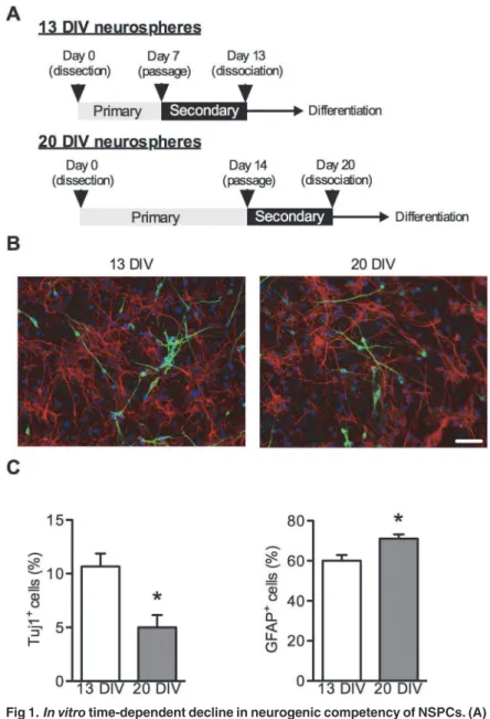
In vitro time-dependent decline of neurogenic potential of NSPCs
NSPCs were obtained from SVZ of rats on embryonic day 16 and cultured in NSPC prolifera-tion medium as floating neurospheres [13,14]. Because NSPCs switch their differentiation po-tency from neurogenic to gliogenic in a time-dependent mannerin vivoandin vitro[6], we prepared 13 DIV and 20 DIV neurospheres as described in theMethodsand as shown in Fig. 1A. Immunocytochemistry showed that the percentage of Tuj1-positive cells differentiated from 20 DIV neurospheres significantly decreased compared with that from 13 DIV neuro-spheres (Fig. 1B, C). In contrast, the percentage of GFAP-positive cells differentiated from 20 DIV neurospheres was increased compared with that from 13 DIV neurospheres (Fig. 1B, D). These data indicate that, in our culture system, long-term-cultured NSPCs temporally lose neu-rogenic potential and obtain gliogenic potential.
Expression of IAP2 mRNA in NSPCs
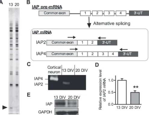
To identify differences in gene expression between 13 DIV and 20 DIV neurospheres, we per-formed fluorescence PCR differential display analysis [15]. Total RNA samples extracted from 13 DIV and 20 DIV neurospheres were subjected to PCR differential display analysis using a fluorescence differential display kit (TAKARA BIO, Inc.). Most of the cDNA bands were iden-tical among individuals; however, one cDNA band was differentially expressed between 13 DIV and 20 DIV neurospheres, when 50-T
nAC-30downstream primer and No.22 (50 -AGC-CAGCGAA-30) upstream primer were used as the primer pairs (Fig. 2A). This sequence was
100% homologous to the 30-end region of rat IAP cDNA (GenBank ID: NM019195.2). IAP is a
oligonucleotides that were present in all 4 forms of IAP and bracketed the region of the mRNA encoding the alternatively spliced cytoplasmic tails (Fig. 2B). Using this PCR strategy, the 4 forms could be distinguished based on the size of the PCR products (Fig. 2B, C). This RT-PCR analysis revealed that NSPCs predominantly expressed form 2 IAP (IAP2) mRNA; however, mature neurons expressed form 4 (Fig. 2C). These data indicate that NSPC populations with high expression of IAP2 tend to differentiate into neurons. In addition, real-time RT-PCR showed that 13 DIV neurospheres had approximately 2-fold higher levels of IAP2 mRNA
Fig 1.In vitrotime-dependent decline in neurogenic competency of NSPCs. (A)Strategy for the functional screening of candidate genes involved in the temporal specification of NSPCs using the neurosphere method. (B) Representative immunofluorescent images of NSPCs differentiated from 13 DIV neurospheres (left) and 20 DIV neurospheres (right). Secondary neurospheres were dissociated in NSPC proliferation medium and plated on poly l-lysine-coated cover glass. After 24 hours, NSPCs were exposed to NSPC differentiation medium, fixed at 10 days after differentiation, stained for a neuron marker (Tuj1; green) and an astrocyte marker (GFAP; red), and counterstained with DAPI (blue). Scale bar: 50μm. Quantification
of Tuj1-positive cells (C) or GFAP-positive cells (D) 10 days after differentiation. Data are shown as the mean±SEM of four independent experiments.*p<0.05.
expression than 20 DIV neurospheres (Fig. 2D), which is consistent with the results of the dif-ferential display analysis. Western blot analysis demonstrated that the protein expression level of IAP was also higher in 13 DIV neurosphere than 20 DIV neurosphere (Fig. 2E). These data suggest that IAP2 is involved in the mechanisms by which NSPCs change their neurogenic and gliogenic potential in a time-dependent manner.
Effect of IAP2 overexpression on NSPC differentiation
To study the role of IAP2 in NSPC differentiation, we infected secondary neurospheres with a retrovirus encoding the rat IAP2 open reading frame and harboring enhanced green fluores-cence protein (EGFP) or EGFP only (control) (Fig. 3A). We confirmed IAP2 overexpression by real-time PCR (Fig. 3B) and western blot analysis (Fig. 3C). The percentage of Tuj1-positive cells was significantly increased when IAP2 was overexpressed in 13 DIV neurospheres (Fig. 3D-G, L). On the other hand, there was no significant change in the percentage of Tuj1-positive cells when IAP2 was overexpressed in 20 DIV neurospheres (Fig. 3H-K, L). These re-sults suggest IAP2 is specifically expressed in neurogenic NSPCs and can promote neuronal
Fig 2. Expression of IAP2 mRNA in NSPCs.(A) Differential display analysis of 13 DIV neurosphere (left lane) and 20 DIV neurosphere (right lane) RNA using No. 22 upstream primer and 50-T
nAC-30downstream
primer sets. Arrowhead indicates the differentially expressed IAP2 cDNA fragment between 13 DIV
neurospheres and 20 DIV neurospheres. (B) Schematic representation of the alternative splicing possibilities for generation of IAP forms. Alternative splice forms of IAP were identified by RT-PCR analysis using primer pairs designed in common exon sequences (arrows). Right arrow indicates the position of the forward primer and the left arrow indicates the position of the reverse primer for RT-PCR as shown in (C). (C) Identification of splicing forms of IAP expressed in NSPCs using RT-PCR analysis. Total RNA was isolated from 13 DIV neurospheres and 20 DIV neurospheres. Alternative splice forms were distinguished by the size of PCR products using primer sets shown in (B). (D) Quantification of IAP2 mRNA expression levels in 13 DIV neurospheres and 20 DIV neurospheres. Total RNA was isolated from 13 and 20 DIV neurospheres. The expression levels of IAP2 and Ppia (internal standard) mRNA were determined by real-time quantitative PCR analysis. (E) Western blot analysis of IAP protein expression at 13 DIV (left lane) and 20 DIV (right lane). Each value represents the mean±SEM from three independent experiments and is expressed in reference
to 13 DIV neurospheres.**p<0.01 compared with 13 DIV neurospheres.
differentiation of short-term-cultured neurogenic NSPCs because IAP2 stimulates neuronal differentiation of young neurogenic NSPCs derived from 13 DIV neurospheres, but long-term-cultured gliogenic NSPCs derived from 20 DIV neurospheres are not reactive to IAP2 stimulation.
Discussion
Cell-fate determination of NSPCs is regulated by cell-intrinsic mechanisms and local envi-ronmental cues. However, the intracellular signaling cascades linking extracellular signals to transcription in NSPCs remain unknown. In this study, we report for the first time that a membrane protein IAP2 induces neuronal differentiation of NSPCs. This conclusion is based on the following results: (i) fluorescent differential display revealed that neurogenic NSPCs expressed higher levels of IAP2 mRNA than gliogenic NSPCs and (ii) immunocytochemical analysis revealed that IAP2 overexpression increased the percentage of Tuj1-positve cells.
In this study, we prepared two distinct populations of NSPCs, which differed in their neuro-genic potential. It has been reported that NSPCs change their differentiation potential from neurogenic to gliogenic in a time-dependent mannerin vivoandin vitro[6,17,18]. We demon-strated that 13 DIV neurospheres differentiate into more neurons than 20 DIV neurospheres suggesting that NSPCs gradually decreased their neurogenic potential in our culture system.
IAP gene expresses four alternative splicing variants and the alternative splicing forms of IAP mRNA are tissue specific [16]. Although previous reports showed that IAP4 is mainly ex-pressed in the adult mouse brain [16,19], it was unknown whether IAP is expressed in NSPCs. Furthermore, as none of the IAP cytoplasmic extensions has any known motif for enzymatic activity or protein interaction, the difference in downstream signaling among the 4 splicing forms is unknown. In this study, RT-PCR analysis showed that NSPCs predominantly express IAP2 mRNA and other forms of IAP were not detected, indicating that the expression of IAP2 mRNA could be a marker for neurogenic NSPCs.
Although we demonstrated that overexpression of IAP2 significantly increased the percent-age of Tuj1-positive cells differentiated from 13 DIV neurospheres, the downstream signaling for IAP that induces neuronal differentiation of NSPCs is unknown. It has been reported that the interacting protein partners of IAP, such as integrins and SIRPαare important for IAP2-mediated downstream signaling [9,20]. IAP-integrin interaction occurs only within a single membrane. On the other hand, IAP-SIRPαinteraction can mediate cell–cell contact. Interest-ingly, in this study, when IAP2 was overexpressed in 13 DIV neurospheres, the percentage of Tuj1-positive cells differentiated from not only EGFP-positive NSPCs, but also EGFP-negative NSPCs that did not overexpress IAP2, was increased (data not shown), suggesting that IAP2-overexpressing cells induce neuronal differentiation of neighboring cells. These data indicates the possible involvement of the IAP-SIRPαsignaling pathway in IAP2-induced neuronal dif-ferentiation of NSPCs.
Importantly, in the present data, IAP2 did not affect the differentiation of 20 DIV neuro-spheres with low expression levels of IAP2, despite the fact that 13 DIV neuroneuro-spheres exhibited neuronal differentiation induced by IAP2 overexpression. Because NSPCs temporally change their neurogenic potential by environmental cues and cell-intrinsic mechanisms [2], it is possi-ble that cell-intrinsic mechanisms, such as epigenetic changes, inhibit differentiation of 20 DIV neurospheres (H-KE–H) were dissociated and harvested asFig. 1. Cells were fixed on day 10 after differentiation and immunostained for a neuronal marker (Tuj1; D, F, H, J). Immunofluorescent images of cells stained for EGFP (green) were merged to images of cells stained for Tuj1 (red; shown in E, G, I, K). Scale bar for all: (in K) 50μm. (L) Quantification of Tuj1-positive cells (expressed as a percentage of EGFP-positive cells). Data represent the mean±SEM from the four independent experiments.*p<0.05.
neurospheres into neurons. Since a previous report showed that neuronal environmental
Wnt-β-catenin signaling could not induce neuronal differentiation of NSPCs in the later stages of brain development, in which induction of neurogenin-1 transcription byβ-catenin is epigeneti-cally inhibited [21], it may be possible that IAP2-mediated neuronal differentiation is also at-tenuated in 20 DIV neurospheres by cell-intrinsic mechanisms.
In conclusion, this is the first report to show that IAP2 promotes neuronal differentiation of NSPCs. Our results provide fundamental insights that may be useful to clarify the molecular mechanisms that regulate cell-fate determination of NSPCs.
Author Contributions
Conceived and designed the experiments: KF TN TK HS TK. Performed the experiments: KF TN YS. Analyzed the data: KF TN YS. Contributed reagents/materials/analysis tools: KF TN YS YI TK HS AA TK. Wrote the paper: KF TN TK.
References
1. Eriksson P, Perfilieva E, Björk-Eriksson T, Alborn A, Nordborg C, Peterson DA, et al. Neurogenesis in the adult human hippocampus. Nat Med. 1998; 4: 1313–1317. PMID:9809557
2. Temple S. The development of neural stem cells. Nature 2001; 414: 112–117. PMID:11689956 3. Lie DC, Song H, Colamarino SA, Ming GL, Gage FH. Neurogenesis in the adult brain: new strategies
for central nervous system diseases. Annu Rev Pharmacol Toxicol. 2004; 44: 399–421. PMID: 14744252
4. Goldman S. Stem and progenitor cell-based therapy of the human central nervous system. Nat Biotech-nol. 2005; 23: 862–871. PMID:16003375
5. Ménard C, Hein P, Paquin A, Savelson A, Yang XM, Lederfein D, et al. An essential role for a MEK-C/ EBP pathway during growth factor-regulated cortical neurogenesis. Neuron 2002; 36: 597–610. PMID: 12441050
6. Miller F, Gauthier A. Timing is everything: making neurons versus glia in the developing cortex. Neuron 2007; 54: 357–369. PMID:17481390
7. Grandbarbe L, Bouissac J, Rand M, Hrabé de Angelis M, Artavanis-Tsakonas S, Mohier E. Delta-Notch signaling controls the generation of neurons/glia from neural stem cells in a stepwise process. Development 2003; 130: 1391–1402. PMID:12588854
8. Ge W, Martinowich K, Wu X, He F, Miyamoto A, Fan G, et al. Notch signaling promotes astrogliogen-esis via direct CSL-mediated glial gene activation. J Neurosci Res. 2002; 69: 848–860. PMID: 12205678
9. Brown E, Frazier W. Integrin-associated protein (CD47) and its ligands. Trends Cell Biol. 2001; 11: 130–135. PMID:11306274
10. Jiang P, Lagenaur CF, Narayanan V. Integrin-associated protein is a ligand for the P84 neural adhesion molecule. J Biol Chem. 1999; 274: 559–562. PMID:9872987
11. Okazawa H, Motegi S, Ohyama N, Ohnishi H, Tomizawa T, Kaneko Y, et al. Negative regulation of phagocytosis in macrophages by the CD47-SHPS-1 system. J Immunol. 2005; 174: 2004–2011. PMID: 15699129
12. Takada T, Matozaki T, Takeda H, Fukunaga K, Noguchi T, Fujioka Y, et al. Roles of the complex forma-tion of SHPS-1 with SHP-2 in insulin-stimulated mitogen-activated protein kinase activaforma-tion. J Biol Chem. 1998; 273: 9234–9242. PMID:9535915
13. Nakanishi M, Niidome T, Matsuda S, Akaike A, Kihara T, Sugimoto H. Microglia-derived interleukin-6 and leukaemia inhibitory factor promote astrocytic differentiation of neural stem/progenitor cells. Eur J Neurosci. 2007; 25: 649–658. PMID:17328769
14. Reynolds B, Weiss S. Generation of neurons and astrocytes from isolated cells of the adult mammalian central nervous system. Science 1992; 255: 1707–1710. PMID:1553558
15. Liang P, Pardee A. Differential display of eukaryotic messenger RNA by means of the polymerase chain reaction. Science 1992; 257: 967–971. PMID:1354393
17. Shen Q, Wang Y, Dimos JT, Fasano CA, Phoenix TN, Lemischka IR, et al. The timing of cortical neuro-genesis is encoded within lineages of individual progenitor cells. Nat Neurosci. 2006; 9: 743–751. PMID:16680166
18. Naka H, Nakamura S, Shimazaki T, Okano H. Requirement for COUP-TFI and II in the temporal specifi-cation of neural stem cells in CNS development. Nat Neurosci. 2008; 11: 1014–1023. doi:10.1038/nn. 2168PMID:19160499
19. Huang AM, Wang HL, Tang YP, Lee EH. Expression of integrin-associated protein gene associated with memory formation in rats. J Neurosci. 1998; 18: 4305–4313. PMID:9592107
20. Matozaki T, Murata Y, Okazawa H, Ohnishi H. Functions and molecular mechanisms of the CD47-SIR-Palpha signalling pathway. Trends Cell Biol. 2009; 19: 72–80. doi:10.1016/j.tcb.2008.12.001PMID: 19144521