Article
Printed in Brazil - ©2012 Sociedade Brasileira de Química0103 - 5053 $6.00+0.00
A
*e-mail: [email protected]
Pterodon pubescens
Benth: Stability Study of Microencapsulated Extract and
Isolated Compounds Monitored by Antinociceptive Assays
Leila Servat,a,b Humberto M. Spindola,a,b Rodney A. F. Rodrigues,a Ilza M. O. Sousa,a
Ana L. T. G. Ruiz,a João E. de Carvalhoa,b and Mary A. Foglio*,a,b
aCentro Pluridisciplinar de Pesquisas Químicas, Biológicas e Agrícolas (CPQBA),
Universidade Estadual de Campinas, CP 6171, 13083-970 Campinas-SP, Brazil
bÁrea de Farmacologia, Anestesiologia e Terapêutica, Departamento de Ciências Fisiológicas,
Faculdade de Odontologia de Piracicaba, Universidade Estadual de Campinas, CP 52, 13414-903 Piracicaba-SP, Brazil
As sementes de Pterodon pubescens Benth. (Pp), disponíveis comercialmente no Brasil, são utilizadas na medicina popular em preparações anti-inflamatórias, analgésicas e anti-reumáticas. O presente estudo demonstrou as propriedades antinociceptivas da mistura de isômeros 6α-hidroxi-7β -acetoxi-vouacapano-17β-oato de metila e 6α-acetoxi-7β-hidroxi-vouacapano-17β-oato de metila (C1) isolada de Pp, empregando modelos experimentais diferentes. Um estudo de estabilidade foi realizado para investigar a relação da microencapsulação por “spray-drying” na manutenção do efeito antinociceptivo. Para isso, amostras de C1 e extrato de Pp foram monitorados no estudo de estabilidade acelerada, avaliando tanto amostras microencapsuladas como não microencapsuladas. Foi observado que a amostra C1 possui atividade antinociceptiva, revelada pelos ensaios das contorções abdominais e de formalina; foi observada também atividade significativa antialodínica, mas não efeito anti-hiperalgésico. A microencapsulação manteve a atividade e integridade de C1 e do extrato bruto de Pp, demonstrando que a microencapsulação por “spray drying” é uma alternativa útil para aumentar o tempo de prateleira.
Pterodon pubescens Benth. (Pp) seeds, commercially available in Brazil, are used in folk medicine in anti-inflammatory, analgesic, and anti-rheumatic preparations. The present study demonstrated the antinociceptive properties of isomers 6α-hydroxy-7β-acetoxy-vouacapan-17β -oate methyl ester and 6α-acetoxy-7β-hydroxy-vouacapan-17β-oate methyl ester (C1), isolated from (Pp), employing different experimental models. A stability study was performed to investigate the relationship of microencapsulation by spray-drying on the maintenance of antinociceptive action. Therefore, C1 and Pp extract samples were monitored in accelerated stability study, evaluating both microencapsulated and non-microencapsulated samples. It was observed that sample C1 possess antinociceptive activity revealed by writhing and formalin tests; C1 showed significantly anti-allodynic, but not antihyperalgesic effect; the microencapsulation maintained the activity and integrity of both, sample C1 and Pp crude extract; microencapsulation by spray drying is a useful alternative to increase shelf life.
Keywords: microcapsules, stability, spray-drying, antinociceptive, Pterodon pubescens
Introduction
Pterodon pubescens Benth. (Leguminosae) seeds are commercially available in Brazil. The plant’s crude alcoholic extract is used in folk medicine in anti-inflammatory, analgesic, and anti-rheumatic preparations.1,2
Phytochemical studies of Pterodon genus have revealed the
presence of alkaloids, isoflavones and diterpenes. Furan
diterpenes were identified and isolated from Pterodon
species.3-7Authors have reported that furanoditerpenes
possessing vouacapan skeleton are involved with the anti-inflammatory, antinociceptive and antiproliferative properties of Pterodon pubescens seed crude extract.7-12
Benth seeds.12 This report gives sequence to that study,
evaluating the antinociceptive properties of isomers 6α-hydroxy-7β-acetoxy-vouacapan-17β-oate methyl ester and 6α-acetoxy-7β-hydroxy-vouacapan-17β-oate methyl ester (C1) isolated from P. pubescens crude extract, as well as the influence of the microencapsulation process as a tool for maintaining stability of both the crystals and extracts. Therefore, we evaluated microencapsualed and free samples of C1 and the crude extract on the antinociceptive assays.
Experimental
General experimental procedures
IR spectra were recorded on a JASCO-FT/IR-410
spectrometer. 1H, 13C NMR and 2D experiments were
conducted on a Varian Inova-500 spectrometer (11 T).
Chemical shifts were recorded in CDCl3 solutions and
quoted relative to TMS (dH 0.0, 1H NMR) and CHCl3
(dH 77.0,
13C NMR). High-resolution electron inpact
ionization mass spectrometry (HREIMS) was recorded on a VG-AutoSpec high resolution mass spectrometer (70 eV) using direct probe. GC/MS was carried out using a HP-6890/5970 system equipped with a J&W Scientific DB-5 fused capillary column (25 m × 0.2 mm × 0.33 µ). Temperature program was 40 (5 oC min-1) - 300 oC (10 min).
Injector 250 oC, detector 300 oC. Helium was used as carrier
gas (0.7 bar, 1 mL min-1). The MS were taken at 70 eV.
Scanning speed was 0.84 scans s-1 from 40 to 550. Sample
volume was 1 µL. Split: 1:40. Column chromatography (CC): silica gel (0.063 × 0.200 mm, Merck). TLC (thin
layer chromatography): precoated plates (775554 Merck),
UV detection and anisaldehyde solution.
Phytochemistry
Plant material
Pterodon pubescens (Pp) seeds were previously collected in Minas Gerais state (Ponto Chique city), in March 2008 and identified by Prof. Dr. Jorge Yoshio Tamashiro from IB-Unicamp (Department of Botany), who identified the plant species. A voucher specimen was deposited at the University of Campinas (Unicamp) Herbarium, under number 1402.
Extracts production and compounds isolation
Freeze-dried seeds (1000 g) were ground in mill (POLI model LS-06) and extracted with dichloromethane three times during 2 h periods, with 5:1 solvent/plant ratio, at room temperature. The extract was dried over anhydrous Na2SO4 to remove residual water, filtered and concentrated
under vacuum (BUCHI model RE 215), providing the crude extract in 40% yield.
The crude extract (240 g) was chromatographed over silica gel (1900 g) and eluted sequentially with n-hexane/ ethyl acetate gradients. The fractions were monitored by thin layer chromatography (TLC). The TLC plates were sprayed with p-anisaldehyde reagent (50 mL acetic acid, 0.5 mL sulfuric acid and 0.5 mL p-anisaldehyde) followed
by heating at 100 oC. Similar fractions were grouped
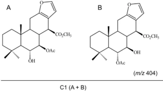
according to their TLC profile. The fractions that were eluted with hexane:ethyl acetate (75:25) providing fraction E, gave crystals (8.4 g; 3.5%). The crystals were analysed by gas chromatography (GC-MS) with identification of an isomers mixture. Comparison of the physical and spectral data (mass,1H NMR, 13C NMR) of sample C1 with reported
data4-6 provided identification of the isomer mixture as a 1:1
ratio of (i) 6α-hydroxy-7β-acetoxy-vouacapan-17β-oate methyl ester and (ii) 6α-acetoxy-7β
-hydroxy-vouacapan-17β-oate methyl ester. The isomers mixture showed a
melting point in the range of 178-182 oC.
Microcapsules production
Pp extract and C1 microencapsulation
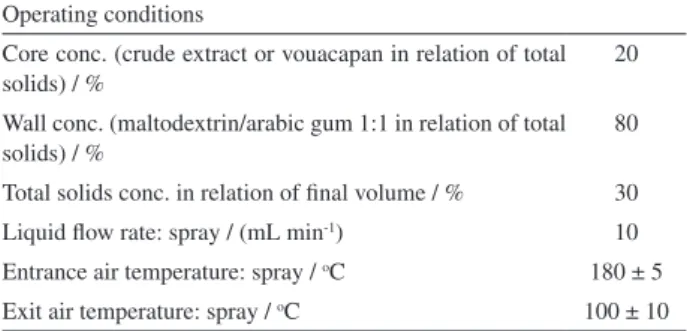
A mini spray-dryer Buchi, model B-290, with inert loop B-295 (Flawil, Switzerland) was used for the microencapsulation process; operating conditions are shown in Table 1.
Samples were homogenized with maltodextrin/arabic gum (1:1) in a ratio of 2:8, using Tween 80® as emulsifier
and IKA® T10Basic-Ultra Turrax®.
Particle morphology
The samples processed by spray-dryer were set at a double-sided adhesive tape in cylindrical aluminum (stubs) according to the procedure described by Rosemberg and Young.13 The cylinders were subjected to metallization by
coating with a thin layer of gold using a current of 40 mA Table 1. Operating conditions of the spray-dryer in the microencapsulation process, using maltodextrin and arabic gum as wall material
Operating conditions
Core conc. (crude extract or vouacapan in relation of total solids) / %
20
Wall conc. (maltodextrin/arabic gum 1:1 in relation of total solids) / %
80
during 180 s under vacuum. This technique, known as sputtering, was performed in an evaporator (Balzer). The morphology of the samples was evaluated using a scanning electronic microscope (JEOL), operating at 10 kV.
Stability evaluation
Microcapsules produced were stored at climate chamber, with constant temperature (40 oC) and humidity
(70%). Samples of microencapsulated C1 were taken at 0, 30, 60, 150 and 180 days and of microencapsulated Pp extract, were taken at 0 and 180 days. The samples were submitted to antinociceptive activity assay. Pp extract and C1 without encapsulation were used as a control.
Pharmacology
Drugs
All drugs and compounds were diluted with Tween80
1% (Sigma-Aldrich, USA) in 0.9% saline solution (NaCl diluted in distilled water), C1 microencapsulated was diluted with saline solution. The following substances were used: pentobarbital, indomethacin, morphine hydrochloride (Cristália, Brazil), acetic acid, complete Freund’s adjuvant (CFA), carrageenan (Sigma- Aldrich, USA) and formaldehyde (Chemco-Brasil).
Animals
Male Swiss mice (25-35 g) and Wistar rats (150-250 g) were kept at 25 ± 2 ºC exposed to 12 h light-dark cycles (the light phase starting at 7:00 am) and maintained in animal facilities (10 and 5 animals per cage, respectively) with water and food ad libitum, for at least 7 days prior to the assays. Separate groups of mice and rats were used only once for each experiment, except in the hyperalgesia and allodynia assays. The intraperitoneal (i.p.) route was used for all experiments, based on previous report.12 Studies were
carried out in accordance with the current guidelines for veterinary care of laboratory animals14 and were performed
under the consent and surveillance of Unicamp’s Institute of Biology Ethics Committee for Animal Research (1951-1).
Evaluation of locomotor activity
The ambulatory behavior was assessed in an open-field test as described previously.12 The apparatus consisted of
a plastic box measuring 45 × 45 × 20 cm, with the floor divided into 9 equal squares (15 × 15 cm). The number of squares crossed with all paws (crossing) was counted in a 3-min session. Mice were 30 min previously treated intraperitoneally (i.p.) with C1 (30 mg kg-1), pentobarbital
(35 mg kg-1) or vehicle. Results were expressed as
mean ± S.E.M. of 4 animals per group.
Abdominal constriction induced by acetic acid (writhing test)
The writhing test was carried out as described previously.12 Groups of mice (n = 6) were treated i.p.
with vehicle (10 mL kg-1), indomethacin (30 mg kg-1)
or sample C1, in a dose-dependent manner: 10, 30, 100, and 300 mg kg-1, to determine the effective dose (ED
50).
Writhing was induced by an i.p. injection of 0.8% acetic acid solution (0.1 mL per 10 kg-1), 30 min after treatment.
After injection of the acetic acid solution, the numbers of writhings (abdominal constrictions) were cumulatively counted over 15 min, for nociception measurement. Data represents the average of the total writhing observed per dose concentration.
Formalin test
The procedure was the same as described by Hunskaar and Hole,15 with few changes. The nociceptive stimulus
is a subplantar (s.pl.) injection of formalin solution (2%
formaldehyde in PBS; 20 µL per paw) into the dorsal
surface of the right hind paw of the mice. Groups of animals (n = 6) were treated with sample C1 (3, 10, 30 and 100 mg kg-1, i.p.) 30 min before the formalin injection.
Indomethacin (30 mg kg-1, i.p.) and morphine (10 mg kg-1,
i.p.) were used as reference drugs. Control animals received only the vehicle. The animals were placed individually in an observation chamber made of transparent acrylic to allow clear observation of the animal’s paws. The amount of time the animal spent licking the injected paw, indicative of pain, was recorded during 45 min after formalin injection. The initial nociceptive scores peaked 5 min after formalin injection (early phase) and 25-45 min after formalin injection (late phase), representing the neurogenic and inflammatory pain responses, respectively.
Mechanical allodynia induced by complete Freund adjuvant (CFA)
The procedures were developed and standardized in our laboratory based on the method previously described16
with changes in protocol and data analysis. Different groups of rats (n = 5) were used, and inflammation was induced with a solution of CFA (1 mg mL-1 of heat killed
0.5 mm) was pushed against the hind paw with ascending force (touch stimulator). The force ranged from 0 to 35 g over a 20 s period. When the animal withdrew the hind paw, the mechanical stimulus was automatically stopped, and the force applied by the animal to withdraw the paw was recorded to the nearest 0.1 g. An allodynia score was determined after four consecutive measurements using the touch stimulator sequentially on the left and right hind paw and calculated considering the formula below determined by the authors:
Left hind paw value / Right hind paw value = Allodynia score
The basal score was measured before CFA injection on day 0, and animals considered for testing were those with a mean value nearest to 1 (demonstrating no significant difference between both paw stimuli). After CFA injection, measurements were carried out considering three different phases, as follows: 4 h on day 0 (acute pain); 24 h on day 1 (sub-acute pain) and on day 14 (chronic pain). Vehicle (10 mL kg-1) or sample C1 were administered (30 mg kg-1,
i.p.) 30 min prior to touch stimulation, in order to evaluate the possible anti-allodynic activity observed for each phase. A positive control was not employed as the aim of this test was to evaluate the activity of compounds using doses equal to those used in the tests described below for evaluating potential antinociceptive mechanisms.
Mechanical hyperalgesia induced by carrageenan
The procedures used for this study were similar to those described previously17with some changes in the protocol
and data analysis. Different groups of rats (n = 6) were submitted to pressure stimulus (0 to 500 g) on the right hind paw using an analgesy-meter (Ugo Basile, model 37215/372116, Italy) prior to carrageenan injection, in order to determine the basal value. The hypernociceptive response was considered when animals vocalized or withdrew the paw from the equipment, demonstrating pain. After this first measurement, animals received a carrageenan (0.1 mL) intraplantar (i.pl.) injection (2.5% in saline) into the right hind paw surface. After 2.5 h, they were submitted to pressure again, to evaluate whether the hypernociceptive state was reached (40% decrease). Animals were than treated with vehicle (10 mL kg-1, i.p. - negative control),
indomethacin (30 mg kg-1, i.p. - positive control) and sample
C1 (30 mg kg-1, i.p.), and the mechanical hyperalgesia
was evaluated after 30 min, 1 h, 2 h and 3 h. The value demonstrating mechanical hyperalgesia was obtained after each measurement, and the results were shown as decreased percentage compared to the pressure tolerated in the basal (100%) value for each group.
Statistical analysis
All results were submitted to one way analysis of variance (ANOVA) with repeated measurements, p ≤ 0.05 was considered the critical level for evaluating significant difference between the control and treated groups, followed by Duncan´s Test, using StatSoft® software. Graphs were
designed using the Origin® software.
Results and Discussion
In the present study, we investigated the antinociceptive activity of vouacapans sample C1 (Figure 1) isolated from P. pubescens (Pp), employing different experimental models. A stability study was performed to investigate the influence of microencapsulation by spray-drying on maintenance of antinociceptive action. For this purpose, were monitored sample C1 and Pp crude extract activity in acelerated stability study (180 days period), comparing both microencapsulated and non-microencapsulated samples.
The most relevant findings of this paper were that sample C1 (i) possess antinociceptive activity revealed by writhing test; (ii) is more effective on the first phase (neurogenic pain) then second phase (inflammatory pain) of formalin test; (iii) showed significantly anti-allodynic, but not antihyperalgesic effect; (iv) the microencapsulation kept the activity and integrity of both, sample C1 and Pp crude extract; (v) microencapsulation by spray drying is a useful alternative to increase shelf life time.
Evaluation of C1 antinociceptive, anti-hypernociceptive and anti-allodynic activities
Initially, the open field test was performed in order to exclude the possibility of the sample C1 action could be related to non-specific disturbances in the locomotor activity of the animals.12 The treatment with sample C1
(30 mg kg-1, i.p.) did not cause any significant change
in the ambulation of mice when tested in the open field. However, pentobarbital (35 mg kg-1, i.p.) significantly
(p < 0.001) reduced the locomotor activity of animals in this test. The mean number of crossings was 48.2 ± 2.2, 13.7 ± 6.1, 40 ± 9 for vehicle, pentobarbital and sample C1, respectively.
With this result, the antinociceptive properties of sample C1 were evaluated using the writhing test. Acetic-acid induced pain is a screening tool for the assessment of analgesic or anti-inflammatory properties of natural or synthetic compounds. Inflammatory pain transmission primarily involves peripheral polymodal receptors around small vessels which signal to the CNS via sensory afferent C-fibers entering the dorsal horn. Acetic acid may act indirectly by inducing the release of endogenous mediators, which stimulate the nociceptive neurons and can be blocked by nonsteroidal anti-inflammatory drugs (NSAIDs) and
opioids.18 This method has good sensitivity but poor
specificity allowing misinterpretation of the results. This problem can be avoided by complementation with other nociception models, as described here.
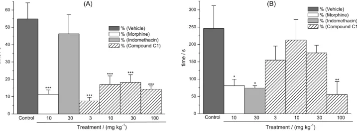
The results presented in Figure 2 showed that sample C1 produced a significant effect, reducing writhings of the mice in a dose-dependent manner, compared to the control group (vehicle only), presenting ED50 = 35.6 mg kg-1.
In order to confirm this property, and distinguish this action between inflammatory and neurogenic pain, the formalin test was performed. The intraplantar injection of formalin produces a biphasic response characterized by an immediate and intense increase in the spontaneous activity followed by a quiescent phase and then a more prolonged increase in cell firing of both primary afferents
as well as dorsal horn neurons.19 Neurogenic pain is
caused by direct activation of nociceptive nerve terminals, while inflammatory pain is mediated by a combination of peripheral input and spinal cord sensitization.15 Intraplantar
injection of formalin in rodents produces significant increases in spinal levels of different mediators, such as excitatory amino acids, PGE2, and nitric oxide, as well as
kinins and other peptides.20 Furthermore, systemic spinal
and supraspinal administration of tachykinin receptor antagonists, NOS inhibitors, NMDA receptor antagonists,
opioids,α2-adrenoceptor agonist, and NSAIDs can all
antagonize formalin induced nociception.21
As shown in Figure 3, treatment with sample C1 produced a marked reduction of the licking time in the first phase. This result suggests that the antinociceptive action Figure 2. Graph demonstrating effects of sample C1 against the acetic acid-induced writhing in mice. Animals were pretreated (30 min) with vehicle (10 mL kg-1), C1 (doses 10, 30, 100 and 300 mg kg-1; i.p.) or indomethacin (30 mg kg-1; i.p.). Each column represents the mean ± S.E.M. for six mice in each group. (*p ≤ 0.05, **p ≤ 0.01, ***p ≤ 0.001).
may be related to both central and peripheral mechanisms. In the second phase (inflammatory pain), sample C1 was effective only at the highest doses (100 mg kg-1). The
reference drug indomethacin (30 mg kg-1) inhibited pain
induced by formalin in the second phase, whereas morphine (10 mg kg-1) significantly attenuated the pain responses of
both phases.
Next, we investigated the anti-hypernociceptive effect of sample C1 induced by carrageenan in rats. Carrageenan-induced hind paw inflammation is a neutrophil-mediated acute inflammatory response that produces hind paw swelling, erythematic and localized hyperthermia.22 This
inflammatory method has become a widely used model for studying acute inflammation, involving macrophages, mast cells, and endothelial cells.23 The results presented
in Figure 4 showed that indomethacin (30 mg kg-1, i.p.)
produced significant anti-hypernociception activity increasing the percentage of stimulus compared to the control group (vehicle), whereas sample C1 (30 mg kg-1, i.p.)
did not present any effect 30 min, 1 h, 2 h and 3 h after carrageenan injection. In this assay, which has essentially inflammatory components, sample C1 was not effective, suggesting that C1 has a higher affinity for neurogenic components, corroborating the results observed in the second phase of the formalin test, in which there was a need to increase dosage to have activity in inflammatory pain.
Finally, the anti-allodynic effect of sample C1 was evaluated throughout a persistent pain model caused by complete Freund adjuvant (CFA). Allodynia (pain-like
responses to normally innocuous stimuli) was induced by CFA intraplantar injection,24 which involves central
sensitization due to the release of multiple inflammatory and pain mediators that account for sensitivity increase of both peripheral sensory afferents at the injury site, and in the central nervous system.25
The results presented in Figure 5 showed that sample C1 (30 mg kg-1, i.p.) produced a significant anti-allodynic
effect reducing the allodynic score compared to the control group during the acute (4 h post CFA) and sub acute phases (24 h post CFA) of the test. Sample C1 exerted no activity during the chronic phase (14 days post CFA). These results demonstrated the activity of sample C1 during the acute and sub acute phases of allodynic response, suggesting activity related to central and/or peripheral pathway.
Evaluation stability study
Spray-drying is the most commonly used method in the herbal processing industries, but there are few reports on the influence of adjuvants and technological processing on the herbal extracts or herbal substances.26 In the
development of herbal medicines, bioguided techniques have been the methods of choice among the technological processes. This approach allows optimizing formulations with a view toward maximizing the therapeutic effect.27
This study evaluated for the first time the contribution of microencapsulation by spray drying in stability of crude extract and isolated compounds, employing antinociceptive activity assays.
Figure 4. Graph demonstrating results of vocalization or paw with drawal related to pressure exerted in the right hind paw in response to the hypernociception caused by injection of carrageenan (2.5%, 0.1 mL) in rats treated with indomethacin or sample C1 (30 mg kg-1, i.p.) compared to the control group (vehicle). Results expressed as reduction percentage means ± S.E.M. of 5 animals for experimental groups compared to the basal values (considered 100%) (**p ≤ 0.01; ***p ≤ 0.001).
Once we demonstrated the potential use of C1 for pain relief, we proceed to evaluate the microencapsulation by spray-drying processes as a tool for maintaining
P. pubescens extracts and sample C1 integrity.
The microcapsules of Pp crude extract or sample C1 were produced by a spray-drying technique using maltodextrin and arabic gum (1:1) as wall material. The excellence properties of arabic gum as emulsifier, good volatile retention and low viscosity added to low cost of maltodextrin, makes this blend a great choice for wall material in the production of microcapsules.28 Images
obtained by scanning electron microscopy (images not show) demonstrated that the microencapsulated material has predominantly rough surface, which is very common for this type of technique due to contraction of the particles during the fast drying.29
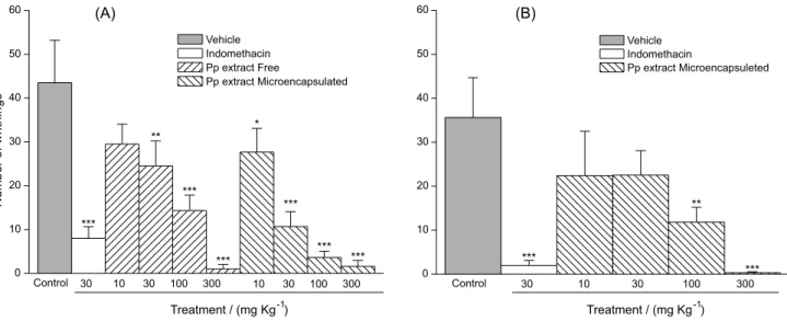
Initially, we evaluated the Pp crude extract stability, employing writhing test for the antinociceptive activity evaluation. This assay was performed with free and microencapsulated Pp extract samples, representative of 0 (T0) and 180 (T180)-day times of the stability study. The results are shown in Figure 6.
The free and microencapsulated extracts demonstrated activity in all the tested dosages (10, 30, 100 and
300 mg kg-1, i.p.). The free and microencapsulated
extracts gave ED50 values of 32.3 mg kg
-1 and 12.8 mg kg-1
respectively, showing that microencapsulation processes maintained the in vivo extract activity. An assay with the extract´s microcapsules was performed after 180 day time, demonstrating that the effect was preserved (ED50 32 mg kg-1). The free extract was not evaluated in this
time due to the glassy appearance of the sample, which is not adequate for the antinociceptive assay.
The results showed that spray-drying microencapsulation enabled the use of de Pp extract after stocking, besides preserving the in vivo effect. This technique showed to be a useful alternative to increase shelf life.
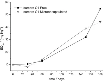
To determine the stability of sample C1, writhing test were performed with free and microencapsulated C1 at T0, T30, T60, T150 and T180 day-times of stability study. The results of dose-response curves are represented in Figure 7.
The ED50 values, calculated from the results of
dose-response curves, were 10, 10.5, 13, 32 and 55 mg kg-1
for the free and 10, 8, 14, 39 and 44 mg kg-1 for the
microencapsulated samples (Figure 8).
In the in vivo assays, using the writhing test in mice, both the free and the microencapsulated sample C1, were effective in the dosages tested, maintaining the original characteristics.
At lower dosages both free and microencapsulated sample C1, decreased activity as stability study time lapsed (Figure 7), resulting in need of higher dosages necessary to reduce the number of contortions to 50% (ED50) (Figure 8).
A gradual decrease in both samples C1 effectiveness was observed, mainly after 60 days, preserving however their activity until the end of the study (T180-day time).
The free and microencapsulated sample C1 maintained stability even when stored in drastic temperature and humidity conditions. The microencapsulation process, not only preserves the content, but also solves the inconvenience of low aqueous solubility.
Figure 8. Graph showing the results of the effective dose (ED50) of free sample C1 and microencapsulated at 0, 30, 60, 150 and 180 days of stability study.
Conclusions
The most relevant findings of this paper were that sample C1 (i) possess antinociceptive activity revealed by writhing test; (ii) is more effective on the first phase (neurogenic pain) then second one (inflammatory pain) of formalin test; (iii) showed significantly anti-allodynic, but not antihyperalgesic effect; (iv) the microencapsulation kept the activity and integrity of both sample C1 and Pp crude extract; (v) microencapsulation by spray drying is a useful alternative to increase shelf life time.
The free and microencapsulated sample C1 maintained stability even when stored in drastic temperature and humidity conditions. Whereas the crude Pp extract only maintained stability with the microencapsulation process, not only preserving the content, but also solving the inconvenience of low aqueous solubility.
Acknowledgments
The authors thank CAPES, FAPESP and CNPq for financial support and research fellowships.
References
1. Pio Correa, M.; Dicionário das Plantas úteis do Brasil e das Exóticas Cultivadas, vol. I, Inst. Bras. Des. Flor.: Rio de Janeiro, 1975.
2. Lorenzi, H.; Árvores Brasileiras. Manual de Identificação e Cultivo de Plantas Arbóreas Nativas do Brasil, vol. 1, 2nd ed.,
Instituto Plantarum de Estudos da Flora: São Paulo, 1998. 3. Mahjan, J. R.; Monteiro, M. B.; J. Chem. Soc. Perkin Trans.
1973, 1, 520.
4. Fascio, M.; Mors, W. B.; Gilbert, B.; Mahajan, J. R.; Monteiro, M. B.; dos Santos Filho, D.; Vichnewski, W.; Phytochemistry
1975, 15, 201.
5. Campos, A. M.; Silveira, E. R.; Braz-Filho, R.; Teixeira, T. C.; Phytochemistry 1994, 36, 403.
6. Arriaga, A. M.; Gomes, G. A.; Braz-Filho, R.; Fitoterapia 2000, 71, 211.
7. Spindola, H. M.; Carvalho, J. E.; Ruiz, A. L. T. G.; Rodrigues, R. A. F.; Denny, C.; Sousa, I. M. O.; Tamashiro, J. Y.; Foglio, M. A.; J. Braz. Chem. Soc. 2009, 20, 569.
8. Nunan, E. A.; Carvalho, M. G.; Piloveloso, D.; Braz. J. Med. Biol. Res.1982, 15, 450.
9. Carvalho, J. C. T.; Sertié, J. A. A.; Barbosa, M. V. J.; Patrício, K. C. M.; Caputo, L. R. G.; Sarti, S. J.; Ferreira, L. P.; Bastos, J. K.; J. Ethnopharmacol. 1999,64, 127.
10. Silva, M. C. C.; Gayer, C. R. M.; Lopes, C. S.; Calixto, N. O.; Reis, P. A.; Passeas, C. P. B.; Paes, M. C.; Dalmau, S. R.; Sabino, K. C. C.; Todeschini, A. R.; Coelho, M. G. P.; J. Pharm. Pharmacol. 2004, 55, 135.
11. Vieira, C. R.; Marques, M. F.; Soares, P. R.; Matuda, L.; De Oliveira, C. M. A. P.; Kato, L.; Da Silva, C. C.; Guillo, L. A.; Phytomedicine 2008, 15, 528.
12. Spindola, H. M.; Servat, L.; Denny, C.; Rodrigues, R. A. F.; Eberlin, M. N.; Cabral, E.; Sousa, I. M. O.; Carvalho, J. E.; Tamashiro, J. Y.; Foglio, M. A.; BMC Pharmacol. 2010, 10, 1. 13. Rosemberg, M.; Young, S. L.; Food Struct.1993, 12, 31.
14. Voipio, H. M.; Baneux, P.; Gomez de Segura, I. A.; Hau, J.; Wolfensohn, S.; Lab. Anim. 2008, 42, 1.
15. Hunskaar, S.; Hole, K.; Pain 1987, 30, 103.
16. Villeti, G.; Bergamarchi, M.; Bassani, F.; Bolzoni, P. T.; Maiorino, M.; Pietra, C.; Rondelli, I.; Chamiot-Clark, P.; Simonato, M.; Barbieri, M.; J. Pharmacol. Exp. Ther. 2003, 306, 804.
17. Randall, R. O.; Selitto, J. J.; Arch. Int. Pharmacodyn. 1957, 111, 409.
18. Collier, H. O.; Dinneen, L. C.; Johnson, C. A.; Schneider. C.; Br. J. Pharmacol. Chemother.1968, 32, 295.
19. Shannon, H. E.; Jones, C. K.; Li, D. L.; Peters, S. C.; Simmons, R. M. A.; Iyengar, S.; Pain 2001, 93, 221.
20. Santos, A. R. S.; Gadotti, V. M.; Oliveira, G. L.; Tibola, D.; Paszuck, A. P.; Neto, A.; Spindola, H. M.; Souza, M. M.; Rodrigues, A. L. S.; Calixto, J. B.; Neuropharmacology 2005, 48, 1021.
21. Santos, A. R. S.; Calixto, J. B.; Neurosci. Lett. 1997, 235, 73.
22. Cunha, M. T.; Verri Jr, W. A.; Silva, J. S.; Poole, S.; Cunha, F. Q.; Ferreira, S. H.; Proc. Natl. Acad. Sci. U. S. A. 2005, 102, 1755.
23. Winter, C. A.; Risley, E. A.; Nuss, G. W.; Proc. Soc. Exp. Biol. Med. 1962, 111, 544.
C. F.; Rubin, M. A.; Ferreira, J.; Bonacorso, H. G.; Life Sci.
2008, 83, 739.
25. Samad, T. A.; Moore, K. A.; Sapirstein, A.; Billet, S.; Allchorne, A.; Poole, S.; Bonventre, J. V.; Woolf, C. J.; Nature 2001, 410, 471.
26. Cunha, A. M.; Menon, S.; Menon, R.; Couto, A. G.; Burger, C.; Biavatti, M. W.; Phytomedicine 2010, 17, 37.
27. De Souza, K. C. B.; Bassani, V. L.; Schapoval, E. E. S.; Phytomedicine 2007, 14, 102.
28. Fernandes, L. P.; Turatti, I. C. C.; Lopes, N. P.; Ferreira, J. C.; Candido, R. C.; Oliveira, W. P.; Drying Technol. 2008, 26, 1534.
29. Rosemberg, M.; Kopelman, I. J.; Talmon, Y.; J. Agric. Food Chem. 1990, 38, 1288.
Submitted: May 9, 2011 Published online: June 14, 2012