TATIANE YUMI NAKAMURA KANNO
SCRATCH2 na diferenciação neural em embriões e em células-tronco
Tese apresentada ao Departamento de Biologia Celular e do Desenvolvimento do Instituto de Ciências Biomédicas da Universidade de São Paulo, para obtenção do Título de Doutor em Ciências.
Área de concentração: Biologia Celular e Tecidual
Orientação: Profa. Dra. Chao Yun Irene Yan
Versão original.
TATIANE YUMI NAKAMURA KANNO
SCRATCH2 in embryonic and stem cell neural differentiation
Thesis submitted in partial fulfillment of the requirements for the degree of Doctor in Science at the Department of Development and Cell Biology of the Biomedical Science Institute.
Research area: Cell and Tissue Biology
Supervisor: Profa. Dra. Chao Yun Irene Yan
Original version.
RESUMO
Kanno, TYN. SCRATCH2 na diferenciação neural em embriões e em células-tronco. [tese (Doutorado em Biologia Celular e Tecidual)]. São Paulo: Instituto de Ciências Biomédicas, Universidade de São Paulo; 2016.
Os genes SCRATCH codificam fatores de transcrição que pertence à superfamília
SNAIL e participam do desenvolvimento neural. Membros desta superfamília têm sido descritos como repressores da transcrição. Estruturalmente, apresentam um domínio altamente conservado presente na região N-terminal chamado de SNAG e o domínio zinc-finger, na região C-terminal. Além disso, os membros da família
SCRATCH apresentam o domínio SCRATCH, que também é altamente conservado. Durante o desenvolvimento de embriões de galinha, SCRATCH2 é expresso em células precursoras neuronais embrionárias em início diferenciação no do tubo neural. Em camundongos, SCRATCH2 é expresso em precursores da glia radial (PGR) e em progenitores intermediários (PI) modulando a migração e diferenciação neuronal. Consistente com o seu papel como fator de transcrição, SCRATCH2 está presente no núcleo das células. A retenção nuclear de SCRATCH2 é controlada pelo domínio zinc-finger, enquanto sua atividade repressora encontra-se na região
N-terminal. Ao contrário dos membros da família SNAIL, a atividade repressora de SCRATCH2 não é mediada pelo domínio SNAG. No caso de SCRATCH2, a atividade repressora é modulada pelo domínio SCRATCH. A análise da sequência de SCRATCH2, através da comparação de homologia, identificou uma sequência conservada presente na região N-terminal que apresenta dois resíduos fosforiláveis, tirosina77 (Y77) e serina78 (S78). A sequência contendo estes resíduos é
completamente conservada entre os homólogos de SCRATCH2 em galinha, humano, rato e camundongo. Mutação em Y77 ou S78 reduz a capacidade repressora de SCRATCH2. A superexpressão destes mutantes em células HEK293T e embriões de galinha detectou a presença da proteína no núcleo, demonstrando que a redução na repressão de transcrição não é devida a uma mudança na localização subcelular. A substituição concomitante de ambos os resíduos, Y77 e S78, resgata a função normal de SCRATCH2. A análise in silico, que prediz o efeito
dos nossas mutações sobre a exposição dos sítios de ligação de proteínas, indica que a região entre os domínios SNAG e SCRATCH apresenta uma grande quantidade de alterações. Isto sugere que a substituição de resíduos Y77 e S78 pode modificar a conformação secundária e terciária da proteína e interferir na sua função. Nós verificamos também, a função de SCRATCH2 no contexto de corticogênese de células-tronco embrionárias de camudongo. Durante este processo, os genes Pax6>Nng>Tbr2>Tbr1 são expressos sequencialmente para
originar PGR, PI e, finalmente, neurônios pós-mitóticos. Scratch2 é gradualmente
expresso durante este processo e seu padrão de expressão se assemelha ao de
Tbr2. A remoção de Scratch2 por edição genômica via CRISPR/Cas9 resulta no
aumento da expressão Pax6, Tbr2 e Ngn1. Em contraste, os níveis de Tbr1
diminuem. Esses dados sugerem que SCRATCH2 atua na manutenção de PI, via
Pax6, e participa do início da diferenciação neural.
Palavras-chave: Células-tronco embrionárias. Corticogênese. Embrião de galinha.
ABSTRACT
Kanno, TYN. SCRATCH2 in embryonic and stem cell neural differentiation. Doctoral thesis (Cell and Tissue Biology)]. São Paulo: Instituto de Ciências Biomédicas, Universidade de São Paulo; 2016.
SCRATCH genes code zinc-finger transcription factors that belongs to the SNAIL
superfamily and participate in neural development. This superfamily members have been describe as transcriptional repressors. Structurally, they presented a highly conserved N-terminal SNAG and C-terminal zinc-finger domains. Additionally, SCRATCH family members present the SCRATCH domain that is also highly conserved. SCRATCH is specifically expressed in neural tissues, being found in early postmitotic neural progenitors. During chicken embryos development, SCRATCH2 is expressed in embryonic neuronal precursor cells in early differentiation in the neural tube. In mice, SCRATCH2 is expressed in radial glia progenitos (RGP) and intermediate progenitors (IP) modulating the neuronal migration and differentiation. Consistent with its role as a transcription factor, SCRATCH2 is expressed in the nucleus of chicken neural tube cells. Nuclear retention is controlled by the zinc-finger domain, while its repressor activity relies on the N-terminal region. Unlike SNAIL family members, SCRATCH2 transcriptional repression is not mediated by SNAG domain. In SCRATCH2 case, the repressor activity is modulated by the SCRATCH domain. An analysis of SCRATCH2 through homology comparison identified a N-terminal conserved sequence containing two phosphorylatable residues, tyrosine77
(Y77) and serine78 (S78). The sequence containing these residues is completely
conserved between the homologues of SCRATCH2 in chicken, human, rat and mice. All single mutants forms reduced SCRATCH2 repressor ability. Overexpression of cSCRATCH2 single point mutants in HEK293T and chicken embryos remain in the nucleus demonstrating that the lack of transcriptional repression is not due a subcellular localization shift. Concomitant replacement of both residues, Y77 and S78, rescue the normal function. An in silico analysis that predicts the effect of our
mutations on the exposure to binding sites demonstrated that the linker region between SNAG and SCRATCH domains presents the higher amount of changes. This suggests that the replacement of residues Y77 and S78 might modify the protein conformation interfering in its function. We also verified the SCRATCH2 function during mouse embryonic stem cell (mESC) differentiation into cortical neurons. During this process, Pax6>Nng>Tbr2>Tbr1 genes are sequentially expressed to form
RGP, IP and finally differentiating postmitotic cells. Scratch2 is gradually expressed
in this process and its expression pattern resembles that of Tbr2. Elimination of Scratch2 expression by editing its genomic locus with the CRISPR/Cas9 system
leads to an upregulation of Pax6, Tbr2 and Nng1. In contrast, Tbr1 levels are
downregulated. This data suggests that SCRATCH2 plays a role maintaining the IP pool, acting via Pax6, thereby regulating the onset of neural differentiation.
Keywords: Chicken embryo. Corticogenesis. Embryonic stem-cells. Neurogenesis.
1 INTRODUCTION
1.1 NEUROGENESIS AND NEURAL DIFFERENTIATION
The developmental process of neurulation involves a series of coordinated morphological movements that result in formation of the neural tube. The neural tube gives rise to vertebrate central nervous system originating the brain and spinal cord. Abnormalities in this process can lead to neural tube defects with devastating effects on nervous system function. The embryonic precursor of the neural tube is the neural plate, or neuroepithelium, a thickened region of ectoderm on the dorsal surface of the early embryo. The neural plate undergoes rostrocaudal lengthening, mediolateral narrowing and further apico–basal thickening, except in the midline where it becomes anchored to the underlying notochord and midline neuroepithelial cells shorten and become wedge-shaped (Colas, Schoenwolf, 2001). After that, the neural plate starts to bend forming the neural folds, which fuses giving rise to the neural tube.
The cephalic portion of the neural tube originates the brain while the caudal portion originates the spinal cord. The neural crest cells, located in the neural plate border, delaminate and migrate to form the peripheral nervous system as well as a variety of other tissues (Figure 1). Neurulation requires both intrinsic and extrinsic forces acting in concert. Intrinsic forces arise within the neural plate and drive neural plate shaping and furrowing, whereas extrinsic forces arise outside the neural plate and drive neural plate folding and neural groove closure.
Figure 1 - Neurulation process. The neural plate bends forming the neural groove
cells. The neural fold fuses giving rise to the neural tube. Near the time of neural tube closure (depending on the species), the neural crest cells delaminate from the neural tube and migrate along defined pathways.
Figure source: Wislet-Gendebien et al., 2011.
The central nervous system contains a wide variety of neuronal and glia types. The neurons are organized in layers and clusters, each presenting different functions and connections. The cell proliferation in the neural tube occurs in the luminal side of the cell layers, in a region known as the ventricular zone (VZ). In the developing spinal cord, after exiting the cells cycle, the postmitotic cells migrate toward the marginal zone (MZ). During the migration, they refine their identity to become mature neurons. Thus, the posterior neural tube can be divided functionally in concentrically arranged zones around the lumen: a VZ with proliferative progenitors, an intermediate zone (IZ) with postmitotic and undifferentiated cells and a MZ, or mantle zone, containing the differentiated neurons.
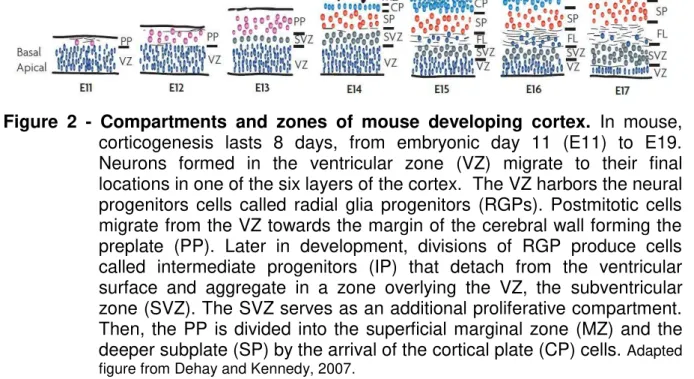
Figure 2 - Compartments and zones of mouse developing cortex. In mouse,
corticogenesis lasts 8 days, from embryonic day 11 (E11) to E19. Neurons formed in the ventricular zone (VZ) migrate to their final locations in one of the six layers of the cortex. The VZ harbors the neural progenitors cells called radial glia progenitors (RGPs). Postmitotic cells migrate from the VZ towards the margin of the cerebral wall forming the preplate (PP). Later in development, divisions of RGP produce cells called intermediate progenitors (IP) that detach from the ventricular surface and aggregate in a zone overlying the VZ, the subventricular zone (SVZ). The SVZ serves as an additional proliferative compartment. Then, the PP is divided into the superficial marginal zone (MZ) and the deeper subplate (SP) by the arrival of the cortical plate (CP) cells. Adapted figure from Dehay and Kennedy, 2007.
Figure 3 - Cortical layers are sequentially generated in an ‘inside-out’ way. The
neocortex is composed by six different layers, each contain a specific neuronal cell types. The layers are grouped from outside (pial surface) to inside (white matter). The newly arriving cells migrate radially past through the existing neurons before reaching the cortical plate. Thus, the youngest neurons are found closer to the pial surface while oldest neurons are located close to the white matter. Adapted figure from Gilmore and Herrup 1997.
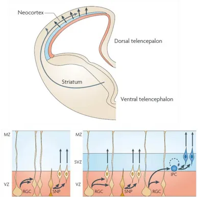
Cortical neurons are generated from three types of precursor cells: radial glial progenitors (RGPs), which are restricted to the VZ, short neural precursors (SNPs) and intermediate progenitor cells (IPs) (Figure 4).
Figure 4 - Mouse cortical progenitor cells. Cortical neurons are generated from
precursors (SNPs) and intermediate progenitor cells (IPs). RGPs and SNPs divide at the apical surface of the ventricular zone (VZ). RGCs can self-renew or undergo asymmetrical division giving rise to the intermediate progenitors (IPs). SNPs are committed neural precursors. IPCs divide away from the ventricular surface in the VZ and in the subventricular zone (SVZ). Most of the IPs undergo neural differentiation, however a small fraction self renew (dotted arrow). Adapted figure from Dehay and Kennedy, 2007.
RGPs and SNPs divide at apical surface of the VZ. SNPs are committed to generate neurons. Through asymmetric cells division, RPGs give rise to IPs. IPs divide in the apical region in VZ and SVZ undergoing few more cell divisions and then centrifugally migrate to their adult location in the cortex (Dehay and Kennedy 2007).
1.2 SCRATCH FAMILY OF TRANSCRIPTION FACTORS
SCRATCH genes code zinc-finger transcription factors that belongs to the SNAIL superfamily. All members of the SNAIL superfamily share a similar organization and also distinct signatures, which classified them in the different families. SNAIL superfamily proteins present a highly conserved C-terminal half and a divergent N-terminal half, with some conserved domains (Nieto, 2002).
In the C-terminal domain is found four to six fingers. These zinc-fingers are C2H2 type presenting two conserved cysteines and histidines that coordinate the zinc ion. The fingers are structurally composed of two β-strands
followed by an α-helix and function as sequence-specific DNA-binding motifs. The
number of zinc-fingers varies among SNAIL superfamily members, which may reflect the variations in the length of DNA being recognized (Chiang, Ayyanathan, 2013; Nieto, 2002). Alignments of the five zinc fingers DNA binding domains identified that the first, second and last zinc fingers are specific of each subgroup, whereas the third and fourth zinc fingers present an overall conservation throughout SNAIL superfamily (Kerner et al., 2009).
(Nakakura et al., 2001). Once binding to the E-box consensus site, SNAIL family members act as transcriptional repressors. The repressor activity depends not only on the zinc-finger region, but also on at least two different motifs that are found in the N-terminal region (Hemavathy et al., 2000; Hou et al., 2008; Lin et al., 2010; Molina-Ortiz et al., 2012; Montoya-Durango et al., 2008).
The N-terminal region of SNAIL superfamily members is more divergent, but all members present a conserved domain called SNAG. SNAG is conserved in all vertebrate SNAIL and SCRATCH proteins. It is composed of eight amino acids (MPRSFLVK) and function as a transcription repressor domain (Ayyanathan et al., 2007; Chiang, Ayyanathan et al., 2013; Ferrari-Amorotti et al., 2013). In SNAIL2, removal of SNAG domain impairs the recruitment of the co-repressor proteins affecting its co-repressor activity (Molina-Ortiz et al., 2012). In other words, the SNAG domain plays a role recruiting co-repressors to regulate transcriptional repressor of SNAIL1 and SNAIL2. (Ayyanathan et al., 2007; Chang et al., 2013; Ferrari-Amorotti et al., 2013; Lin et al., 2010; Molina-Ortiz et al., 2012; Nieto, 2002).
The SNAIL superfamily can be subdivided into two related and independent groups: the SNAIL and SCRATCH families (Manzanares et al., 2001). SNAIL superfamily was divided because new family members in other organisms emerged. This new genes were more similar to Drosophila SCRATCH (dSCRATCH) and its ortholog in Caenorhabditis elegans (CES-1) than any SNAIL gene (Nieto, 2002). According to SCRATCH family phylogenic history proposed by Barrallo-Gimeno and Nieto (2002) based on phylogentic analysis from placozoans to humans, the duplication of a single SNAIL gene in the metazoan ancestor originated two other
genes, SNAIL and SCRATCH (Barrallo-Gimeno, Nieto 2005; Barrallo-Gimeno, Nieto
Figure 5 - Origin and evolution of SCRATCH2 gene. An ancestor gene was
duplicated during Metazoan evolution giving rise to SCRATCH and SNAIL genes. Later, independent duplications in Cnindarian and
Bilaterian originated several family members in each group. The whole genome duplication in the vertebrate lineage gave rise to SCRATCH1 and SCRATCH2. Ancestral genes are represented in grey. Figure source: Barrallo-Gimeno, Nieto, 2009.
The founding member of the SNAIL family in vertebrates was isolated from Drosophila melanogaster. In vertebrates, the whole genome went through two
duplications. The first genome duplication gave rise to the SNAIL1/SNAIL2 and SNAIL3 ancestor. This event also originated two novel member of SCRATCH family: SCRATCH1 and SCRATCH2. SNAIL 1 and SNAIL 2 (also called SLUG - Barrallo-Gimeno, Nieto, 2005) arose from a subsequently genome duplication of the SNAIL1/SNAIL2 and SNAIL3 ancestor (Barrallo-Gimeno, Nieto, 2009). An additional genome duplication in the teleost lineage originated a duplicated SNAIL and SCRATCH genes, SCRATCH1a/b and SNAIL1a/b (Figure 5).
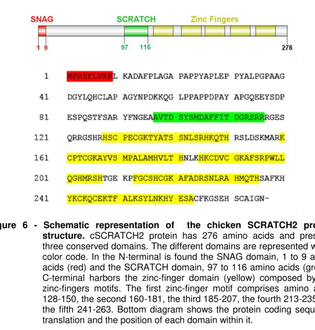
Figure 6 - Schematic representation of the chicken SCRATCH2 protein structure. cSCRATCH2 protein has 276 amino acids and presents
three conserved domains. The different domains are represented with a color code. In the N-terminal is found the SNAG domain, 1 to 9 amino acids (red) and the SCRATCH domain, 97 to 116 amino acids (green). C-terminal harbors the zinc-finger domain (yellow) composed by five zinc-fingers motifs. The first zinc-finger motif comprises amino acids 128-150, the second 160-181, the third 185-207, the fourth 213-235 and the fifth 241-263. Bottom diagram shows the protein coding sequence translation and the position of each domain within it.
In hSCRATCH1, the SNAG domain is not necessary to the repressor activity (Nakakura et al., 2001). However, little is known about SNAG domain function in other SCRATCH family proteins.
SNAIL genes plays multiple roles during development controlling neural crest specification and delamination, mesoderm specification, left-right asymmetry and triggering the epithelial to mesenchymal transition (EMT). They are also implicated in cancer metastasis (Barralo-Gimeno, Nieto 2005; Isaac et al., 1997; Manzanares et al., 1993; Mayor et al., 1995; Nieto et al., 1994; Nieto, 2002; Swami 2009). On the other hand, SCRATCH proteins are exclusively expressed in the developing nervous system and are involved with neurogenesis.
1.3 SCRATCH EXPRESSION PATTERN AND ITS ROLE IN NEUROGENESIS
SCRATCH gene was first identified and described in Drosophila
(dSCRATCH), where it is expressed in dividing neuroblasts and persists in postmitotic neurons (Roark et al., 1995). The gene is called SCRATCH due to the scratched eyes phenotype resulting from the gene mutation. Drosophila SCRATCH null mutant presents a reduced number of photoreceptors in the eye and deformations in the ommatidia, suggesting that SCRATCH expression promotes neurogenesis. dSCRATCH ectopic expression in this experimental model results in formation of extra neurons. Furthermore, increased expression of SCRATCH ortholog of Caenorhabditis elegans (CES-1) expression blocked apoptosis of specific
neuronal populations: the sister cells of serotoninergic neurosecretory motor neurons and I2, leading to the appearance of supranumerary neurons, further suggesting that it regulates the genes required for programmed cell deaths (Ellis, Horvitz, 1991). Likewise, SCRATCH2 knockdown increased the rate of apoptosis in neural tube in zebrafish embryos. SCRATCH2 is required to newly differentiated neurons survival by repressing Puma expression (Rodriguez-Aznar, Nieto, 2011).
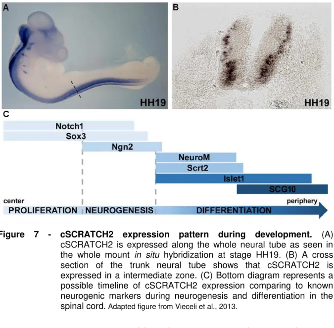
NGN2, a neural progenitor cell marker (Figure 7, Vieceli et al., 2013). Together, these data define cSCRATCH-positive cells as a subset of immediately postmitotic neural progenitors.
Figure 7 - cSCRATCH2 expression pattern during development. (A)
cSCRATCH2 is expressed along the whole neural tube as seen in the whole mount in situ hybridization at stage HH19. (B) A cross
section of the trunk neural tube shows that cSCRATCH2 is expressed in a intermediate zone. (C) Bottom diagram represents a possible timeline of cSCRATCH2 expression comparing to known neurogenic markers during neurogenesis and differentiation in the spinal cord. Adapted figure from Vieceli et al., 2013.
In mice, two forms of SCRATCH were identified: Scratch1 (mScrt1) and Scratch2 (mScrt2). Both are predominantly expressed in the brain and spinal cord
appearing in newly differentiating postmitotic neurons. Its expression persists since the onset of postmitotic cells differentiation until the first postnatal day, when mScrt2
is became absent in areas where there is no neurogenesis. During mouse development, high levels of SCRATCH start to be detected around E10.5-E11.5 (Itoh et al., 2013; Marín, Nieto, 2006). In these embryonic days mScrt is expressed
layer. mScrt is not detected in premigratory neural crest cells, however both forms
are found in some crest derivatives, such as the cranial and dorsal root ganglia as well as in the Schwann cells of the vomeronasal nerve. As development progresses, its expression is significantly downregulated. In mouse adult brain, mScrt expression
is restricted to a few isolated neurons in the dentate gyrus of the hippocampus (Marín, Nieto, 2006).
In the mouse developing cortex, mScrt2 is expressed in subsets of
mitotic and neurogenic RGP at the apical surface of the VZ, IPs at SVZ/IZ and differentiating neurons located in the upper SVZ and IZ border. It is not detected in proliferating cells present in VZ. In VZ, mScrt2 expression is relatively low, as
expected, since it is expressed in cells that have already exited the cell cycle. In this region, mScrt2 expression co-localizes with the expression of PAX6 while in the
SVZ/IZ, it co-localizes with the expression of NEUROD1, a marker of early differentiating neurons and partially with the IP marker TBR2. Conditional activation of transgenic mScrt2 in mice cortical progenitors promotes neuronal differentiation by
favoring the direct mode of neurogenesis of RGPs. In other words, mScrt2 favors the
generation of neurons directly from RGPs. The high levels of mScrt2 induce
premature cell cycle exit leading to neurogenic divisions of the progenitors, and diminishing the IP pool. Thus, neuronal amplification via indirect IP neurogenesis is extenuated, leading to a mild postnatal reduction of cortical thickness (Figure 8) (Paul et al., 2012). Corroborating this data, in vivo overexpression of mScrt2 suppressed
the generation of IPs from RGPs and caused a delay in the radial migration of upper layer neurons toward the CP (Itoh et al., 2013). Once RGPs cells commit to becoming neurons or IPs, they delaminate and start migration toward the pial surface. During migration, the cells express different genes and acquire their identity. In this context, mScrt induces the delamination by repressing E-cadherin promoter
(Itoh et al., 2013). Together, these expression data strongly suggest that mScrt2 may
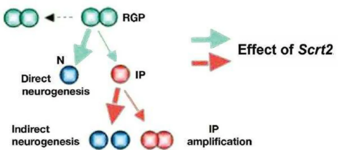
Figure 8 - Schematic illustration of mScrt2 overexpression effect in neural progenitors cells. High levels of mScrt2 favor the asymmetric divisions
of RGPs (thick green arrow) generating excess of neurons, while it does not affect the symmetric division (green cells). The pool of IPs reduces and the cells preferentially undergo differentiation (thick red arrow). Figure source: Paul et al., 2012.
mScrt1 expression is also detected in mouse P19 embryonal carcinoma
cells induced to neural differentiation. Additionally, overexpression of hSCRATCH1 in this cell line leads to neural differentiation (Nakakura et al., 2001). This indicate that SCRATCH1 might influence neuronal differentiation.
5 CONCLUSION
Our findings highlight the role of distinct conserved domains relevant to SCRATCH2 subcellular localization and function. We demonstrated that SCRATCH2 nuclear retention is controlled by its zinc-finger domain, present in the C-terminal region. In the SCRATCH2 branch of the SCRATCH family, the SNAG domain role as a repressor domain was lost. Here, we show that the transcriptional repression function of SCRATCH2 is mediated by the SCRATCH domain. In addition, our study identified two phosphorylatable amino acids present in a conserved sequence at the N-terminal portion, Y77 and S78. Single mutations in these residues reduced the repressor capacity of SCRATCH2 while double mutation rescued its function. Thus, we suggest that the residues identity is important for SCRATCH2 normal activity.
In this study, we also investigated SCRATCH2 function during mESC differentiation into cortical neurons. First, we compared Scratch2 temporal expression
profile with known markers of cortical development. Our results point that Scratch2
expression profile is similar to Tbr2, an intermediate progenitor marker. Then, to
better understand its role in mESC cortical differentiation, we removed Scratch2
expression by editing its genomic locus with the CRISPR/Cas9 system. We observed that the elimination of Scratch2 expression leads to an upregulation of the early
neurogenic markers Pax6, Tbr2 and Ngn1. In contrast, the levels of the postmitotic
neurons marker, Tbr1, is downregulated. Therefore, we suggest that Scratch2 plays
a role maintaining the intermediate progenitos pool, acting via Pax6, thereby
REFERENCES
Abranches E, Silva M, Pradier L, Schulz H, Hummel O, Henrique D, Bekman E. Neural differentiation of embryonic stem cells in vitro: a road map to neurogenesis in the embryo. PLoS One. 2009;4(7):e6286.
Afonso C, Henrique D. PAR3 acts as a molecular organizer to define the apical domain of chick neuroepithelial cells. J Cell Sci. 2006;119(Pt 20):4293-304.
Alev C, Wu Y, Nakaya Y, Sheng G. Decoupling of amniote gastrulation and streak formation reveals a morphogenetic unity in vertebrate mesoderm induction. Development. 2013;140(13):2691-6.
Arnold SJ, Huang GJ, Cheung AF, Era T, Nishikawa S, Bikoff EK, Molnár Z, Robertson EJ, Groszer M. The T-box transcription factor Eomes/Tbr2 regulates neurogenesis in the cortical subventricular zone. Genes Dev. 2008;15;22(18):2479-84.
Ayyanathan K, Peng H, Hou Z, Fredericks WJ, Goyal RK, Langer EM, Longmore GD, Rauscher FJ 3rd The Ajuba LIM domain protein is a corepressor for SNAG domain mediated repression and participates in nucleocytoplasmic Shuttling. Cancer Res. 2007; 67(19):9097-106.
Banda E, McKinsey A, Germain N, Carter J, Anderson NC, Grabel L. Cell Polarity and Neurogenesis in Embryonic Stem Cell-Derived Neural Rosettes. Stem Cells Dev. 2014;Dec 3. [Epub ahead of print]
Barrallo-Gimeno A, Nieto MA. The Snail genes as inducers of cell movement and survival: implications in development and cancer. Development. 2005;132(14):3151-61.
Barrallo-Gimeno A, Nieto MA. Evolutionary history of the Snail/Scratch superfamily. Trends Genet. 2009;25(6):248-52.
Bedogni F, Hodge RD, Elsen GE, Nelson BR, Daza RA, Beyer RP, Bammler TK, Rubenstein JL, Hevner RF. Tbr1 regulates regional and laminar identity of postmitotic neurons in developing neocortex. Proc Natl Acad Sci U S A. 2010;20;107(29):13129-34.
Berry M, Rogers AW. The migration of neuroblasts in the developing cerebral cortex. J Anat. 1965;99(Pt 4):691-709.
Bonaguidi MA, Peng CY, McGuire T, Falciglia G, Gobeske KT, Czeisler C, Kessler JA. Noggin expands neural stem cells in the adult hippocampus. J Neurosci. 2008;28(37):9194-204.
International Comitee of Medical Journal Editors. Uniform requirements for manuscripts submitted to
Britanova O, de Juan Romero C, Cheung A, Kwan KY, Schwark M, Gyorgy A, Vogel T, Akopov S, Mitkovski M, Agoston D, Sestan N, Molnár Z, Tarabykin V. Satb2 is a postmitotic determinant for upper-layer neuron specification in the neocortex. Neuron. 2008;57(3):378-92.
Britz O, Mattar P, Nguyen L, Langevin LM, Zimmer C, Alam S, Guillemot F, Schuurmans C. A role for proneural genes in the maturation of cortical progenitor cells. Cereb Cortex. 2006;16 1:i138-51.
Carpenter MK, Inokuma MS, Denham J, Mujtaba T, Chiu CP, Rao MS. Enrichment of neurons and neural precursors from human embryonic stem cells. Exp Neurol. 2001;172(2):383-97.
Chang C, Yang X, Pursell B, Mercurio AM. Id2 complexes with the SNAG domain of Snai1 inhibiting Snai1-mediated repression of integrin β4. Mol Cell Biol. 2013; 33(19):3795-804.
Chen Y-X, Krull CE, Reneker LW. Targeted gene expression in the chicken eye by in ovo electroporation. 2004;10:874–83.
Chiang C, Ayyanathan K. Snail/Gfi-1 (SNAG) family zinc finger proteins in transcription regulation, chromatin dynamics, cell signaling, development, and disease. Cytokine Growth Factor Rev. 2013;24(2):123-31.
Choi S, Yamashita E, Yasuhara N, Song J, Son SY, Won YH, Hong HR, Shin YS, Sekimoto T, Park IY, Yoneda Y, Lee SJ. Structural basis for the selective nuclear import of the C2H2 zinc-finger protein Snail by importin β. Acta Crystallogr D Biol Crystallogr. 2014;70(Pt 4):1050-60.
Colas JF, Schoenwolf GC. Towards a cellular and molecular understanding of neurulation. Dev Dyn. 2001;221(2):117-45.
Compagnucci C, Nizzardo M, Corti S, Zanni G, Bertini E In vitro neurogenesis: development and functional implications of iPSC technology. Cell Mol Life Sci. 2014;71(9):1623-39.
Dehay C, Kennedy H. Cell-cycle control and cortical development. Nat Rev Neurosci. 2007;8(6):438-50.
Domínguez D1, Montserrat-Sentís B, Virgós-Soler A, Guaita S, Grueso J, Porta M, Puig I, Baulida J, Francí C, García de Herreros A. Phosphorylation regulates the subcellular location and activity of the snail transcriptional repressor. Mol Cell Biol. 2003;23(14):5078-89.
Ellis RE, Horvitz HR. Two C. elegans genes control the programmed deaths of specific cells in the pharynx. Development. 1991;112(2):591-603.
Englund C, Fink A, Lau C, Pham D, Daza RA, Bulfone A, Kowalczyk T, Hevner RF. Pax6, Tbr2, and Tbr1 are expressed sequentially by radial glia, intermediate progenitor cells, and postmitoticneurons in developing neocortex. J Neurosci. 2005;25(1):247-51.
Estivill-Torrus , Pearson H, van Heyningen V, Price DJ, Rashbass P. Pax6 is required to regulate the cell cycle and the rate of progression from symmetrical to asymmetrical division in mammalian cortical progenitors. Development. 2002;129(2):455-66.
Evans MJ, Kaufman MH. Establishment in culture of pluripotential cells from mouse embryos. Nature. 1981;292(5819):154-6.
Evans M. Discovering pluripotency: 30 years of mouse embryonic stem cells.Nat Rev Mol Cell Biol. 2011;12(10):680-6.
Ferrari-Amorotti G, Fragliasso V, Esteki R, Prudente Z, Soliera AR, Cattelani S, Manzotti G, Grisendi G, Dominici M, Pieraccioli M, Raschellà G, Chiodoni C, Colombo MP, Calabretta B. Inhibiting interactions of lysine demethylase LSD1 with snail/slug blocks cancer cell invasion. Cancer Res. 2013;3(1):235-45.
Frantz GD, Weimann JM, Levin ME, McConnell SK.Otx1 and Otx2 define layers and regions in developing cerebral cortex and cerebellum.
J Neurosci. 1994;14(10):5725-40.
Gaspard N, Bouschet T, Hourez R, Dimidschstein J, Naeije G, van den Ameele J, Espuny-Camacho I, Herpoel A, Passante L, Schiffmann SN, Gaillard A,Vanderhaeghen P. An intrinsic mechanism of corticogenesis from embryonic stem cells. Nature. 2008;455(7211):351-7.
Germain N, Banda E, Grabel L. Embryonic stem cell neurogenesis and neural specification. J Cell Biochem. 2010;111(3):535-42.
Glover JC, Boulland JL, Halasi G, Kasumacic N. Chimeric animal models in human stem cell biology. ILAR J. 2009;51(1):62-73.
Götz M, Huttner WB. The cell biology of neurogenesis. Nat Rev Mol Cell Biol. 2005;6(10):777-88.
Hamburger V, Hamilton HL. A series of normal stages in the development of the chick embryo. 1951 Dev Dyn. 1992;195(4):231-72.
Hevner RF, Hodge RD, Daza RA, Englund C. Transcription factors in glutamatergic neurogenesis: conserved programs in neocortex, cerebellum, and adult hippocampus. Neurosci Res. 2006;55(3):223-33.
Hill RE, Favor J, Hogan BL, Ton CC, Saunders GF, Hanson IM, Prosser J, Jordan T, Hastie ND, van Heyningen V. Mouse small eye results from mutations in a paired-like homeobox-containing gene. Nature. 1991;354:522–525.
Hodge RD, Kowalczyk TD, Wolf SA, Encinas JM, Rippey C, Enikolopov G, Kempermann G, Hevner RF. Intermediate progenitors in adult hippocampalneurogenesis: Tbr2 expression and coordinate regulation ofneuronal output. J Neurosci. 2008;28(14):3707-17.
Holm PC, Mader MT, Haubst N, Wizenmann A, Sigvardsson M, Götz M. Loss- and gain-of-function analyses reveal targets of Pax6 in the developing mouse telencephalon. Mol Cell Neurosci. 2007;34(1):99-119. Epub 2006 Dec 8.
Hong S, Kang UJ, Isacson O, Kim KS. Neural precursors derived from human embryonic stem cells maintain long-term proliferation without losing the potential to differentiate into all three neural lineages, including dopaminergic neurons. J Neurochem. 2008;104:316–24
Hou Z, Peng H, Ayyanathan K, Yan KP, Langer EM, Longmore GD, et al. The LIM protein AJUBA recruits protein arginine methyltransferase 5 to mediate SNAIL-dependent transcriptional repression.Mol Cell Biol. 2008;28:3198–3207.
Itoh Y, Moriyama Y, Hasegawa T, Endo TA, Toyoda T, Gotoh Y. Scratch regulates neuronal migration onset via an epithelial-mesenchymal transition-like mechanism. Nat Neurosci. 2013;16(4):416-25. doi:
Kageyama R, Ohtsuka T, Kobayashi T. Roles of Hes genes in neural development. Dev Growth Differ. 2008;50 Suppl 1:S97-103.
Kageyama R, Ohtsuka T. The Notch-Hes pathway in mammalian neural development. Cell Res. 1999;9(3):179-88.
Keirstead HS, Nistor G, Bernal G, Totoiu M, Cloutier F, Sharp K, Steward O. Human embryonic stem cell-derived oligodendrocyte progenitor cell transplants remyelinate and restore locomotion after spinal cord injury. J Neurosci. 2005;25(19):4694-705.
Kerner P, Hung J, Béhague J, Le Gouar M, Balavoine G, Vervoort M. Insights into the evolution of the snail superfamily from metazoan wide molecular phylogenies and expression data in annelids. BMC Evol Biol. 2009;9:94.
Kim JY, Kim YM, Yang CH, Cho SK, Lee JW, Cho M. Functional regulation of Slug/Snail2 is dependent on GSK-3β-mediated phosphorylation. FEBS J. 2012; 279(16):2929-39.
Kowalczyk T, Pontious A, Englund C, Daza RA, Bedogni F, Hodge R, Attardo A, Bell C, Huttner WB, Hevner RF. Intermediate neuronal progenitors (basal progenitors) produce pyramidal-projection neurons for all layers of cerebral cortex. Cereb Cortex. 2009;19(10):2439-50.
Lang KJ, Rathjen J, Vassilieva S, Rathjen PD. Differentiation of embryonic stem cells to a neural fate: a route to re-building the nervous system? J Neurosci Res. 2004;76(2):184-92.
Langer EM, Feng Y, Zhaoyuan H, Rauscher FJ 3rd, Kroll KL, Longmore GD. Ajuba LIM proteins are snail/slug corepressors required for neural crest development in Xenopus. Dev Cell. 2008;14(3):424-36.
Le Dréau G, Martí E. Dorsal-ventral patterning of the neural tube: a tale of three signals. Dev Neurobiol. 2012;72(12):1471-81.
Lee KJ, Jessell TM. The specification of dorsal cell fates in the vertebrate central nervous system. Annu Rev Neurosci. 1999;22:261-94.
Lin Y, Wu Y, Li J, Dong C, Ye X, Chi YI, Evers BM, Zhou BP. The SNAG domain of Snail1 functions as a molecular hook for recruiting lysine-specific demethylase 1. EMBO J. 2010;29(11):1803-16.
Manzanares M, Locascio A, Nieto MA. The increasing complexity of the Snail gene superfamily in metazoan evolution. Trends Genet. 2001;17(4):178-81.
Marín F, Nieto MA. The expression of Scratch genes in the developing and adult brain. Dev Dyn. 2006;235(9):2586-91.
Martynoga B, Drechsel D, Guillemot F. Molecular control of neurogenesis: a view from the mammalian cerebral cortex. Cold Spring Harb Perspect Biol. 2012;4(10).
Mauhin V, Lutz Y, Dennefeld C, Alberga A. Definition of the DNA-binding site repertoire for the Drosophila transcription factor SNAIL. Nucleic Acids Res. 1993;21(17):3951-7.
Megason SG, McMahon AP. A mitogen gradient of dorsal midline Wnts organizes growth in the CNS. Development. 2002;129(9):2087-98.
Mingot JM, Vega S, Maestro B, Sanz JM, Nieto MA. Characterization of Snail nuclear import pathways as representatives of C2H2 zinc finger transcription factors. J Cell Sci. 2009;122(Pt 9):1452-60.
Miyata, T., Kawaguchi, A., Saito, K., Kawano, M., Muto, T. & Ogawa, M. Asymmetric production of surface-dividing and non-surface-dividing cortical progenitor cells. Development. 2004;131, 3133–3145.
Molina-Ortiz P, Villarejo A, MacPherson M, Santos V, Montes A, Souchelnytskyi S, Portillo F, Cano A. Characterization of the SNAG and SLUG domains of Snail2 in the repression of E-cadherin and EMT induction:modulation by serine 4 phosphorylation. PLoS One. 2012;7(5):e36132.
Molyneaux BJ, Arlotta P, Menezes JR, Macklis JD. Neuronal subtype specification in the cerebral cortex. Nat Rev Neurosci. 2007;8(6):427-37.
Montoya-Durango DE, Velu CS, Kazanjian A, Rojas ME, Jay CM, Longmore GD, Grimes HL. Ajuba functions as a histone deacetylase-dependent co-repressor for autoregulation of the growth factor-independent-1 transcription factor. J Biol Chem. 2008;283(46):32056-65.
Morgan R. Conservation of sequence and function in the Pax6 regulatory elements. Trends Genet. 2004;20(7):283-7.
Muqbil I, Wu J, Aboukameel A, Mohammad RM, Azmi AS. Snail nuclear transport: the gateways regulating epithelial-to-mesenchymal transition? Semin Cancer Biol. 2014;27:39-45.
Nakakura EK, Watkins DN, Schuebel KE, Sriuranpong V, Borges MW, Nelkin BD, Ball DW. Mammalian Scratch: a neural-specific Snail family transcriptional repressor. Proc Natl Acad Sci U S A. 2001;98(7):4010-5.
Nakakura EK, Watkins DN, Sriuranpong V, Borges MW, Nelkin BD, Ball DW. Mammalian Scratch participates in neuronal differentiation in P19 embryonal carcinoma cells. Brain Res Mol Brain Res. 2001;95(1-2):162-6.
Nieto MA. The snail superfamily of zinc-finger transcription factors. Nat Rev Mol Cell Biol. 2002;3(3):155-66.
Noctor SC, Martínez-Cerdeño V, Ivic L, Kriegstein AR. Cortical neurons arise in symmetric and asymmetric division zones and migrate through specific phases. Nat Neurosci. 2004;7(2):136-44.
Paridaen JT, Huttner WB. Neurogenesis during development of the vertebrate central nervous system. EMBO Rep. 2014;15(4):351-64.
Paul V, Tonchev AB, Henningfeld KA, Pavlakis E, Rust B, Pieler T, Stoykova A. Scratch2 modulates neurogenesis and cell migration through antagonism of bHLH proteins in the developing neocortex. Cereb Cortex. 2014;24(3):754-72.
Pierfelice T, Alberi L, Gaiano N. Notch in the vertebrate nervous system: an old dog with new tricks. Neuron. 2011;10;69(5):840-55.
Pilaz LJ, Silver DL. Post-transcriptional regulation in corticogenesis: how RNA-binding proteins help build the brain. Wiley Interdiscip Rev RNA. 2015;6(5):501-15.
Pinson J, Simpson TI, Mason JO, Price DJ. Positive autoregulation of the transcription factor Pax6 in response to increased levels of either of its major isoforms, Pax6 or Pax6(5a), in cultured cells. BMC Dev Biol. 2006;25;6:25.
Pontious A, Kowalczyk T, Englund C, Hevner RF. Role of intermediate progenitor cells in cerebral cortex development. Dev Neurosci. 2008;30(1-3):24-32.
Rakic P. Neurons in rhesus monkey visual cortex: systematic relation between time of origin and eventual disposition. Science. 1974;183(4123):425-7.
Rakic P.Principles of neural cell migration. Experientia. 1990;46(9):882-91.
Renier N, Wu Z, Simon DJ, Yang J, Ariel P, Tessier-Lavigne M. iDISCO: a simple, rapid method to immunolabel large tissue samples for volume imaging. Cell. 2014;159(4):896-910.
Roark M, Sturtevant MA, Emery J, Vaessin H, Grell E, Bier E. scratch, a pan-neural gene encoding a zinc finger protein related to snail, promotes neuronal development. Genes Dev. 1995;9(19):2384-98.
Rodríguez-Aznar E, Barrallo-Gimeno A, Nieto MA. (2013) Scratch2 prevents cell cycle re-entry by repressing miR-25 in postmitotic primary neurons. J Neurosci. 33(12):5095-105.
Rogers CD, Moody SA, Casey ES. Neural induction and factors that stabilize a neural fate. Birth Defects Res C Embryo Today. 2009 Sep;87(3):249-62.
Scardigli R, Bäumer N, Gruss P, Guillemot F, Le Roux I. Direct and concentration-dependent regulation of the proneural gene Neurogenin2 by Pax6. Development. 2003;130(14):3269-81.
Sessa A, Mao CA, Hadjantonakis AK, Klein WH, Broccoli V. Tbr2 directs conversion of radial glia into basal precursors and guides neuronal amplification by indirect neurogenesis in the developing neocortex. Neuron. 2008;60(1):56-69.
Shin S, Dalton S, Stice SL. Human motor neuron differentiation from human embryonic stem cells. Stem Cells Dev. 2005;14:266–9.Thomson JA, Itskovitz-Eldor J, Shapiro SS, Waknitz MA, Swiergiel JJ, Marshall VS, Jones JM. Embryonic stem cell lines derived from human blastocysts. Science. 1998;282(5391):1145-7.
Suter DM, Tirefort D, Julien S, Krause KH. A Sox1 to Pax6 switch drives neuroectoderm to radial glia progression during differentiation of mouseembryonic stem cells. Stem Cells. 2009;27(1):49-58.
Thomson JA, Itskovitz-Eldor J, Shapiro SS, Waknitz MA, Swiergiel JJ, Marshall VS, Jones JM. Embryonic stem cell lines derived from human blastocysts. Science. 1998;282(5391):1145-7.
Vandromme M, Gauthier-Rouvière C, Lamb N, Fernandez A. Regulation of transcription factor localization: fine-tuning of gene expression. Trends Biochem Sci. 1996;21(2):59-64.
Vieceli FM, Simões-Costa M, Turri JA, Kanno T, Bronner M, Yan CY. The transcription factor chicken Scratch2 is expressed in a subset of early postmitotic neural progenitors. Gene Expr Patterns. 2013;13(5-6):189-96.
Ware M, Hamdi-Rozé H1, Dupé V.Notch signaling and proneural genes work together to control the neural building blocks for the initial scaffold in the hypothalamus. Front Neuroanat. 2014;8:140.
Wislet-Gendebien S, Laudet E, Neirinckx V, Rogister B. Adult bone marrow: which stem cells for cellular therapy protocols in neurodegenerative disorders? J Biomed Biotechnol. 2012;2012:601560.
Yachdav G, Kloppmann E, Kajan L, Hecht M, Goldberg T, Hamp T, Hönigschmid P, Schafferhans A, Roos M, Bernhofer M, Richter L, Ashkenazy H,Punta M, Schlessinger A, Bromberg Y, Schneider R, Vriend G, Sander C, Ben-Tal N, Rost B. PredictProtein--an open resource for online prediction of protein structural and functional features. Nucleic Acids Res. 2014;42(Web Server issue):W337-43.
Yamasaki H, Sekimoto T, Ohkubo T, Douchi T, Nagata Y, Ozawa M, Yoneda Y. Zinc finger domain of Snail functions as a nuclear localization signal for importin beta-mediated nuclear import pathway. Genes Cells. 2005;10(5):455-64.