through the Direct Transcriptional Regulation of Msx2 by
FOXC1
Farideh Mirzayans1, Rotem Lavy2, Jonathan Penner-Chea1, Fred B. Berry1,2*
1Department of Medical Genetics, University of Alberta, Edmonton, Alberta, Canada,2Department of Surgery, University of Alberta, Edmonton, Alberta, Canada
Abstract
Hierarchal transcriptional regulatory networks function to control the correct spatiotemporal patterning of the mammalian skeletal system. One such factor, the forkhead box transcription factor FOXC1 is necessary for the correct formation of the axial and craniofacial skeleton. Previous studies have demonstrated that the frontal and parietal bones of the skull fail to develop in mice deficient for Foxc1. Furthermore expression of the Msx2 homeobox gene, an essential regulator of calvarial bone development is absent in the skull mesenchymal progenitors of Foxc1 mutant mice. Thus we sought to determine whether Msx2 was a direct target of FOXC1 transcriptional regulation. Here, we demonstrate that elevated expression of FOXC1 can increase endogenous Msx2 mRNA levels. Chromatin immunoprecipitation experiments reveal that FOXC1 occupies a conserved element in theMSX2promoter. Using a luciferase reporter assay, we demonstrate that FOXC1 can stimulate the activity of the both human and mouseMSX2promoters. We also report that reducingFOXC1levels by RNA interference leads to a decrease in MSX2 expression. Finally, we demonstrate that heterologous expression ofFoxc1 in C2C12 cells results in elevated alkaline phosphatase activity and increased expression of Runx2 and Msx2. These data indicate thatFoxc1expression leads to a similar enhanced osteogenic differentiation phenotype as observed with Msx2
overexpression. Together these findings suggest that aFoxc1-.Msx2regulatory network functions in the initial stages of osteoblast differentiation.
Citation:Mirzayans F, Lavy R, Penner-Chea J, Berry FB (2012) Initiation of Early Osteoblast Differentiation Events through the Direct Transcriptional Regulation of Msx2 by FOXC1. PLoS ONE 7(11): e49095. doi:10.1371/journal.pone.0049095
Editor:Tsutomu Kume, Feinberg Cardiovascular Research Institute, Northwestern University, United States of America
ReceivedMay 28, 2012;AcceptedOctober 7, 2012;PublishedNovember 7, 2012
Copyright:ß2012 Mirzayans et al. This is an open-access article distributed under the terms of the Creative Commons Attribution License, which permits unrestricted use, distribution, and reproduction in any medium, provided the original author and source are credited.
Funding:Funding was provided by the Canadian Institutes of Health Research MOP-245378 (http://www.cihr.ca/) and The Shriners Hospital for Children. The funders had no role in study design, data collection and analysis, decision to publish, or preparation of the manuscript.
Competing Interests:The authors have declared that no competing interests exist. * E-mail: fberry@ualberta.ca
Introduction
The development of the skeleton can proceed via two distinct mechanisms: endochondral and intramembranous ossification [1,2]. Endochondral ossification involves the prior establishment of a cartilaginous template, formed from osteochondral mesen-chyme progenitor cells differentiating into chondrocytes, that is subsequently replaced by bone-forming osteoblasts. Intramem-branous ossification involves the direct differentiation of chondral mesenchyme progenitor cells into bone-forming osteo-blasts without a cartilage intermediate. The long bones of the limbs and other components of the appendicular skeletal system arise through endochondral ossification, whereas bones in the skull arise through intramembranous ossification events.
The regulation of bone formation events needed for correct growth and patterning of the skeletal system is controlled by a network of transcriptional regulatory proteins [3]. The forkhead box transcription factor FOXC1 is required for normal de-velopment and patterning of bones originating from both endochondral and intramembranous origins [4,5,6,7]. Targeted deletion of theFoxc1gene in mice results in numerous defects in the axial skeleton. The dorsal neural arches of the vertebrae do not ossify and the lateral arches and vertebral bodies are reduced in size. [6]. The rib cage and sternum also display severe ossification defects in the Foxc1homozygous null embryos with many fused,
misshapen and fragile ribs [5,6]. The craniofacial skeleton is also severely affected inFoxc12/2mutant mice, as there is a complete absence of the skull vault. The bones that form the skull vault, or calvarium, are comprised of the frontal, parietal and intraparietal bones. In Foxc1null mice only rudimentary calvarial bones are observed near the sites of initial mesenchyme cell condensations [6,7]. In addition to the skull vault phenotypes, patterning defects to the base of the skull, the basiooccipital bone, and to the hyoid bones are observed in Foxc1 mutant mice [6]. Furthermore, expression of two genes critical in the formation of the mouse craniofacial skeleton,Alx4andMsx2, are reduced inFoxc1mutant mice [7,8,9]. The above data indicate an important role for FOXC1 in the formation of the axial and craniofacial skeleton. However, how FOXC1 functions in these processes is not entirely known.
The MSX2 transcription factor is also a critical regulator required for bone formation and development of the craniofacial skeleton. In humans, gain of function mutations in the MSX2
genes results in Craniostynostosis, Boston Type, a premature fusion of the cranial sutures [9]. In contrast,MSX2loss of function mutations cause delays in the formation of cranial sutures [10,11].
of the phenotype in humans [12]. As described above, levels of Msx2 expression are greatly reduced in the developing skull vault of Foxc1 mutant mice, suggesting this gene is under direct regulation by Foxc1 [7]. In this report we demonstrate that expression ofMsx2 is directly regulated by FOXC1 through the binding of a conserved FOXC1 consensus regulatory element in the promoters of the mouse and human Msx2 genes. Furthermore, we demonstrate that heterologous expression ofFoxc1in C2C12 cells results in a similar enhanced osteogenic differentiation phenotype to that observed with Msx2overexpression. Together these findings suggest that a Foxc1-.Msx2 regulatory network functions in early stages of osteoblast differentiation.
Materials and Methods
Plasmids
FOXC1 expression plasmids have been described previously [13,14]. The human and mouseMSX2promoters were amplified from genomic DNA using the following primers: HumanMSX2
forward 59-gctagcgaacttattctggcggtagagg-39; Human MSX2 re-verse 59-aaggcttcatgacttctctgccctagc-39; MouseMsx2 forward 59 -gctagcgcagatttccaacattctcagg-39; MouseMsx2reverse 59 -agatcttcc-gacgaaaacaagtcacc-39. DNA fragments were cloned into the NheI and HindIII (human) or BglII (mouse) sites of pGL3-Basic. The vector pBABE-FOXC1 was created by inserting the full length human FOXC1 cDNA into the EcoRI and SalI sites of pBABEpuro.
Cell Culture
U2OS, CH310T1/2 (herein referred to as 10T1/2, MDA MB231, HEK293T and C2C12 cells (obtained from ATCC) were cultured in Dulbecco’s Modified Eagle Media (DMEM) supple-mented with 10% Fetal Bovine Serum (FBS). For transient transfections cells were plated 24 hours before transfection at a concentration of 46104cells per ml. The next day, cells were
transfected with cDNA expression vectors using FuGENE6 reagent. We typically transfect cells with a ratio of 3ml of FuGENE6 per 1mg DNA. Forty-eight hours after transfection
cells were harvested for protein, RNA, or luciferase assays.
RNA Isolation and q-RT PCR
RNA was isolated from cells using the RNAeasy Mini Kit as described by the manufacturer (Qiagen). Five hundred nanograms of RNA were used in reverse transcription reactions. RT reactions were subsequently diluted 1:50 and used for quantitative RT PCR reactions. Kapa SYBR-FAST qPCR kits were used as per the manufacturer’s protocols to detect changes in gene expression using the DDCT method. Samples were run in triplicate on an ABI Prism 7900HT thrermocycler or BIO-RAD CFX96 Touch real time PCR detection system. Primers for qRT-PCR were selected from the Primer bank database [15]. Statistical analysis of real-time PCR data was determined by Student T-test or Mann-Whitney U test using SigmaPlot version 12.
Chromatin Immunoprecipitation (ChIP)
ChIP assays were performed as described previously [16]. Briefly, sheared, cross-linked chromatin from 10T1/2 cells was incubated with 1mg anti-FOXC1 antibody (Abcam) or with normal goat IgG overnight at 4uC. Magnetic Protein-G beads (25ml) that were blocked with BSA and salmon sperm DNA were
added and reactions continued for a further 2 hours. After four washes with ChIP Wash Buffer (20 mM Tris-Cl, pH 8.0; 150 mM NaCl; 2 mM EDTA; 0.1% SDS and 1% Triton X-100) and a single wash in ChIP Final Wash Buffer (20 mM Tris-Cl, pH 8.0;
500 mM NaCl; 2 mM EDTA; 0.1% SDS and 1% Triton X-100), the DNA was eluted from the beads and the crosslinking was reversed. Qiagen PCR cleanup kit was used to isolate ChIP DNA. A region flanking the putative FOXC1 binding site in the mouse Msx2 promoter was isolated with the following primers: 59 -gcagatttccaacattctcagg-39 (forward) and 59 -gcagagttgttattaggc-gatcc-39 (reverse). As a negative control, primers corresponding to exon 2 of mouse Msx2 gene were used (forward 59 gcacaccctt-caccacatcc-39; reverse 59-agggaagggcagactgaagc-39). ChIP prod-ucts, along with input chromatin fraction (1%), were amplified on a BIORAD CFX96 touch. Ct values for anti-FOXC1 ChIP and mock (IgG) ChIP were analyzed for statistical significance using T-test. Data were presented as percentages of Input DNA amplification signals.
Luciferase Reporter Assays
Reporter gene assays were performed in 10T1/2 cells. Cells were seeded into 24 well plates 24 hours prior to transfection at a density of 46104cells per well. The following day, cells in each well were transfected with 50 ng of human or mouse MSX2 reporter, along with 250 ng of pcFOXC1 or empty pcDNA4 expression vectors and 10 ng of RL-TK. Dual luciferase assays were performed as per the manufacturer’s instructions (Promega). Each experiment was performed in triplicate and all experiments were repeated three times.
Electrophoretic Mobility Shift Assays (EMSA)
We utilized a non radioactive EMSA to detect FOXC1-DNA interactions using 59-IR700 labeled oligonucleotides. Protein lysates (25mg) from U2OS cells transfected with Xpress-tagged FOXC1 or un-transfected cells were incubated with a 46EMSA
binding buffer (40 mM Hepes, 20% glycerol, 100 mM NaCl, 8 mM DTT, 0.4 mM EDTA, 0.2mg/ml poly dI-dC) for 10 minutes at room temperature. Probe was then added (50 nM final concentration) to each reaction and incubated for 20 minutes at room temperature. A 6% poly-acrylamide Tris-glycine-EDTA gel was pre-run at 105 V for 15 minutes. Samples were then loaded onto the gel, and run at 95 V for 1 hour. The gel was scanned using a Li-Cor Odyssey Infrared Imaging System.
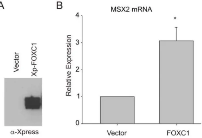
Figure 1. FOXC1 inducesMSX2expression. (A).U2OS cells were transiently transfected with Xpress-taggedFOXC1expression vectors. Levels of exogenous FOXC1 protein were determined by immunoblot-ting with anti-Xpress antibody. (B)MSX2mRNA levels were measured by qRT-PCR in FOXC1-over expressing cells. Error bars represent the standard error of the mean. *, p value,0.0001.
doi:10.1371/journal.pone.0049095.g001
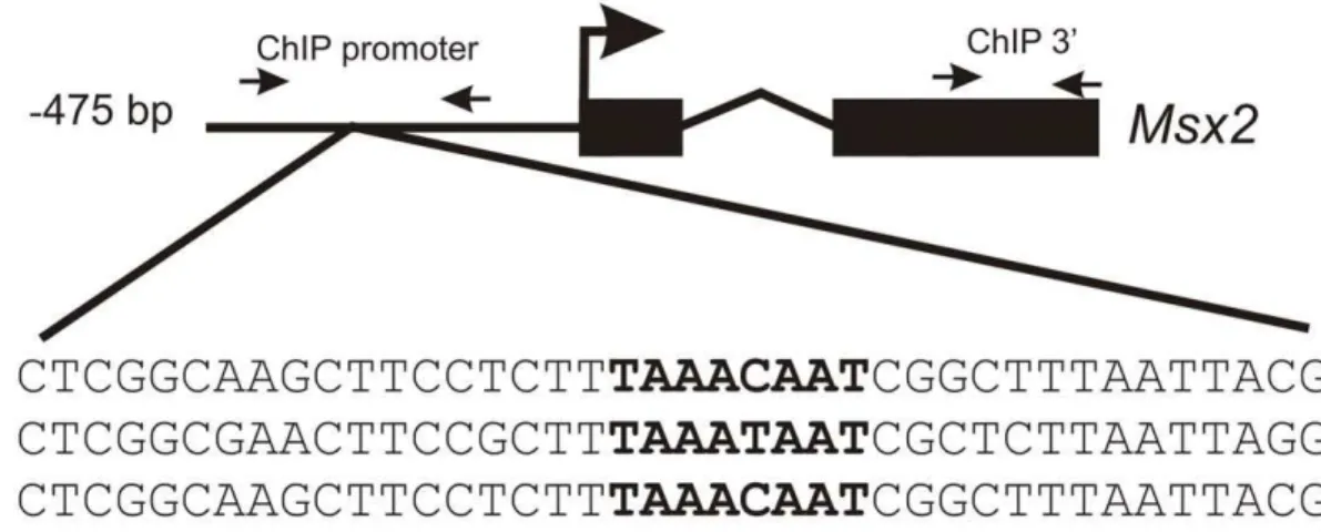
Figure 2. FOXC1 binds to theMsx2promoter in vivo.(A) Sequence analysis of upstream regulatory elements reveals the presence of a FOXC1 binding motif, indicated in bold, (TAAAT/CAAT) located in a conserved motif near the predictedMsx2transcription start site of the mouse, rat and human genes. Small arrows correspond to the position of ChIP primers located in the promoter region or the coding region of mouseMsx2. Nucleotide sequences of the Electrophoretic mobility shift assay (EMSA) probes for wild type (WT) and mutated (MUT) FOXC1 binding sites are indicated. (B) Chromatin immunoprecipitation assays confirm the binding of FOXC1 to theMsx2promoterin vivo. Quantitative PCR (qPCR) was conducted on ChiP products isolated from 10T1/2 cells using antibodies recognizing FOXC1 or normal immunoglobulins (IgG). Primers were designed amplify regions in the promoter flanking the putative FOXC1 binding site or exon 2 of the mouse Msx2 gene. Amplification signals are presented a percentage compared to input chromatin fraction. (C) EMSAs demonstrate FOXC1 binding to DNA elements in the Msx2 promoter. Extracts from U2OS cells or cells transfected with FOXC1 were incubated with IR700-labeled oligonucleotides correspond to the WT or MUT FOXC1 binding sites. FOXC1-DNA complexes are indicated by the arrow.
Virus Production and Transduction
Lentiviral vectors (pLKO.1) expressing shRNAs targeting human FOXC1 were purchased from Open Biosystems. The vector pLKO1-EGFP, which produces shRNAs targeting en-hanced green fluorescent protein (EGFP) was used as a negative control. HEK-293T cells (1.56105cells) were transfected with psPAX2 (900 ng), pMD2.G and pLKO1-FOXC1 (1mg) or pLKO1-EGFP (1mg) in a 60 mm tissue culture plate. Eighteen hours following transfection the media was removed and replaced with 6 ml of high serum (30%) DMEM. Media was collected every 12 hours for 48 hours. Pooled media collections were centrifuged at 5006g to remove any remaining packaging cells. For lentiviral infection, MDA-MB231 cells were subcultured at a density of 56105cells in a 60 mm tissue culture plate the day prior to infection. The following day, the media was removed and replaced with 6 ml fresh growth media containing polybrene (8mg/ml final concentration) and 500ml of lentiviral supernatant. Twenty-four hours later the media was removed and replaced with fresh growth media containing 0.5mg/ml puromycin. After 4 days of selection, resistant colonies (,200) were pooled and expanded. For retrovirus production, HEK293T cells were transfected with pBABE (1mg) or pBABE-FOXC1 (1mg) along with pCL-ECO (1mg) as above. Virus containing supernatants were collected as above and filtered through a 0.45m filter. C2C12 cells were infected and selected in a fashion similar to MDA-MB231 cells.
Osteogenic Differentiation
Wild type C2C12, C2C12-BABE and C2C12-FOXC1 cells were seeded at a density of 2.56105cells per well of a 6 well plate. Four days later, the cells were prepared for RNA isolation or fixed in 4% parformaldehyde and washed with PBS. To detect alkaline phosphatase activity, cells were incubated with BCIP/NBT liquid substrate system overnight.
Results
FOXC1 Positively Regulates MSX2 mRNA Expression
Msx2 mRNA expression is reduced in the mesenchyme progenitor cells that form the skull in Foxc1 mutant mice, suggestingMsx2as a putative target gene for FOXC1 regulation [7]. In order to test whether FOXC1could regulate the expression of endogenous MSX2, we over expressed FOXC1 in U2OS human osteosarcoma cells and measuredMSX2mRNA expression by qRT-PCR. As indicated in Fig. 1, levels of endogenousMSX2
mRNA were elevated by 3 fold compared to control transfected cells. These data suggest that FOXC1 positively regulates expression of endogenousMSX2mRNA.
FOXC1 Binds to and Activates the MSX2 Promoter
To assess whether Msx2 is indeed a direct target of FOXC1 transcriptional regulation, the upstream regulatory regions of human and mouse genes were surveyed for the presence of a FOXC1 binding motif using a position weight matrix for the
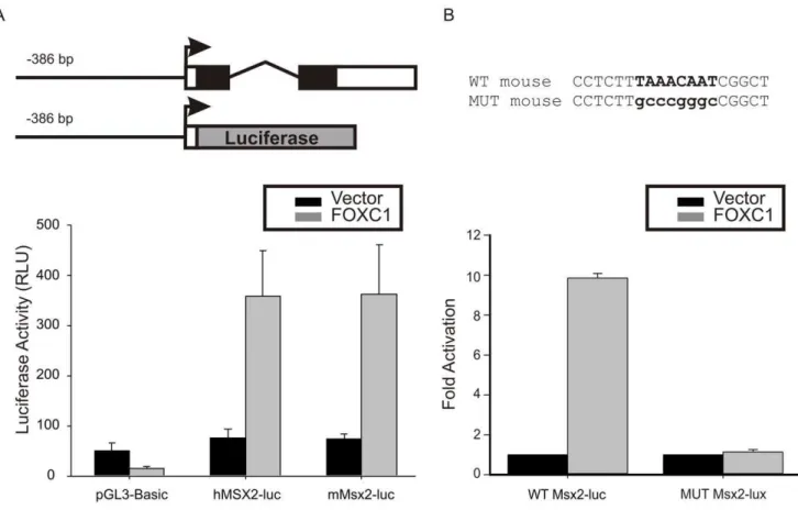
Figure 3. FOXC1 activates theMSX2promoter.(A) Luciferase (luc) reporter vectors consisting of the human (h) or mouse (m)MSX2promoter which contained the conserved FOXC1 binding element were created. Transfection of 10T1/2 cells withFOXC1expression vectors lead to a robust activation of both human and mouseMSX2promoters. (B) The putative FOXC1 binding sites was mutated in the mouse Msx2-luc reporter. Wild type and mutated Msx2-luciferase vectors were co-transfected with empty pcDNA4 (vector) or Xpress tagged-FOXC1. Error bars correspond the standard error of the mean.
doi:10.1371/journal.pone.0049095.g003
FOXC1 recognition site. FOXC1 recognizes a core 59-TAAAT/ CAA-39consensus sequence [14,17] and such sequence was found approximately 280 bp upstream of the transcription start site in the mouse, human and rat Msx2 genes (Fig. 2). We designed primers flanking this putative FOXC1 binding site in the mouse gene and performed chromatin immunoprecipitation experiments in 10T1/2 mouse mesenchymal cells. As indicated in Figure 2B, a DNA fragment corresponding to the FOXC1-binding element described above was successfully recovered by ChIP with a -FOXC1 antibodies, indicating that -FOXC1 was in fact bound to
this region. No amplification signal was detected with primers amplifying the 39 coding region of the Msx2 gene. Finally, we performed EMSAs to confirm binding of FOXC1 to Msx2
regulatory regions. Cell extracts expressing FOXC1 were able to bind an oligonucleotide probe corresponding the putative FOXC1 binding site (Fig. 2C), but not a probe containing a mutated sequence.
Next we cloned the promoter elements of mouse and human
Msx2genes into the pGL3 basic luciferase reporter and tested for promoter activity. We created luciferase reporters containing the
Figure 4.FOXC1shRNA expression reducesMSX2mRNA levels.(A) MB231 cells were transduced with lentiviral particles containing EGFP or FOXC1 shRNA. Pooled puromycin resistant colonies were expanded and FOXC1 expression analyzed semiquatitative rt-PCR. (B). qRT-PCR data indicating reduced expression ofMSX2mRNA in MB231 cells whenFOXC1levels are reduced by RNA interference.
proximal promoter along with 475 and 482 bp region of the mouse and human Msx2 genes, respectively, containing the
FOXC1 binding site. We tested whether FOXC1 could activate expression from these reporters in CH310T1/2 cells. As indicated in Fig. 3A, robust activation of both the human and mouse reporter construct was observed upon cotransfection of a FOXC1 cDNA. Together, these data suggest that Msx2 is indeed a target of FOXC1 transcriptional regulation in mesenchymal cells. We then mutated the FOXC1 binding site in the mouseMsx2-luc reporter and observed that FOXC1 was unable to activate this reporter (Fig. 3B).
Loss of FOXC1 Expression Reduces MSX2 mRNA Levels
Given that Msx2 expression was markedly reduced in the developing skull of Foxc1 mutant mice [7], we wished to test whether loss ofFOXC1expression in human cells would reduce levels of endogenous MSX2 mRNA. FOXC1 and MSX2 are expressed in MDA-MB231 human mammary adenosarcoma cells (Fig. 4). MDA-MB231 cells were transduced with lentiviral particles containing shRNAs targeting humanFOXC1or EGFP, as a control. Expression of bothFOXC1andMSX2mRNAs were reduced by over 60% in cells transduced with theFOXC1shRNA (Fig. 4B), indicating that, like inFoxc1mutant mice,MSX2mRNA levels are reduced in response to decreased levelsFOXC1.
Figure 5. FOXC1 overexpression induced ectopic osteoblast transdifferentiation of C2C12 cells.C2C12 myoblasts were transduced with retroviruses containing pBABE (empty vector control) or pBABE-FOXC1. Cells were grown to 95% confluence and stained for alkaline phosphatase activity after 4 days.
doi:10.1371/journal.pone.0049095.g005
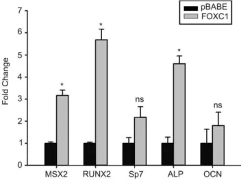
Figure 6. Increased expression of osteogenic marker genes in FOXC1-expressing C2C12 cells.Levels ofMsx2,Runx2,Sp7(Osterix), Alkaline Phosphatase (Alp) and Osteocalcin (Ocn) mRNA levels were determined by qRT-PCR from C2C12 cells transduced with empty pBABE or FOXC1 retroviral particles. * p,0.05; ns, not significant. doi:10.1371/journal.pone.0049095.g006
Activation of the Osteogenic Differentiation Pathway in the Presence of FOXC1 Expression
Overexpression ofMsx2into C2C12 myoblasts cells results in ectopic osteoblast transdifferentiation of this cell type [18]. To test the biological significance regulation of Msx2 expression by FOXC1 we created stable C2C12 cells lines constitutively expressing the human FOXC1 cDNA using retrovirus trans-duction. When FOXC1-expressing C2C12 cells (FOXC1-C2C12) reach confluence, we observe increased staining of alkaline phosphatase (ALP) activity (an early marker of osteoblast differentiation). No alkaline phosphatase activity is observed in confluent parental C2C12 cells or those transduced with empty pBABE retroviruses (Fig. 5). A similar increase in ALP activity has been observed when C2C12 cells are transduced with MSX2
expressing viruses [18]. Finally, we monitored the expression of genes that regulate early and mid osteogenic differentiation events. As indicated in Figure 6, levels of Msx2, Runx2 and Alkaline phosphatasemRNA levels are all increased in FOXC1-C2C12 cells compared to pBABE-transduced control cells. No significant changes in mRNA levels were detected forOsterix (Sp7), Osteocalcin (Ocn) or Dlx5(Figure 6, data not shown). Together these data indicate the elevation of FOXC1 expression can activate expression of osteoblast differentiation regulatory factors.
Discussion
Networks of transcriptional regulatory proteins are critical for the correct coordination of cellular events that drive formation of the skeleton. Here we describe that theMsx2transcription factor gene is a direct target of transcription regulation by the forkhead box transcription factor FOXC1. We demonstrate that FOXC1 elevates endogenous expression levels ofMSX2mRNA, stimulates activity of a MSX2 luciferase promoter construct and binds to a conserved FOXC1 binding element in the Msx2 promoter region. Furthermore, we demonstrate that reducing FOXC1
expression through shRNA concomitantly reducesMSX2mRNA expression. Finally we demonstrate that enforcedFOXC1 expres-sion in C2C12 cells results in the precocious initiation of osteoblast differentiation, similar to what has been observed in with ectopic
Msx2expression [18]. Together these data indicate thatMSX2is abona fidetarget gene of FOXC1 transcriptional regulation.
We have identified a FOXC1 binding element that is conserved in the proximal promoter of mouse, rat and humanMsx2genes. Using chromatin immunoprecipitation assays, we demonstrate that FOXC1 protein was bound to this region in mouse mesenchymal cells. Furthermore, luciferase reporter gene assays revealed that this promoter region from both human and mouse genes was activated in the presence of Foxc1. A growing list of FOXC1 target genes is beginning to emerge [16,19,20,21,22,23,24]. The identification of such target genes in bone-forming cells will aid in our understanding of how FOXC1 contributes to skeletal growth and patterning.
Both Foxc1 and Msx2 are important regulators of skeletal development, especially in the formation of the craniofacial and
axial skeleton [6,7,9,10,11,12,25,26,27,28,29]. In Foxc12/2 mu-tant mice, craniofacial skeleton displays rudimentary calvarial bones (frontal, parietal and intra parietal bones) that fail to grow apically [7,30]. In the developing calvarium,Foxc1is expressed in mesenchymal condensations at E11. As development proceeds (E12–E15), expression of Foxc1 remains elevated in the mesen-chyme of the growing calvarial bones and later in the suture mesenchyme, consistent with a role for FOXC1 in early osteoblast differentiation events. Mutations in eitherFoxc1orMsx2results in defects in the neural arches of the developing vertebral bones in mice [5,28]. In humans, FOXC1 mutations result in Axenfeld-Rieger Malformations, an autosomal dominant disorder charac-terized by craniofacial, ocular and dental anomalies [31,32]. Given the roles for Msx2 in craniofacial skeleton and tooth formation [33,34], we hypothesize that impaired expression of
MSX2 caused by FOXC1 loss of function mutations, may contribute, in part, to the craniofacial and dental phenotypes of Axenfeld Reiger malformations.
We report that FOXC1 is a factor that may function during the initial stages of osteoblast differentiation. FOXC1 expression can be readily detected in mesenchyme condensations prior to the onset of osteogenic differentiation events and expression levels decrease as osteoblast differentiation commences [6,7], consistent with a role for FOXC1 in early differentiation events. Heterol-ogous expression ofFOXC1in mouse C2C12 myoblasts resulted in the ectopic transdifferentiation of these cells into osteoblasts with a concomitant increased expression of Msx2 and Runx2, early markers of osteogenesis. We did not observe a significant increase inSp7(Osterix) expression orDlx5, two transcription factors that act later in osteogenic differentiation events. Nor did we observe any significant increase inOsteocalcinmRNA levels, suggesting FOXC1 function is limited to early events. Similarly, transduction of C2C12 cells withMsx2 adenovirus will lead to the induction of
Runx2 expression and the initiation of osteogenic differentiation events [18]. The mechanisms in which FOXC1 participates to regulate bone formation are not known. Here we demonstrate that FOXC1 directly regulates expression ofMsx2, a key regulator of early osteogenic events. Treatment of C2C12 myoblasts with bone morphogenetic proteins will alter the differentiation capacity of these cells from myogenic to osteogenic fates [35]. In our studies, heterologous expression ofFOXC1in these cells is also sufficient to induce transdifferentiation into osteoblasts cells. Thus, our data suggest that FOXC1 may be involved in regulating the initial differentiation events that direct mesenchymal cells to osteoblasts.
Acknowledgments
F.B.B. holds the Shriner’s Hospital Children Endowed Chair in Pediatric Scoliosis Research.
Author Contributions
Conceived and designed the experiments: FM FBB. Performed the experiments: FM RL JP-C FBB. Analyzed the data: FM JP-C FBB. Wrote the paper: FM FBB.
References
1. Lefebvre V, Bhattaram P (2010) Vertebrate skeletogenesis. Curr Top Dev Biol 90: 291–317.
2. Karsenty G, Kronenberg HM, Settembre C (2009) Genetic control of bone formation. Annu Rev Cell Dev Biol 25: 629–648.
3. Karsenty G (2008) Transcriptional control of skeletogenesis. Annu Rev Genomics Hum Genet 9: 183–196.
4. Gruneberg H (1951) Genetical studies on the skeleton of the mouse. VII. Congenital hydrocephalus. J Genet 45: 327–358.
5. Hong HK, Lass JH, Chakravarti A (1999) Pleiotropic skeletal and ocular phenotypes of the mouse mutation congenital hydrocephalus (ch/Mf1) arise
from a winged helix/forkhead transcriptionfactor gene. Hum Mol Genet 8: 625– 637.
6. Kume T, Deng KY, Winfrey V, Gould DB, Walter MA, et al. (1998) The forkhead/winged helix gene Mf1 is disrupted in the pleiotropic mouse mutation congenital hydrocephalus. Cell 93: 985–996.
9. Jabs EW, Muller U, Li X, Ma L, Luo W, et al. (1993) A mutation in the homeodomain of the human MSX2 gene in a family affected with autosomal dominant craniosynostosis. Cell 75: 443–450.
10. Wuyts W, Reardon W, Preis S, Homfray T, Rasore-Quartino A, et al. (2000) Identification of mutations in the MSX2 homeobox gene in families affected with foramina parietalia permagna. Hum Mol Genet 9: 1251–1255. 11. Wilkie AO, Tang Z, Elanko N, Walsh S, Twigg SR, et al. (2000) Functional
haploinsufficiency of the human homeobox gene MSX2 causes defects in skull ossification. Nat Genet 24: 387–390.
12. Satokata I, Ma L, Ohshima H, Bei M, Woo I, et al. (2000) Msx2 deficiency in mice causes pleiotropic defects in bone growth and ectodermal organ formation. Nat Genet 24: 391–395.
13. Berry FB, Saleem RA, Walter MA (2002) FOXC1 transcriptional regulation is mediated by N- and C-terminal activation domains and contains a phosphor-ylated transcriptional inhibitory domain. J Biol Chem 277: 10292–10297. 14. Saleem RA, Banerjee-Basu S, Berry FB, Baxevanis AD, Walter MA (2001)
Analyses of the effects that disease-causing missense mutations have on the structure and function of the winged-helix protein FOXC1. Am J Hum Genet 68: 627–641.
15. Wang X, Seed B (2003) A PCR primer bank for quantitative gene expression analysis. Nucleic Acids Res 31: e154.
16. Berry FB, Skarie JM, Mirzayans F, Fortin Y, Hudson TJ, et al. (2008) FOXC1 is required for cell viability and resistance to oxidative stress in the eye through the transcriptional regulation of FOXO1A. Hum Mol Genet 17: 490–505. 17. Pierrou S, Hellqvist M, Samuelsson L, Enerback S, Carlsson P (1994) Cloning
and characterization of seven human forkhead proteins: binding site specificity and DNA bending. Embo J 13: 5002–5012.
18. Ichida F, Nishimura R, Hata K, Matsubara T, Ikeda F, et al. (2004) Reciprocal roles of MSX2 in regulation of osteoblast and adipocyte differentiation. J Biol Chem 279: 34015–34022.
19. Ittner LM, Wurdak H, Schwerdtfeger K, Kunz T, Ille F, et al. (2005) Compound developmental eye disorders following inactivation of TGFbeta signaling in neural-crest stem cells. J Biol 4: 11.
20. Seo S, Fujita H, Nakano A, Kang M, Duarte A, et al. (2006) The forkhead transcription factors, Foxc1 and Foxc2, are required for arterial specification and lymphatic sprouting during vascular development. Dev Biol 294: 458–470. 21. Seo S, Kume T (2006) Forkhead transcription factors, Foxc1 and Foxc2, are
required for the morphogenesis of the cardiac outflow tract. Dev Biol 296: 421– 436.
22. Sommer P, Napier HR, Hogan BL, Kidson SH (2006) Identification of Tgf beta1i4 as a downstream target of Foxc1. Dev Growth Differ 48: 297–308.
23. Tamimi Y, Skarie JM, Footz T, Berry FB, Link BA, et al. (2006) FGF19 is a target for FOXC1 regulation in ciliary body-derived cells. Hum Mol Genet 15: 3229–3240.
24. Yamagishi H, Maeda J, Hu T, McAnally J, Conway SJ, et al. (2003) Tbx1 is regulated by tissue-specific forkhead proteins through a common Sonic hedgehog-responsive enhancer. Genes Dev 17: 269–281.
25. De Coster PJ, Mortier G, Marks LA, Martens LC (2007) Cranial suture biology and dental development: genetic and clinical perspectives. J Oral Pathol Med 36: 447–455.
26. Ishii M, Merrill AE, Chan YS, Gitelman I, Rice DP, et al. (2003) Msx2 and Twist cooperatively control the development of the neural crest-derived skeletogenic mesenchyme of the murine skull vault. Development 130: 6131– 6142.
27. Song HM, Fong KD, Nacamuli RP, Warren SM, Fang TD, et al. (2004) Mechanisms of murine cranial suture patency mediated by a dominant negative transforming growth factor-beta receptor adenovirus. Plast Reconstr Surg 113: 1685–1697.
28. Hosokawa R, Urata M, Han J, Zehnaly A, Bringas P Jr, et al. (2007) TGF-beta mediated Msx2 expression controls occipital somites-derived caudal region of skull development. Dev Biol 310: 140–153.
29. Zarbalis K, Siegenthaler JA, Choe Y, May SR, Peterson AS, et al. (2007) Cortical dysplasia and skull defects in mice with a Foxc1 allele reveal the role of meningeal differentiation in regulating cortical development. Proc Natl Acad Sci U S A 104: 14002–14007.
30. Rice DP, Rice R (2008) Locate, condense, differentiate, grow and confront: developmental mechanisms controlling intramembranous bone and suture formation and function. Front Oral Biol 12: 22–40.
31. Mears AJ, Jordan T, Mirzayans F, Dubois S, Kume T, et al. (1998) Mutations of the forkhead/winged-helix gene, FKHL7, in patients with Axenfeld-Rieger anomaly. Am J Hum Genet 63: 1316–1328.
32. Nishimura DY, Swiderski RE, Alward WL, Searby CC, Patil SR, et al. (1998) The forkhead transcription factor gene FKHL7 is responsible for glaucoma phenotypes which map to 6p25. Nat Genet 19: 140–147.
33. Bei M, Stowell S, Maas R (2004) Msx2 controls ameloblast terminal differentiation. Dev Dyn 231: 758–765.
34. Green PD, Hjalt TA, Kirk DE, Sutherland LB, Thomas BL, et al. (2001) Antagonistic regulation of Dlx2 expression by PITX2 and Msx2: implications for tooth development. Gene Expr 9: 265–281.
35. Katagiri T, Yamaguchi A, Komaki M, Abe E, Takahashi N, et al. (1994) Bone morphogenetic protein-2 converts the differentiation pathway of C2C12 myoblasts into the osteoblast lineage. J Cell Biol 127: 1755–1766.