A Yoga Strengthening Program Designed to
Minimize the Knee Adduction Moment for
Women with Knee Osteoarthritis: A
Proof-Of-Principle Cohort Study
Elora C. Brenneman1☯, Alexander B. Kuntz2☯, Emily G. Wiebenga1☯, Monica R. Maly1,2☯*
1School of Rehabilitation Sciences, McMaster University, Hamilton, Ontario, Canada,2Department of Kinesiology, McMaster University, Hamilton, Ontario, Canada
☯These authors contributed equally to this work. *mmaly@mcmaster.ca
Abstract
People with knee osteoarthritis may benefit from exercise prescriptions that minimize knee loads in the frontal plane. The primary objective of this study was to determine whether a novel 12-week strengthening program designed to minimize exposure to the knee adduc-tion moment (KAM) could improve symptoms and knee strength in women with symptom-atic knee osteoarthritis. A secondary objective was to determine whether the program could improve mobility and fitness, and decrease peak KAM during gait. The tertiary objective was to evaluate the biomechanical characteristics of this yoga program. In particular, we compared the peak KAM during gait with that during yoga postures at baseline. We also compared lower limb normalized mean electromyography (EMG) amplitudes during yoga postures between baseline and follow-up. Primary measures included self-reported pain and physical function (Knee injury and Osteoarthritis Outcome Score) and knee strength (extensor and flexor torques). Secondary measures included mobility (six-minute walk, 30-second chair stand, stair climbing), fitness (submaximal cycle ergometer test), and clinical gait analysis using motion capture synchronized with electromyography and force measure-ment. Also, KAM and normalized mean EMG amplitudes were collected during yoga pos-tures. Forty-five women over age 50 with symptomatic knee osteoarthritis, consistent with the American College of Rheumatology criteria, enrolled in our 12-week (3 sessions per week) program. Data from 38 were analyzed (six drop-outs; one lost to co-intervention). Participants experienced reduced pain (mean improvement 10.1–20.1 normalized to 100; p<0.001), increased knee extensor strength (mean improvement 0.01 Nm/kg; p = 0.004),
and increased flexor strength (mean improvement 0.01 Nm/kg; p = 0.001) at follow-up com-pared to baseline. Participants improved mobility on the six-minute walk (mean improve-ment 37.7 m; p<0.001) and 30-second chair stand (mean improvement 1.3; p = 0.006) at follow-up compared to baseline. Fitness and peak KAM during gait were unchanged between baseline and follow-up. Average KAM during the yoga postures were lower than that of normal gait. Normalized mean EMG amplitudes during yoga postures were up to
OPEN ACCESS
Citation:Brenneman EC, Kuntz AB, Wiebenga EG,
Maly MR (2015) A Yoga Strengthening Program Designed to Minimize the Knee Adduction Moment for Women with Knee Osteoarthritis: A Proof-Of-Principle Cohort Study. PLoS ONE 10(9): e0136854. doi:10.1371/journal.pone.0136854
Editor:Thomas Bandholm, Copenhagen University
Hospital, Hvidovre, DENMARK
Received:February 27, 2015
Accepted:August 3, 2015
Published:September 14, 2015
Copyright:© 2015 Brenneman et al. This is an open access article distributed under the terms of the
Creative Commons Attribution License, which permits unrestricted use, distribution, and reproduction in any medium, provided the original author and source are credited.
Data Availability Statement:All relevant data files are available on the McMaster University Libraries Institutional Repository (MacSphere) (https:// macsphere.mcmaster.ca/).
Funding:This study was funded by Labarge Optimal Aging Initiative—Opportunities Fund (MRM) (http://
optimalaging.mcmaster.ca/opportunities_fund.html), Natural Sciences and Engineering Research Council of Canada—Discovery (#353715; MRM) (http://www.
31.0% of maximum but did not change between baseline and follow-up. In this cohort study, the yoga-based strengthening postures that elicit low KAMs improved knee symptoms and strength in women with knee OA following a 12 week program (3 sessions per week). The program also improved mobility, but did not improve fitness or reduce peak KAM during gait. The KAM during the yoga postures were lower than that of normal gait. Overall, the pro-posed program may be useful in improving pain, strength, and mobility in women with knee osteoarthritis. Clinical efficacy needs to be assessed using a randomized controlled trial design.
Trial Registration
ClinicalTrials.govNCT02146105
Introduction
The prevalence of knee osteoarthritis (OA) will increase in parallel with increasing age and obesity in Westernized countries [1]. While joint replacement is the gold standard treatment for knee OA, escalating demands in Canada, the United States, the United Kingdom, and Aus-tralia result in long wait times [2–5]. Implementing conservative treatments that improve symptoms, slow progression, and/or delay or prevent the need for surgery is critical. Systematic reviews and clinical practice guidelines confirm that strengthening exercise is the cornerstone of knee OA management [6–9].
Exercise reduces symptoms, comorbidity, and improves physical function [7–9] in people with knee OA. A meta-analysis of 32 studies of knee OA demonstrated a beneficial effect for pain (n = 3,616) and physical function (n = 3,719) [10]. Effect sizes for both were equivalent to those produced by pain medications [10]. Strength training also improved bone mass [11], aer-obic capacity [12], and psychological health [13] in this population. Studies have implemented a variety of exercises, such as weight-bearing, non-weight-bearing, and neuromuscular exer-cises [6,14–16]. While there are numerous exercise programs for knee OA, the ideal type of strengthening for knee OA is unknown [17]. As a result, a need remains for studies investigat-ing the ideal types and dosages for exercise prescription in knee OA [18]. It is interesting to note that no exercise program has been designed for knee OA based on biomechanical loads.
Mechanical loading is a probable contributor to cartilage degradation in knee OA [19–21] through mechanisms of cell death and alterations in the structure of cartilage [22,23]. The external knee adduction moment (KAM) reflects the relative medial-to-lateral distribution of force across the tibial plateau [24,25]. While the KAM is no substitute for contact forces in the medial knee, it is implicated in the progression of radiographic knee OA [22]. Over a 6 year period, a 25% increase in the peak KAM during gait increased the risk of radiographic progres-sion by 6.46 times among 106 participants with knee OA [26]. More recent studies using mag-netic resonance imaging provide similar findings over follow-up periods between 1 and 5 years [27,28]. We propose utilizing the KAM to identify appropriate exercise for people with knee OA. Given the growing body of evidence showing that exposure to large KAMs is associated with structural progression of knee OA, we developed an exercise intervention that minimized exposure to KAM. Our goal was to explore whether yoga postures that minimized the KAM could benefit clinical outcomes in people with symptomatic knee OA.
The literature has identified several principles that minimize the peak KAM during gait in knee OA: body centre of mass placed over the knee [29–31]; barefoot rather than shod weight-Research and Innovation—Leaders Opportunities
Fund (#27501; MRM) (http://roads.mcmaster.ca/ funding/initiatives/cfi-lof). The funders had no role in study design, data collection and analysis, decision to publish, or preparation of the manuscript.
Competing Interests:The authors have declared
bearing activity [32]; external rotation of the foot [33,34]; low speed [35]; and low repetition [36]. While these strategies are specific to gait, the same principles likely apply to exercise. An exercise program for knee OA that takes advantage of this combination of biomechanics may reduce KAM and therefore abnormal loading at the knee joint. Some exercise programs focus on ideal lower extremity alignment, however these utilize footwear [15,37,38]. We identified that some, but not all, yoga postures combine elements of ideal lower extremity alignment, barefoot, foot alignment in external rotation, relatively slow speed, and minimal repetition. A few studies have investigated yoga as an appropriate intervention for knee OA, but these stud-ies had limited sample sizes, limited exposure to exercise, and did not focus on lower limb bio-mechanics [39–41]. One study examined biomechanical loads associated with seven yoga exercises in healthy older adults [42]. This study found certain exercises (namely single leg stance poses) elicited KAMs greater or close to those experienced during normal gait. However, muscle activations in this previous paper were expressed relative to the maximum achieved during level walking rather than a maximal effort contraction, making it somewhat challenging to identify ideal strengthening exercises [42].
We previously identified postures that maximized quadriceps muscle activation to enable strengthening, while exposing the joint to relatively low magnitude KAMs [43]. Squat and lunge variations generated quadriceps muscle activations reaching 40% of the maximum vol-untary isometric contraction (MVIC) and low magnitude KAMs (-0.29 to 0.18 Nm/kg), where negative values indicating a knee abduction moment; while positive values indicate a knee adduction moment [44]. These KAMs were below peak values experienced during walking, (0.3–0.5 Nm/kg) [45], a common exercise prescription for knee OA [46,47]. Single limb stance exercises produced higher KAMs and lower quadriceps muscle activations, suggesting these may not to be ideal for knee OA [42]. This previous work involved healthy young women; the generalizability of the actual KAM values to older women with knee OA would be unclear. Nonetheless, we expected that the patterns of exercises yielding high and low KAM exposures would be consistent between the samples. We used these data, along with previous literature, to design a strengthening program for knee OA. The impact of these low KAM postures on (1) symptoms and knee strength, (2) mobility and fitness, as well as (3) KAM and muscle activity, is unknown.
The purpose of this study was three-fold. The first objective was to identify whether a 12-week yoga-inspired strengthening program designed to minimize the KAM could improve symptoms and knee strength in women with symptomatic knee OA. Improvement in symp-toms and strength are primary targets for conservative knee OA interventions [17,48]. A sec-ondary objective was to evaluate whether the yoga program could improve mobility
Materials and Methods
To establish proof-of-principle, measurements were recorded on a single-group before (base-line) and after (follow-up) intervention. Because this study was a single-group, blinding (of participants or assessors) was not conducted. We made a modification to the original protocol. We added a third objective to investigate whether postures produced a lower KAM to that of level walking; and to determine if normalized mean EMG amplitudes of lower limb muscles decreased as a result of the program. The postures investigated in the current study were evalu-ated in previous work in our laboratory with young healthy women and found that these pos-tures produced a lower KAM to that of gait [43]. The added aim was to ensure the same results were observed in a knee OA population. As well, EMG was added to the protocol May 2014 (prior to baseline data collection) as there is limited information on the effect of a yoga-based exercise intervention on EMG muscle amplitude.
Participants
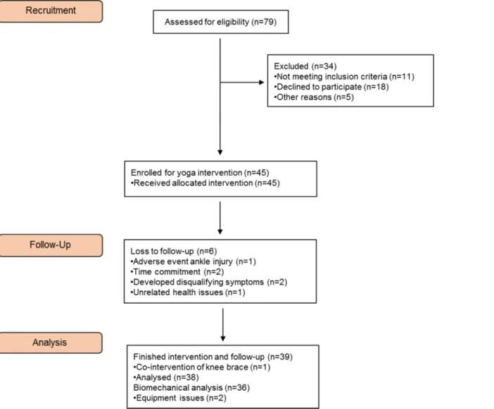
Participants were recruited through rheumatology, orthopaedic, and physical therapy clinics, as well as by word-of-mouth in the Hamilton, Ontario, Canada community. Community dwelling older women meeting the American College of Rheumatology (ACR) criteria for symptomatic knee OA participated. Screening was conducted by one rheumatologist and/or one trained research assistant. These clinical ACR criteria include being 50 years of age or older and answering“yes”on three of the following six criteria: having knee pain on most days of the week, crepitus, bony tenderness or enlargement, no warmth to the touch, or knee stiff-ness lasting longer than 30 minutes [50]. Exclusion criteria included alternate forms of arthritis (e.g., rheumatoid arthritis) or non-arthritic joint disease in the knee, previous knee surgeries, use an assistive walking aid on a regular basis, lower limb trauma within the past 3 months, ipsilateral ankle, knee or hip condition including pain under the knee cap, pregnancy, or any conditions that may have been exacerbated by the protocol (e.g., unstable angina). The partici-pant flowchart is displayed inFig 1.
Ethics Statement
This study was approved by the Hamilton Integrated Research Ethics Board and all partici-pants provided written, informed consent (REB#13–510).
Study Setting
The study was conducted at the MacMobilize Laboratory at McMaster University, Hamilton, Ontario, Canada. The intervention was implemented at a private yoga studio in downtown Hamilton, Ontario, Canada.
Intervention: Yoga for Lower Limb Muscle Strengthening
focus on quadriceps strengthening, postures including supine bridges and heel raises were incorporated to target the hamstrings and ankle plantarflexors respectively. Throughout the program, participants were educated and encouraged to position the knee over the 2ndtoe when weight-bearing in knee flexion. Exercise progressions are included inTable 1. Partici-pants were encouraged to use any modification that allowed them to exert at a 7 on the Borg Perceived Exertion Scale (0–10 scale with 0 = no exertion and 10 = maximal exertion) [51]. If knee pain increased by 2/10 points from pre-exercise pain on a Visual Analog Scale (VAS) dur-ing any exercise, participants were instructed to ask for a modification to alleviate discomfort. Exercise modifications, which entailed alterations in knee range of motion and balance/weight-bearing support, were available for every posture. During the cool-down, participants com-pleted stretches focusing on hip, knee and ankle musculature in supine.
Participants were asked to attend three of four available one-hour classes per week for twelve weeks. Incentives for retention included a physical activity log, a yoga mat, light refresh-ments after each class, rewards for best attendance at the six week point (e.g., movie pass), a $100 stipend at follow-up, and a graduation certificate. The researchers monitored compliance
Fig 1. CONSORT diagram of participant flow through the recruitment, intervention allocation, and analysis of this study.
by taking attendance at each class (ECB, ABK, EGW) and adherence was monitored and facili-tated by the yoga class instructor.
Primary Outcomes: Self-Report and Knee Strength
All primary outcome measures reflected the most symptomatic knee. Self-reported symptoms and physical function were evaluated using the Knee injury and Osteoarthritis Outcome Score (KOOS). The KOOS consists of 42 questions on a five-point Likert scale in five subscales: pain, other symptoms, activities of daily living, function in sport and recreation, and knee-related quality of life. Subscale scores were normalized out of 100, with 0 = extreme symptoms and 100 = no symptoms [52]. The KOOS produces highly reliable [53] and valid [54] data in knee OA.
Knee extensor and flexor strength were represented by the peak torques obtained during maximum voluntary isometric contractions (MVIC) of knee extension and flexion, expressed relative to body mass (Nm/kg). Participants were positioned on a dynamometer (Biodex Medi-cal Systems, Inc., Shirley, NY, USA) such that the knee was in 65° of knee flexion and the knee joint centre was in line with the dynamometer axis. The ankle cuff (point of force application) was placed immediately proximal to the medial and lateral malleoli (average distance to
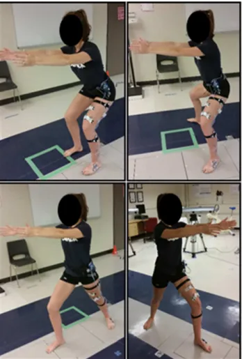
Fig 2. Yoga postures used in this lower limb strengthening program for knee osteoarthritis.The four postures investigated included the legs-together squat (top left), wide-legged squat (top right), and the lunge (bottom; the involved and uninvolved limb were analysed separately).
dynamometer axis = 29.3±2.5 cm). Additional straps across the chest and pelvis minimized extraneous motions. After a submaximal isotonic warm-up and practice, participants com-pleted five knee extensor MVICs and five knee flexor MVICs. Verbal encouragement was used to maximize effort during these contractions. The peak value from each of the five MVICs for extension and flexion efforts was extracted and normalized to body mass to represent strength.
Secondary Outcomes: Mobility, Fitness, and Peak KAM
Mobility was captured using a Six-Minute Walk Test (6MWT), a 30-second chair stand test (30-s CST), and a stair-climbing protocol. The 6MWT measures the furthest distance a partici-pant can walk in six minutes in an indoor, well-lit, rectangular hallway. The 6MWT was com-pleted as per the published protocol and produces reliable data in knee OA [55]. The 30-s CST measured the number of times the participant could rise and sit back down from a standard chair in 30 seconds. The total number of completed stand-and-sit attempts before the trial ended was the score. This test produces reliable and valid data in people with knee OA [56]. Finally, the stair-climbing protocol assessed the time to ascend and descend (separately) a 9-step staircase as quickly and safely as possible, without running or skipping stairs. Recording time started when the participant’s lead foot left the ground and recording time stopped when both feet were planted firmly on the final step. A handrail was available. For each of stair ascending and descending, the mean of two attempts was recorded as a score [55]. In 30 healthy adults, these stair ascent and descent tasks produced reliable data (ICC 0.72–0.88, SEM <0.4s) in our laboratory.
Participants were asked to complete a cycle ergometer submaximal test to yield an estimated maximal oxygen consumption score (mL/kg/min). The YMCA cycle ergometer submaximal test estimates maximal oxygen consumption from submaximal data points [57]. This test is ideal for assessing the cardiovascular fitness of individuals not accustomed to maximal inten-sity exercise. The protocol included three or more consecutive 3-minute stages at a given work-load. The target was to raise the participant’s heart rate (measured with a heart rate monitor) to a value between 110 beats per minute and approximately 85% of the age predicted maximum
Table 1. Levels 1 through 4 of the progressions in the yoga strengthening program.Please refer toS1 Appendixfor further information on the exercises.
Yoga Posture Level 1 Level 2 Level 3 Level 4
Squat (legs together)
Hands on hips, bend knees to 30°flexion
Hands on hips, bend knees to 60°flexion
Shouldersflexed to 90° with elbows straight, bend knees to 60°flexion
Arms over head, bend knees to 80°flexion, look up to ceiling for added challenge
Squat (wide-legged)
Hands on hips, bend knees to 30°flexion
Hands on hips, bend knees to 60°flexion
Shouldersflexed to 90° with elbows straight, bend knees to 60°flexion
Arms over head, bend knees to 80°flexion, look up to ceiling for added challenge
Supported lunge
Hands on hips, the hip of the trail leg is in neutral (noflexion, no extension)
Hands on hips, the hip of the trail leg is in extension
Shouldersflexed to 90° with elbows straight, the hip of the trail leg is in extension
Arms over head, the hip of the trail leg is in extension
Lunge (lateral trunk)
Elbow on knee of the lead leg, place other hand on the hip
Elbow on knee of the leading leg,flex other shoulder to 180°
Hand beside the foot instep of the lead leg,flex other shoulder to 180°
Hand beside lateral side of foot of the lead leg,flex other shoulder to 180°
Lunge (upright trunk 1)
Hands on hips Shouldersflexed to 90° with elbows straight
Arms over head Arms over head, look up to the ceiling for added challenge Lunge (upright
trunk 2)
Hands on hips Arms abducted to 90° with elbows straight
Arms abducted to 90° with elbows straight
Arms abducted to 90° with elbows straight
Bridge Arms by side Clasp hands together, increase height of bridge
Clasp hands together, increase height of bridge
Clasp hands together, increase height of bridge
heart rate for two consecutive stages. The initial stage consisted of a 25 Watt workload at a ped-aling rate of 50 revolutions per minute. The workload of subsequent stages was determined by heart rate during the final minute of the first stage. When the target heart rate was achieved for two consecutive stages, the test was terminated. Oxygen cost was estimated for the final two stages using a formula encompassing workload, body mass, and derived constants (S2 Appen-dix). The predicted values from submaximal tests have been cross-validated to the traditional gold standard methods of achieving a true maximal oxygen consumption (via maximal tread-mill running) [57].
The KAM was calculated from motion and force data and expressed relative to body mass (Nm/kg) from the most symptomatic knee. The peak KAM during gait was captured. Each participant was instrumented with four clusters of infrared emitting diode (IRED) markers. These clusters were placed on the lateral foot, shank, and thigh segments of the involved limb and the sacrum. Three-dimensional motion of these clusters during gait and the yoga exercises was collected at 100 Hz with a 9-camera (3 banks) motion capture system (Optotrak Certus, Northern Digital Inc., Waterloo, ON). Synchronized with this kinematic collection, kinetic data were recorded using four floor-embedded force plates (OR6-7, Advanced Mechanical Technologies Inc, Watertown, MA) at 1000 Hz. For the gait analysis, each participant was instructed to walk down a 10-metre walkway barefoot at a self-selected pace. Trials were considered acceptable when the participant completed a full heel-strike and toe-off on a force plate with the involved limb. Five complete trials were collected for each participant.
Tertiary Outcomes: Biomechanical Measures
All tertiary outcomes were captured from the limb of the most symptomatic knee. Kinematic and kinetic data were collected as described above. Analyses of four yoga postures (squat and lunge exercises) were completed barefoot. We selected four postures to ensure neuromuscular fatigue did not confound the measures. We selected these four postures because these formed the basic position of multiple postures used in the intervention that required variations on arm placement. The squats were“legs-together”(feet hip width apart) and“wide-legged”(feet externally rotated and shoulder-width apart). Both squats involved as much knee and hip flex-ion as possible while maintaining balance and comfort. Alignment instructflex-ions included the following: with the involved limb on one force plate, align knees over the 2ndtoes, maintain a neutral lumbar lordosis, and flex shoulders to 90° (elbows extended). The lunges involved a staggered stance. The aim was to flex the forward or“leading”knee to 90°. In the“trailing”leg, the knee was extended and the foot was externally rotated to 45° (as able) to maintain heel-floor contact. Alignment instructions were as follows: ensure heels align in the sagittal plane, position the leading knee over the 2ndtoe, maintain pelvic alignment in the frontal plane, and flex shoulders to 90° (elbows extended). Participants were asked to flex the leading knee as much as possible while maintaining balance and comfort. Lunges were repeated such that the involved limb was in each the leading and trailing position, with the involved limb placed on the force plate during both postures. For all squats and lunges, each position was held for ten seconds and repeated three times.
value was chosen for analysis. Mean KAM during this window was calculated and an average of two completed trials were computed (Mathworks Inc., Natick, MA).
Normalized mean muscle amplitude was captured using electromyography (EMG) during the yoga postures and presented as a percentage of MVIC (%MVIC). After preparing skin by shaving and wiping with isopropyl alcohol, silver-silver chloride dual electrodes (Natus Neu-rology Inc., Middleton, WI) were affixed to the skin over the biceps femoris, rectus femoris, semitendinosus, vastus lateralis, and vastus medialis muscles along the orientation of the mus-cle fibers (www.seniam.org, Enschede, Netherlands). Time-synchronized with force and motion data, raw EMG signals were bandpass filtered at 10–500 Hz and sampled at 1500 Hz (Desktop Telemyo DTS, Noraxon, Scottsdale, AZ). Each muscle was collected with a wire-min-imal EMG sensor equipped with a reference electrode (dual differential amplifier,
CMRR>100 dB, input impedance>100 MO).
For each trial, the mean amplitude of EMG signal during each exercise was calculated from the same analysis window selected for the KAM (Mathworks Inc., Natick, MA). All EMG data had bias removed, were full-wave rectified, and linear-enveloped using a 2ndorder low-pass zero lag Butterworth filter with a cut-off of 6 Hz (Mathworks, Inc., Natick, MA). Peak activa-tions from MVICs were used to normalize EMG amplitude. EMG data represented the average of two completed trials for each static exercise.
Sample Size Estimation
Sample size estimation for a t-test between two time points in one group was determineda pri-orifor the primary outcomes of self-reported symptoms and knee strength. Systematic reviews of the effectiveness of resistance training for improving self-reported symptoms including pain and isometric knee extensor and flexor strength in knee OA had average effect sizes of -2.11 [9] and 0.41 [60] respectively. Given the lower effect size (0.41) and a Type I error = 0.05 on a 1-tailed test (based on strong evidence of improvement in symptoms [61] and strength [48]), a sample of 39 participants was chosen to yield 80% power to detect a significant effect. We aimed to recruit 45 participants to account for drop-outs and losses to follow-up.
Statistical Analyses
Descriptive statistics were calculated. Normality of the outcomes was assessed using the Sha-piro-Wilk test. Missing data were treated using case-wise deletion. Statistical analyses were completed in SPSS (IBM, Versions 21 and 22).
The primary analyses evaluated whether the yoga strengthening program improved self-reported outcomes (KOOS subscales) and knee strength (extensor, flexor torques) from base-line to follow-up using 1-tailed paired t-tests for normally distributed data. The secondary analyses evaluated whether the yoga strengthening program improved mobility performance (6MWT, 30-s CST, stair-climbing) and fitness, and decreased peak KAM during gait from baseline to follow-up using 2-tailed paired t-tests for normally distributed data. A 2-tailed paired t-test was used for a more conservative statistical approach as mobility performance, fit-ness, and peak KAM are less frequently reported in the literature. If data were not normally dis-tributed, related-samples Wilcoxon signed rank test was used. Multiple comparisons were adjusted using a Bonferroni correction.
Results
Participants
Recruitment took place in May-June 2014 and follow-up data collection was completed in August 2014. A total of 79 people were assessed for eligibility. People were excluded for the fol-lowing reasons: did not meet the inclusion criteria (n = 11), declined to participate (n = 18), and other (n = 5). Forty-five participants enrolled, 39 completed follow-up, and data from 38 participants were analyzed (Fig 1). Of the six participants that did not complete follow-up, one was an adverse event (undiagnosed ankle injury) unrelated to the intervention. Two partici-pants developed disqualifying knee symptoms (patellofemoral pain), which may be a potential harm of the intervention. Two participants dropped out due to time commitment, and one par-ticipant dropped out due to a chronic respiratory illness. One parpar-ticipant completed the study but commenced wearing a knee brace during the intervention. Data from this participant were excluded.Table 2outlines participant baseline characteristics for the 38 participants whose data were included in the analyses. The average number of classes attended per week was 2.6 (0.4). The average absolute number of classes attended was 31.3 (8.7), or an average adherence rate of 87.1%.
No statistical differences were observed for any demographic variables between those that did and did not complete follow-up (p>0.05). The drop-out group had a significantly lower KOOS Quality of Life score than the group that completed follow-up suggesting the drop-out group had a decreased knee-related quality of life at baseline. No other primary or secondary outcome measures were statistically different between the two groups. No tertiary outcome measures were statistically different between those that completed and those that did not com-plete follow-up (p>0.05).
All participants completed the fitness test; however we were unable to calculate a score for five participants at baseline and six participants at follow-up. Two factors precluded calcula-tion. First, some participants were unable to maintain the required cadence. Second, based on heart rate responses, the test was terminated before two stages of the test were completed. Two stages were necessary to calculate a score. Finally, KAM and raw EMG data were analysed for 36 participants due to equipment issues with two participants (kinematic calibration issue).
Primary Outcomes: Self-Report and Knee Strength
The Shapiro-Wilk test showed that data for all self-report outcome measures were normally distributed.Table 3summarizes the outcome measures at baseline and follow-up for self-reported symptoms and physical function. Improvements were noted in all subscales of the KOOS (p<0.001). The pain, symptoms, activities of daily living, and quality of life subscales improved 16.3–20.3%. The greatest improvement occurred in the sports and recreation sub-scale, with a 42.5% improvement at follow-up compared to baseline.
Table 2. Baseline characteristics of 38 women with symptomatic knee OA who completed the yoga intervention.
Variable Mean (Standard Deviation) [Minimum, Maximum]
Age (years) 60.3 (6.5) [50, 77]
Body Mass Index (kg/m2) 29.5 (5.3) [21.1, 43.9]
Height (m) 1.63 (0.06) [1.50, 1.72]
Body mass (kg) 78.1 (14.8) [51.2, 115.4]
Waist Circumference (cm) 93.9 (11.5) [70, 123]
Classes Attended (/week) 2.6 (0.7) [0.8, 3.6]
Table 4summarizes the baseline and follow-up results for isometric strength. Knee extensor strength data were normally distributed. Knee extensor strength increased 5.6% from baseline to follow-up (1.8±0.6 to 1.9±0.6 Nm/kg; p = 0.004). A related-samples Wilcoxon signed rank test was used to examine change in knee flexor strength. Knee flexor strength increased from 0.7±0.3 Nm/kg to 0.8±0.3 Nm/kg from baseline to follow-up (14.3% increase; p = 0.001).
Secondary Outcomes: Mobility, Fitness, and Peak KAM
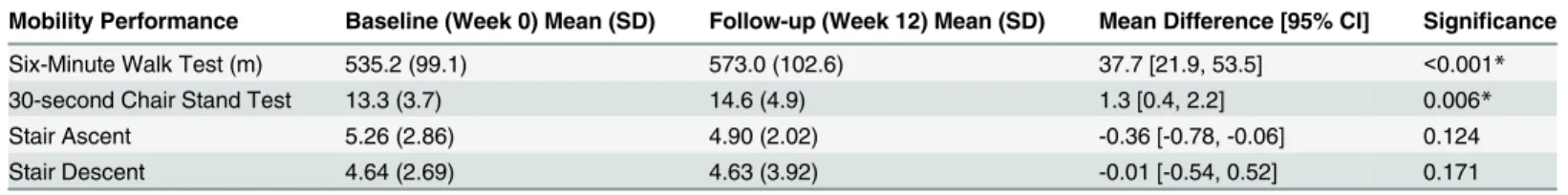
The Shapiro-Wilk test demonstrated that of the mobility performance and fitness measures, only the 6MWT data were normally distributed.Table 5summarizes the outcome measures at baseline and follow-up for mobility and fitness. Participants demonstrated a 7.1% improve-ment in 6MWT scores (535.2 to 573.0 m, p<0.001) and a 9.8% improvement in the 30-s CST (13.3 to 14.6 repetitions, p = 0.006). Stair ascent and descent time did not change between time points.
Table 6shows results of fitness and peak KAM. The estimated maximal oxygen consump-tion was the same at baseline and follow-up (p = 0.372) for the subsample (n = 32) for which estimated values were calculated. The peak KAM during gait was unchanged at follow-up (p = 0.524).
Tertiary Outcomes: Biomechanical Measures
Baseline KAM data were not normally distributed. Average KAM during the four yoga pos-tures are presented inTable 7and were nominal at both baseline and follow-up. Results from the Kruskal-Wallis one-way ANOVA revealed that average KAM values during all four exer-cises were lower than that of peak KAM during gait at baseline (p<0.001).
Data were not normally distributed for the hamstrings normalized mean EMG amplitudes across yoga postures. Vastus medialis normalized mean EMG amplitude was not normally
Table 4. Mean (Standard Deviation) at baseline and follow-up for strength.Mean difference scores (follow-up–baseline) with 95% confidence intervals (CI) are also given, with positive change indicating improvement in self-reported outcomes and strength. Multiple comparisons for all outcome measures were corrected using a Bonferroni correction (new alpha level was set to p = 0.007). Significant comparisons are denoted with an asterisk (*).
Strength (Nm/kg) Baseline (Week 0) Mean (SD) Follow-up (Week 12) Mean (SD) Mean Difference [95% CI] Significance
Knee Extensor Torque 1.8 (0.6) 1.9 (0.6) 0.1 [0.0, 0.2] 0.004*
Knee Flexor Torque 0.7 (0.3) 0.8 (0.3) 0.1 [0.0, 0.1] 0.001*
doi:10.1371/journal.pone.0136854.t004
Table 3. Mean (Standard Deviation) at baseline and follow-up for self-reported physical function (Knee injury and Osteoarthritis Outcome Score). Mean difference scores (follow-up–baseline) with 95% confidence intervals (CI) are also given, with positive change indicating improvement in self-reported outcomes and strength. Multiple comparisons for all outcome measures were corrected using a Bonferroni correction (new alpha level was set to p = 0.007). Significant comparisons are denoted with an asterisk (*). ADL–Activities of Daily Living
Self-Reported Outcomes [KOOS (/100)]
Baseline (Week 0) Mean (SD)
Follow-up (Week 12) Mean (SD)
Mean Difference [95% CI]
Significance
Pain 67.7 (15.4) 79.4 (12.7) 12.0 [7.9, 16.1] <0.001*
ADL 74.9 (15.8) 87.1 (11.1) 11.3 [6.6, 16.1] <0.001*
Symptom 63.5 (17.5) 74.6 (16.6) 12.5 [8.6, 16.4] <0.001*
Sports and Recreationɸ
48.9 (25.0) 69.6 (24.0) 20.1 [13.2, 27.0] <0.001*
Quality of Lifeǂ 49.0 (19.8) 59.0 (20.0) 101. [5.8, 14.3] <0.001*
ǂMissing due to incomplete questionnaire (n = 1) ɸMissing due to incomplete questionnaire (n = 3)
distributed for the lunge postures. Rectus femoris normalized mean EMG amplitude was not normally distributed for the leading leg of a lunge only. Normalized mean EMG amplitudes ranged from 7.3 to 31.0% at both baseline and follow up. Following an adjustment for multiple comparisons, normalized mean EMG amplitude in the five lower limb muscles did not change from baseline to follow-up for all four postures.
Discussion
We explored whether a novel strengthening exercise program for knee OA, designed specifi-cally to minimize the KAM, would be useful to pursue. Exposure to large KAM has been impli-cated in the structural progression of this disease [26–28,62]. It is important to ensure that treatment options for knee OA promote physical activity but do not incorporate exposures to this risk factor for worsening the disease. The proposed yoga-based strengthening program aims to enable people with knee OA to exercise without contributing a cumulative exposure to large KAM. After completing this yoga strengthening program in this sample of women with knee OA, we observed improvements in all subscales of the KOOS (pain, symptoms, activities of daily living, sports and recreation, and quality of live) and knee extensor and flexor strength compared to baseline. Further, two of three mobility performance measures improved at fol-low-up compared to baseline; however fitness and peak KAM during gait were unchanged. Consistent with our tertiary hypothesis, the average KAM during the yoga postures was lower than the peak KAM experienced during gait. However, inconsistent with our initial hypothesis, the mean normalized EMG amplitudes during yoga postures were also unchanged between baseline and follow-up. Normalized mean EMG amplitudes during the strengthening exercises ranged between 7.3 and 31.0% of maximum effort contractions. These data suggest this magni-tude of stimulus related to improved strength. These data show promise that an innovative
“low KAM”yoga strengthening program could improve self-reported outcomes, knee strength, and mobility performance in women with knee OA.
Table 5. Mean (Standard Deviation) at baseline and follow-up for mobility performance.Mean difference scores (follow-up—baseline) with 95% confi-dence intervals (CI) are also given, with positive change indicating improvement in mobility performance and fitness. Multiple comparisons for all outcome measures were corrected using a Bonferroni correction (new alpha level was set to p = 0.008). Significant comparisons are denoted with an asterisk (*).
Mobility Performance Baseline (Week 0) Mean (SD) Follow-up (Week 12) Mean (SD) Mean Difference [95% CI] Significance
Six-Minute Walk Test (m) 535.2 (99.1) 573.0 (102.6) 37.7 [21.9, 53.5] <0.001*
30-second Chair Stand Test 13.3 (3.7) 14.6 (4.9) 1.3 [0.4, 2.2] 0.006*
Stair Ascent 5.26 (2.86) 4.90 (2.02) -0.36 [-0.78, -0.06] 0.124
Stair Descent 4.64 (2.69) 4.63 (3.92) -0.01 [-0.54, 0.52] 0.171
doi:10.1371/journal.pone.0136854.t005
Table 6. Mean (Standard Deviation) at baseline and follow-up for fitness and peak KAM during gait.Mean difference scores (follow-up—baseline) with 95% confidence intervals (CI) are also given, with positive change indicating improvement in mobility performance and fitness. Multiple comparisons for all outcome measures were corrected using a Bonferroni correction (new alpha level was set to p = 0.008). Significant comparisons are denoted with an aster-isk (*).
Fitness and Peak KAM Baseline (Week 0) Mean (SD)
Follow-up (Week 12) Mean (SD)
Mean Difference [95% CI]
Significance
Estimated Maximal Oxygen Consumption (mL/ kg/min)˩
26.3 (5.4) 26.8 (5.1) 0.5 [-0.6, 1.7] 0.372
Peak KAM 0.42 (0.16) 0.43 (0.15) 0.01 [-0.02, 0.04] 0.524
˩
Participants completed the protocol, but, in some cases, their cadence or heart rate responses precluded calculation of estimated values at baseline (n = 5) and follow-up (n = 6). Nofitness data from these participants are included in the table.
The ideal type of strengthening for knee OA is unknown [17]. Strengthening intervention studies have implemented a variety of exercises. Examples include simple quadriceps exercises (e.g., straight leg raises); non-weight bearing exercises using cuff-weights, Therabands, and dynamometers; weight-bearing exercises; and neuromuscular programs to enhance sensorimo-tor function [6,14–16]. Many chose non-weight-bearing exercises to minimize the knee com-pressive load, though these exercises have little functional relevance [17]. The exercise program proposed in the current study marks the first attempt that we are aware of to design a strength-ening program with functional relevance based on the biomechanical loads.
While we cannot comment on the efficacy of the program without a RCT design, it is unlikely that improvements across several domains, including self-reported symptoms and physical function, strength, and mobility were spontaneous. Clinical improvements observed in the current sample are equivalent to improvements reported after traditional exercise pro-grams. The magnitude of improvement in KOOS, strength, and mobility were similar to that found in other studies [14,15,38,63]. For example, an 18-month RCT of 454 participants with knee OA compared three groups receiving diet, exercise, or both. The exercise-only group demonstrated 28% and 24% improvements on pain and function subscales of the Western Ontario and McMaster Universities Osteoarthritis Index respectfully [63]. Despite our smaller sample size and shorter intervention, findings from the current proof-of-principle study dem-onstrated similar improvements. Further, we observed improvements greater than minimal detectable change (MDC) data for the KOOS symptoms and sports and recreation subscales [64]. We were unable to identify MDC or minimal clinically important difference (MCID) val-ues for mobility performance or strength outcomes based on exercise interventions in knee OA. Improvements observed for the 6MWT [55] and strength [65] were approximately half of the estimated MDC determined from data before and after arthroplasty. While these improve-ments in strength appear relatively small and the clinical relevance of these strength increases are unclear, the 5.6–14.3% increase in strength values found in the current study are within the range expected in strengthening programs in knee OA [46]. A need exists for MDC and MCID data relevant to exercise. Nonetheless, clinical improvements in symptoms, strength and mobility performance following this novel program for knee OA must be compared to change observed in a control group, using a RCT design, to establish efficacy.
While mobility performance was improved during the walking and sit-to-stand tasks, no improvement in stair-climbing was noted after participants completed this intervention. It is pos-sible that this negative finding reflects training specificity. While participants completed yoga postures such as squats that likely translate into improvements in sit-to-stand activities, the inter-vention did not incorporate exercises that reflected stair-climbing. It is important to note, at base-line, that the participants ascended and descended nine stairs, on average, between 4s and 5s. These scores leave little room for improvement. A ceiling effect is likely responsible for the lack
Table 7. Knee adduction moment (KAM) normalized to body mass during four yoga postures to strengthen knee muscles (Nm/kg).Biomechanical data are presented on n = 36 participants.
Yoga Posture Baseline (Week 0) Mean (SD) [Min, Max]
Follow-up (Week 12) Mean (SD) [Min, Max]
Legs-together Squat
-0.04 (0.14) [-0.48, 0.34] -0.07 (0.12) [-0.31, 0.20]
Wide-legged Squat -0.02 (0.14) [-0.36, 0.28] -0.03 (0.11) [-0.28, 0.25] Lunge (Leading
Leg)
0.08 (0.13) [-0.22, 0.38] 0.05 (0.10) [-0.25, 0.25]
Lunge (Trailing Leg)
-0.07 (0.14) [-0.29, 0.33] -0.12 (0.12) [-0.53, 0.23]
of change noted before and after the intervention [56]. The stair-climbing task may produce greater variability in a sample with greater mobility limitations, such as pre-surgical candidates.
Inconsistent with our hypothesis, the fitness test selected for this study demonstrated no dif-ferences between baseline and follow-up. It is not surprising that an exercise program tailored primarily to strengthen the quadriceps muscles was not a stimulus for improvement in fitness; though previous work has documented strength training can improve fitness in older adults [12]. It is important to note that the submaximal cycle ergometer test is likely not appropriate for people with knee OA. We initially chose this protocol to eliminate the influence of pain during weight-bearing; for example on a treadmill test. However, the cycle ergometry test used here required a pedaling cadence some participants could not complete. Also, the criteria for heart rate to terminate the test resulted in the inability to estimate a maximal oxygen consump-tion value for several participants. Thus, it is difficult to form conclusions about fitness changes between baseline and follow-up in this study.
We propose here that the KAM may be useful in setting guidelines for exercises for knee OA. While we initially hypothesized that the strengthening intervention would decrease peak KAM during gait, several studies show that exercise has no impact on the KAM during gait [37,38,66–69]. For example, a RCT of lower limb strengthening in 89 participants with knee OA compared the peak KAM during gait, self-reported pain and function, stair-climbing per-formance, and maximum isometric strength before and after 12 weeks of either a strengthening program or no intervention [37]. While these previous findings might initially appear discour-aging, it is possible that, rather than attempt to change the KAM, we could use the KAM to tai-lor exercise. Due to high KAM values identified in previous work [44], the strengthening program used in the current study did not include single leg stance exercises shown to improve hip strength [14,67] and balance [70]. Indeed, in the current sample of women with symptom-atic knee OA, analyses of the basic yoga postures (specifically squats and lunges, upon which variations of arm positions were used) revealed these exercises produced nominal mean KAMs. Thus, utilizing these exercises can promote physical activity among women with symp-tomatic knee OA without concerns of increasing cumulative exposures to large KAMs, which has been implicated in worsening structural knee OA.
Inconsistent with our initial hypothesis, we did not observe a reduction in normalized mean EMG amplitudes during the yoga postures between baseline and follow-up. We anticipated that increased torque output from knee extensors and flexors as a result of the strengthening program would result in concomitant decreases in normalized mean EMG amplitudes to com-plete the same exercise task. During the strengthening exercises, lower limb normalized mean EMG amplitudes ranged between 10.1–28.4% MVIC at baseline and 7.3–31.0% MVIC at fol-low-up. We observed relatively shallow knee flexion during the squats and lunges in this sam-ple (e.g., 9.7–46.6 degrees at baseline and 7.1–50.8 degrees at follow-up) which may account for the low muscle amplitudes. Future work should explore whether increases in peak torque were due to neural changes versus hypertrophy of muscle fibre tissue.
The intervention appeared to be well-accepted by the study participants. Mean weekly attendance was 2.6±0.7 classes and adherence was 87.1% across the 12-week intervention period. All but one participant attended at least one class per week. Our target was 2.5 classes per week because clinical outcomes in knee OA improve after one supervised class per week; however the effect size increases with greater number of contacts [6].
Second, the KAM represents the net reaction moment that does not account for muscle contri-butions and thus may underestimate internal joint loads. Therefore, while the KAM may be low during the yoga exercises presented here, other contributors to joint load such as agonist-antagonist muscle co-activity may contribute to substantial load on the medial knee compart-ment. Nonetheless, KAM was related to joint contact forces directly measured in the knee (R2 = 0.77, p<0.001) [71]. Since an increase in KAM has been related to disease progression [26], designing an exercise program that incorporates low KAM exercises may aid in disease man-agement while improving self-reported outcomes, strength, and mobility. Third, the program is likely inappropriate for individuals who experience patellofemoral pain symptoms. While the proposed strengthening program minimizes KAM, it demands large patellofemoral loads. Fourth, the YMCA submaximal cycle ergometer test was not appropriate for this sample. Fifth, the findings may not be generalizable to men with knee OA. Finally, participants in this study may not have radiographic disease, or disease confined to the medial compartment. Ultimately the goal of the OA-specific strengthening program is to improve clinical outcomes, suggesting an emphasis on symptomatic disease is required.
This study showed that a strengthening program designed to minimize exposure to large magnitude KAMs had promise in improving self-reported symptoms and physical function, and strength among women with clinical knee OA. Two of the three mobility performance tests (walking and sit-to-stand) improved at follow-up compared to baseline, however fitness and peak KAM during gait did not change following the intervention. Unfortunately, the assessment of fitness using the YMCA submaximal cycle ergometry protocol resulted in miss-ing data due to incomplete tests. Also, the stair-climbmiss-ing measurement used likely yielded a ceiling effect in this clinical sample. Finally, it is important to note that the average KAMs dur-ing the yoga postures were lower than that durdur-ing normal gait, providdur-ing confidence that these yoga postures may be useful as a treatment strategy for knee OA. However, strengthening the knee extensors and flexors did not lower muscle activation amplitudes as a result of this strengthening program. It seems reasonable that an exercise program designed with low KAM exercises may aid in disease management while improving self-reported outcomes, strength, and mobility. Further investigation using an RCT design is needed to make conclusions regard-ing the efficacy of this OA-specific strengthenregard-ing program in people with knee OA.
Supporting Information
S1 Checklist. The TIDieR (Template for Intervention Description and Replication) Check-list.
(PDF)
S1 Protocol. Submitted ethics protocol. (DOCX)
S1 Appendix. Biomechanics exercise program with instructions and progressions. (DOCX)
S2 Appendix. YMCA submaximal VO2 cycle ergometry test protocol and data collection package.
(DOCX)
Author Contributions
References
1. Bombardier C, Hawker G, Mosher D. The impact of arthritis in Canada: Today and over the next 30 years. Arthritis Alliance Can. 2011; 1–52.
2. Wait times for priority procedures in Canada. Canadian Institute for Health Information; 2014 pp. 1–10. 3. The NJR Editorial Board. National joint registry for England, Wales and Northern Ireland [Internet].
2013 pp. 1–240. Report No.: 10th Annual Report. Available:www.njrcentre.org.uk
4. Australian Orthopaedic Association National Joint Replacement Registry. Hip and knee arthroplasty. [Internet]. 2014 pp. 1–235. Available:www.aoa.org.au
5. Kurtz S. Projections of primary and revision hip and knee arthroplasty in the united states from 2005 to 2030. J Bone Jt Surg Am. 2007; 89: 780.
6. Fransen M, McConnell S. Exercise for osteoarthritis of the knee. In: The Cochrane Collaboration, editor. Cochrane Database of Systematic Reviews. Chichester, UK: John Wiley & Sons, Ltd; 2008. Available:
http://doi.wiley.com/10.1002/14651858.CD004376.pub2
7. Van Baar ME, Dekker J, Oostendorp RAB, Bijl D, Voorn TB, Bijlsma JWJ. Effectiveness of exercise in patients with osteoarthritis of hip or knee: nine months’follow up. Ann Rheum Dis. 2001; 60: 1123–
1130. PMID:11709454
8. Zhang W, Moskowitz RW, Nuki G, Abramson S, Altman RD, Arden N, et al. OARSI recommendations for the management of hip and knee osteoarthritis, Part I: Critical appraisal of existing treatment guide-lines and systematic review of current research evidence. Osteoarthritis Cartilage. 2007; 15: 981–
1000. PMID:17719803
9. Lange AK, Vanwanseele B, Fiatarone Singh MA. Strength training for treatment of osteoarthritis of the knee: A systematic review. Arthritis Rheum. 2008; 59: 1488–1494. doi:10.1002/art.24118PMID:
18821647
10. Fransen M, McConnell S. Land-based exercise for osteoarthritis of the knee: a metaanalysis of random-ized controlled trials. J Rheumatol. 2009; 36: 1109–1117. doi:10.3899/jrheum.090058PMID:
19447940
11. Kelley GA, Kelley KS, Tran ZV. Resistance training and bone mineral density in women: a meta-analy-sis of controlled trials. Am J Phys Med Rehabil. 2001; 80: 65–77. PMID:11138958
12. Ades PA, Ballor DL, Ashikaga T, Utton JL, Nair KS. Weight training improves walking endurance in healthy elderly persons. Ann Intern Med. 1996; 124: 568–572. PMID:8597320
13. Ruuskanen JM, Ruoppila I. Physical activity and psychological well-being among people aged 65 to 84 years. Age Ageing. 1995; 24: 292–296. PMID:7484485
14. Ageberg E, Nilsdotter A, Kosek E, Roos EM. Effects of neuromuscular training (NEMEX-TJR) on patient-reported outcomes and physical function in severe primary hip or knee osteoarthritis: a con-trolled before-and-after study. BMC Musculoskelet Disord. 2013; 14: 232. doi: 10.1186/1471-2474-14-232PMID:23924144
15. Bennell KL, Egerton T, Wrigley TV, Hodges PW, Hunt M, Roos EM, et al. Comparison of neuromuscu-lar and quadriceps strengthening exercise in the treatment of varus malaligned knees with medial knee osteoarthritis: a randomised controlled trial protocol. BMC Musculoskelet Disord. 2011; 12: 276. doi:
10.1186/1471-2474-12-276PMID:22141334
16. Roos EM, Dahlberg L. Positive effects of moderate exercise on glycosaminoglycan content in knee car-tilage: A four-month, randomized, controlled trial in patients at risk of osteoarthritis. Arthritis Rheum. 2005; 52: 3507–3514. PMID:16258919
17. Bennell KL, Wrigley TV, Hunt MA, Lim B- W, Hinman RS. Update on the role of muscle in the genesis and management of knee osteoarthritis. Rheum Dis Clin N Am. 2013; 39: 145–176.
18. Bennell KL, Buchbinder R, Hinman RS. Physical therapies in the management of osteoarthritis: current state of the evidence. Curr Opin Rheumatol. 2015; 27: 304–311. doi:10.1097/BOR.
0000000000000160PMID:25775185
19. Andriacchi TP, Mündermann A, Smith RL, Alexander EJ, Dyrby CO, Koo S. A framework for the in vivo pathomechanics of osteoarthritis at the knee. Ann Biomed Eng. 2004; 32: 447–457. PMID:15095819
20. Andriacchi TP, Koo S, Scanlan SF. Gait mechanics influence healthy cartilage morphology and osteo-arthritis of the knee. J Bone Jt Surg Am. 2009; 91: 95.
21. Wluka AE, Forbes A, Wang Y, Hanna F, Jones G, Cicuttini FM. Knee cartilage loss in symptomatic knee osteoarthritis over 4.5 years. Arthritis Res Ther. 2006; 8: R90. PMID:16704746
22. Franciozi CES, Tarini VAF, Reginato RD, Gonçalves PRS, Medeiros VP, Ferretti M, et al. Gradual strenuous running regimen predisposes to osteoarthritis due to cartilage cell death and altered levels of glycosaminoglycans. Osteoarthritis Cartilage. 2013; 21: 965–972. doi:10.1016/j.joca.2013.04.007
23. Horisberger M, Fortuna R, Valderrabano V, Herzog W. Long-term repetitive mechanical loading of the knee joint by in vivo muscle stimulation accelerates cartilage degeneration and increases chondrocyte death in a rabbit model. Clin Biomech. 2013; 28: 536–543.
24. Schipplein OD, Andriacchi TP. Interaction between active and passive knee stabilizers during level walking. J Orthop Res. 1991; 9: 113–119. PMID:1984041
25. Andriacchi TP. Editorial: Valgus alignment and lateral compartment knee osteoarthritis: A biomechani-cal paradox or new insight into knee osteoarthritis? Arthritis Rheum. 2013; 65: 310–313. doi:10.1002/ art.37724PMID:23203607
26. Miyazaki T, Wada M, Kawahara H, Sato M, Baba H, Shimada S. Dynamic load at baseline can predict radiographic disease progression in medial compartment knee osteoarthritis. Ann Rheum Dis. 2002; 61: 617–622. PMID:12079903
27. Bennell KL, Bowles K-A, Wang Y, Cicuttini F, Davies-Tuck M, Hinman RS. Higher dynamic medial knee load predicts greater cartilage loss over 12 months in medial knee osteoarthritis. Ann Rheum Dis. 2011; 70: 1770–1774. doi:10.1136/ard.2010.147082PMID:21742637
28. Chehab EF, Favre J, Erhart-Hledik JC, Andriacchi TP. Baseline knee adduction and flexion moments during walking are both associated with 5 year cartilage changes in patients with medial knee osteoar-thritis. Osteoarthritis Cartilage. 2014; 22: 1833–1839. doi:10.1016/j.joca.2014.08.009PMID:
25211281
29. Mündermann A, Asay JL, Mündermann L, Andriacchi TP. Implications of increased medio-lateral trunk sway for ambulatory mechanics. J Biomech. 2008; 41: 165–170. PMID:17678933
30. Chang A, Hayes K, Dunlop D, Song J, Hurwitz D, Cahue S, et al. Hip abduction moment and protection against medial tibiofemoral osteoarthritis progression. Arthritis Rheum. 2005; 52: 3515–3519. PMID:
16255022
31. Hunt MA, Birmingham TB, Bryant D, Jones I, Giffin JR, Jenkyn TR, et al. Lateral trunk lean explains var-iation in dynamic knee joint load in patients with medial compartment knee osteoarthritis. Osteoarthritis Cartilage. 2008; 16: 591–599. doi:10.1016/j.joca.2007.10.017PMID:18206395
32. Keenan GS, Franz JR, Dicharry J, Croce UD, Kerrigan DC. Lower limb joint kinetics in walking: The role of industry recommended footwear. Gait Posture. 2011; 33: 350–355. doi:10.1016/j.gaitpost.2010.09. 019PMID:21251835
33. Chang A, Hurwitz D, Dunlop D, Song J, Cahue S, Hayes K, et al. The relationship between toe-out angle during gait and progression of medial tibiofemoral osteoarthritis. Ann Rheum Dis. 2007; 66: 1271–1275. PMID:17267516
34. Rutherford DJ, Hubley-Kozey CL, Deluzio KJ, Stanish WD, Dunbar M. Foot progression angle and the knee adduction moment: a cross-sectional investigation in knee osteoarthritis. Osteoarthritis Cartilage. 2008; 16: 883–889. doi:10.1016/j.joca.2007.11.012PMID:18182310
35. Mündermann A, Dyrby CO, Hurwitz DE, Sharma L, Andriacchi TP. Potential strategies to reduce medial compartment loading in patients with knee osteoarthritis of varying severity: Reduced walking speed. Arthritis Rheum. 2004; 50: 1172–1178. PMID:15077299
36. Robbins S, Birmingham TB, Callaghan JP, Jones GR, Chesworth BM, Maly MR. Association of pain with frequency and magnitude of knee loading in knee osteoarthritis. Arthritis Care Res. 2011; 63: 991–
997.
37. Bennell KL, Hunt MA, Wrigley TV, Hunter DJ, McManus FJ, Hodges PW, et al. Hip strengthening reduces symptoms but not knee load in people with medial knee osteoarthritis and varus malalignment: a randomised controlled trial. Osteoarthritis Cartilage. 2010; 18: 621–628. doi:10.1016/j.joca.2010.01. 010PMID:20175973
38. Messier SP, Mihalko SL, Beavers DP, Nicklas BJ, DeVita P, Carr JJ, et al. Strength training for arthritis trial (START): design and rationale. BMC Musculoskelet Disord. 2013; 14: 208. doi: 10.1186/1471-2474-14-208PMID:23855596
39. Bukowski E, Conway A, Glentz L, Kurland K, Galantino M. The Effect of Iyengar Yoga and Strengthen-ing Exercises for People LivStrengthen-ing with Osteoarthritis of the Knee: A Case Series. Int Q Community Health Educ. 2006; 26: 287–305. PMID:17827096
40. Kolasinski SL, Garfinkel M, Tsai AG, Matz W, Dyke AV, Schumacher HR Jr. Iyengar yoga for treating symptoms of osteoarthritis of the knees: a pilot study. J Altern Complement Med. 2005; 11: 689–693. PMID:16131293
42. Wang M- Y, Sean SY, Hashish R, Samarawickrame SD, Kazadi L, Greendale GA, et al. The bio-mechanical demands of standing yoga poses in seniors: The yoga empowers seniors study (YESS). BMC Complement Altern Med. 2013; 13: 8. doi:10.1186/1472-6882-13-8PMID:23302513
43. Longpré HS, Brenneman EC, Johnson ALM, Maly MR. Identifying yoga-based knee strengthening exercises using the knee adduction moment. Clin Biomech.
44. Longpré HS. Muscle contributions to knee loads in healthy young women. PhD Dissertation, McMaster University. 2015.
45. Moisio KC, Sumner DR, Shott S, Hurwitz DE. Normalization of joint moments during gait: a comparison of two techniques. J Biomech. 2003; 36: 599–603. PMID:12600350
46. Bennell KL, Hunt MA, Wrigley TV, Lim B-W, Hinman RS. Role of Muscle in the Genesis and Manage-ment of Knee Osteoarthritis. Rheum Dis Clin N Am. 2008; 34: 731–754.
47. Zhang W, Moskowitz RW, Nuki G, Abramson S, Altman RD, Arden N, et al. OARSI recommendations for the management of hip and knee osteoarthritis, Part II: OARSI evidence-based, expert consensus guidelines. Osteoarthritis Cartilage. 2008; 16: 137–162. doi:10.1016/j.joca.2007.12.013PMID:
18279766
48. Runhaar J, Luijsterburg P, Dekker J, Bierma-Zeinstra S. Identifying potential working mechanisms behind the positive effects of exercise therapy on pain and function in osteoarthritis; a systematic review. Osteoarthritis Cartilage. 2015;
49. Larose J, King J, Brosseau L, Wells GA, Reid R, Maetzel A, et al. The effect of walking on cardiorespira-tory fitness in adults with knee osteoarthritis. Appl Physiol Nutr Metab. 2013; 38: 886–891. doi:10. 1139/apnm-2012-0487PMID:23855277
50. Altman R, Asch E, Bloch D, Bole G, Borenstein D, Brandt K, et al. Development of criteria for the classi-fication and reporting of osteoarthritis: classiclassi-fication of osteoarthritis of the knee. Arthritis Rheum. 1986; 29: 1039–1049. PMID:3741515
51. Borg GA. Psychophysical bases of perceived exertion. Med Sci Sports Exerc. 1982; 14: 377–381. PMID:7154893
52. KOOS. KOOS Scoring 2012 [Internet]. 2012. Available:http://koos.nu/KOOSscoring2012.pdf
53. Alviar M, Olver J, Brand C, Hale T, Khan F. Do patient-reported outcome measures used in assessing outcomes in rehabilitation after hip and knee arthroplasty capture issues relevant to patients? Results of a systematic review and ICF linking process. J Rehabil Med. 2011; 43: 374–381. doi:10.2340/ 16501977-0801PMID:21448553
54. Collins NJ, Misra D, Felson DT, Crossley KM, Roos EM. Measures of knee function: International Knee Documentation Committee (IKDC) Subjective Knee Evaluation Form, Knee Injury and Osteoarthritis Outcome Score (KOOS), Knee Injury and Osteoarthritis Outcome Score Physical Function Short Form (KOOS-PS), Knee Outcome Survey Activities of Daily Living (KOS-ADL), Lysholm Knee Scoring Scale, Oxford Knee Score (OKS), Western Ontario and McMaster Universities Osteoarthritis Index
(WOMAC), Activity Rating Scale (ARS), and Tegner Activity Score (TAS). Arthritis Care Res. 2011; 63: S208–S228.
55. Kennedy DM, Stratford PW, Wessel J, Gollish JD, Penney D. Assessing stability and change of four performance measures: a longitudinal study evaluating outcome following total hip and knee arthro-plasty. BMC Musculoskelet Disord. 2005; 6: 3. PMID:15679884
56. Dobson F, Hinman RS, Hall M, Terwee CB, Roos EM, Bennell KL. Measurement properties of perfor-mance-based measures to assess physical function in hip and knee osteoarthritis: a systematic review. Osteoarthritis Cartilage. 2012; 20: 1548–1562. doi:10.1016/j.joca.2012.08.015PMID:22944525
57. Beekley MD, Brechue WF, Dehoyos DV, Garzarella L, Werber-Zion G, Pollock ML. Cross-validation of the YMCA submaximal cycle ergometer test to predict VO2max. Res Q Exerc Sport. 2004; 75: 337–
342. PMID:15487296
58. Winter DA. Biomechanics and motor control of human movement. John Wiley & Sons; 2009. 59. Cole G, Nigg B, Ronsky J, Yeadon M. Application of the joint coordinate system to three-dimensional
joint attitude and movement representation: a standardization proposal. J Biomech Eng. 1993; 115: 344–349. PMID:8309227
60. Roddy E. Aerobic walking or strengthening exercise for osteoarthritis of the knee? A systematic review. Ann Rheum Dis. 2005; 64: 544–548. PMID:15769914
61. Fransen M, McConnell S, Harmer AR, Van der Esch M, Simic M, Bennell KL. Exercise for osteoarthritis of the knee. Cochrane Libr. 2015;
63. Messier SP, Mihalko SL, Legault C, Miller GD, Nicklas BJ, DeVita P, et al. Effects of intensive diet and exercise on knee joint loads, inflammation, and clinical outcomes among overweight and obese adults with knee osteoarthritis: the IDEA randomized clinical trial. JAMA. 2013; 310: 1263–1273. doi:10.1001/ jama.2013.277669PMID:24065013
64. Monticone M, Ferrante S, Salvaderi S, Motta L, Cerri C. Responsiveness and minimal important changes for the knee injury and osteoarthritis outcome score in subjects undergoing rehabilitation after total knee arthroplasty. Am J Phys Med Rehabil. 2013; 92: 864–870. doi:10.1097/PHM.
0b013e31829f19d8PMID:23900017
65. Kean CO, Birmingham TB, Garland SJ, Bryant DM, Giffin JR. Minimal detectable change in quadriceps strength and voluntary muscle activation in patients with knee osteoarthritis. Arch Phys Med Rehabil. 2010; 91: 1447–1451. doi:10.1016/j.apmr.2010.06.002PMID:20801266
66. Foroughi N, Smith RM, Lange AK, Baker MK, Fiatarone Singh MA, Vanwanseele B. Lower limb muscle strengthening does not change frontal plane moments in women with knee osteoarthritis: A randomized controlled trial. Clin Biomech. 2011; 26: 167–174.
67. Sled EA, Khoja L, Deluzio KJ, Olney SJ, Culham EG. Effect of a home program of hip abductor exer-cises on knee joint loading, strength, function, and pain in people with knee osteoarthritis: A clinical trial. Phys Ther. 2010; 90: 895–904. doi:10.2522/ptj.20090294PMID:20378679
68. Lim B, Hinman RS, Wrigley TV, Sharma L, Bennell KL. Does knee malalignment mediate the effects of quadriceps strengthening on knee adduction moment, pain, and function in medial knee osteoarthritis? A randomized controlled trial. Arthritis Rheum. 2008; 59: 943–951. doi:10.1002/art.23823PMID:
18576289
69. Thorstensson CA, Henriksson M, Porat A von, Sjödahl C, Roos EM. The effect of eight weeks of exer-cise on knee adduction moment in early knee osteoarthritis–a pilot study. Osteoarthritis Cartilage. 2007; 15: 1163–1170. PMID:17466541
70. Messier SP, Royer TD, Craven TE, O’Toole ML, Burns R, Ettinger WH Jr. Long-term exercise and its effect on balance in older, osteoarthritic adults: results from the fitness, arthritis, and seniors trial (FAST). J Am Geriatr Soc. 2000; 48: 131–138. PMID:10682941
71. Zhao D, Banks SA, Mitchell KH, Lima DD D’, Colwell CW, Fregly BJ. Correlation between the knee adduction torque and medial contact force for a variety of gait patterns. J Orthop Res. 2007; 25: 789–