Serum 25-hydroxyvitamin D levels are associated with
functional capacity but not with postural balance in
osteoporotic postmenopausal women
Guilherme Carlos Brech,I,II,*Emmanuel Gomes Ciolac,III Mark D. Peterson,IVJu´lia Maria D’Andre´a GreveV
IHospital das Clı´nicas da Faculdade de Medicina da Universidade de Sa˜o Paulo, Instituto de Ortopedia e Traumatologia, Laborato´rio de Estudos do
Movimento, Sa˜o Paulo/SP, Brazil.IIUniversidade Ibirapuera, Fisioterapia, Sa˜o Paulo/SP, Brazil.IIIUniversidade Estadual Paulista Ju´lio de Mesquita Filho – UNESP, Faculdade de Cieˆncias, Departamento de Educac¸a˜o Fı´sica, Bauru/SP, Brazil. IVUniversity of Michigan-Medicine, Michigan, United States. VFaculdade de Medicina da Universidade de Sa˜o Paulo, Instituto de Ortopedia e Traumatologia, Sa˜o Paulo/SP, Brazil.
OBJECTIVES:In post-menopausal women with osteoporosis, insufficient vitamin D levels decrease calcium fixa-tion in the bones and calcium transport in the sarcoplasmic reticulum, which impairs muscle strength, possibly leading to detrimental consequences for the preservation of functional capacity and postural balance, fall prevention, and fracture risk. The aim of this study was to evaluate the association between vitamin D levels and knee muscle strength, postural balance and functional mobility among postmenopausal women with osteoporosis.
METHODS:This cross-sectional study included 63 osteoporotic older women (aged 60.6±3.1 years). The subjects
completed the Timed Up and Go Test to measure functional mobility, and postural balance was assessed on the AccuSway Plus portable force platform. Maximal strength was tested using an isokinetic dynamometer for knee flexion and extension. The subjects were assessed as a group and were divided into quartiles according to their vitamin D levels. Clinicaltrials.gov: NCT02771834.
RESULTS:Vitamin D status was independently associated with the normalized peak torque of the knee extensors (b=0.59;p=0.04) and Timed Up and Go Test (b=-0.07;po0.001). No between-group differences were observed in
the demographic and clinical variables or postural balance; however, significant differences were observed in the Timed Up and Go Test, and the group with the highest vitamin D levels exhibited better performance than the group with the lowest vitamin D levels (po0.001).
CONCLUSION: The serum vitamin D levels were independently associated with normalized knee extension strength and functional mobility in postmenopausal women with osteoporosis.
KEYWORDS: Elderly; Muscle Strength; Postural Balance; Sarcopenia; Timed Up and Go Test; Vitamin D.
Brech GC, Ciolac EG, Peterson MD, D´ Andrea Greve JM. Serum 25-hydroxyvitamin D levels are associated with functional capacity but not with postural balance in osteoporotic postmenopausal women. Clinics. 2017;72(1):11-16
Received for publication onMay 12, 2016;First review completed onAugust 9, 2016;Accepted for publication onSeptember 30, 2016
*Corresponding author. E-mail: [email protected]
’ INTRODUCTION
Osteoporosis is a disease characterized by reductions in the bone mineral density (BMD) and deterioration of bone tissue, with increased fragility and risk of fractures. It is one of the largest public health problems, mainly due to the increasing age of the population (1). Osteoporosis affects approximately 55% of the population over the age of 50 years in the United States (2). In Brazil, the national health system (Sistema Único de Saúde - SUS) is also increasing its expenditures
on treatments for fractures among older individuals, result-ing in 20,778,000 hospital admissions in 2006 (3). Women with osteoporosis often present a vitamin D deficiency, which further increases the risk of falls (4-6).
Optimal vitamin D levels are necessary for musculoskeletal health and function; however, over a billion people worldwide have a deficiency in their circulating vitamin D levels (5). Vitamin D is associated with muscular health (7) and con-tributes to the maintenance of muscle strength during ageing (5,8). Higher serum vitamin D concentrations have also been associated with greater muscle strength in healthy older individuals (4,5) who have suffered falls (9) and in those who have osteoporosis (6). However, these findings are currently being debated in the literature (10,11).
In postmenopausal women with osteoporosis, insufficient vitamin D levels reduce calcium fixation in the bones and calcium transport in the sarcoplasmic reticulum, which impairs muscle strength, possibly leading to detrimental consequences
DOI:10.6061/clinics/2017(01)03
Copyright&2017CLINICS–This is an Open Access article distributed under the terms of the Creative Commons License (http://creativecommons.org/licenses/by/ 4.0/) which permits unrestricted use, distribution, and reproduction in any medium or format, provided the original work is properly cited.
for the preservation of functional capacity and postural balance (5,6,12), fall prevention, and fracture risk (12). How-ever, there is no consensus regarding the association of the serum vitamin D levels with muscle strength, functional capa-city and postural balance in this specific population. Vitamin D supplementation improves muscle strength and postural balance but may have a limited influence on walking ability in older people without osteoporosis; however, subsequent studies are needed to examine these factors in postmenopausal women with osteoporosis (13).
Therefore, the aim of this study was to assess whether the serum vitamin D levels were associated with knee muscle strength, postural balance and functional mobility in post-menopausal women with osteoporosis.
’ METHODS
Study Location and Ethical Issues
The study was conducted at the Institute of Orthopedics and Traumatology, Hospital das Clínicas, School of Medicine, University of São Paulo, with approval granted by the Ethics Committee of the University of São Paulo (CaPPesq no
320/09).
Study Design
This cross-sectional study included 63 osteoporotic older women aged 55 to 65 years. The patients’ baseline
char-acteristics are provided in Table 1. None of the subjects had pre-existing conditions related to the vestibular system, proprioceptive, auditory or neurological conditions, had not been prescribed any antipsychotic medication, had no restrictions on vigorous physical activity, had not been under-went surgery, and had not presented any injury to the legs in the previous six months. The subjects could have participated in physical exercises at a maximum of twice a week but not in strength exercises to remain eligible to participate in the study. According to the International Physical Activity Questionnaire (IPAQ), these subjects were considered to be irregularly active or active. Potential subjects were excluded from the study if they presented alterations in blood pressure or if they were not able to perform the test. The volunteers were analyzed together, and then divided into four groups, according to their serum vitamin D levels (Table 2). After the subjects had been provided an explanation of the study and signed the consent form, they were assessed in accordance with the evaluation protocol.
Measurements
The following data were collected during the preliminary interviews and examinations: age, body mass, height, body mass index (BMI), ethnicity, race, age at the start of meno-pause, smoking status, and used of any hormonal replacement therapy (HRT) or calcium supplement. Moreover, dual-energy X-ray absorptiometry (DXA) values were recorded during each subject’s clinical examination.
All subjects were evaluated at the Laboratory of Kinesiology in the morning by the same professionals. The subjects were instructed not to perform any physical activity 24 hours prior to the tests, to wear comfortable and lightweight clothing, and to consume a low-calorie meal 2 hours before the visit.
The subjects then underwent the Timed Up and Go Test (TUGT) to measure functional mobility (time in seconds). The postural balance assessment (posturography) was per-formed on a portable force platform (AccuSway Plus, AMTIs,
MA, USA). For data acquisition, the force platform was con-nected to a signal-amplifying interface box (PJB-101) that was linked to a computer using an RS-232 cable. The data were collected and stored using Balance Clinicssoftware; the data
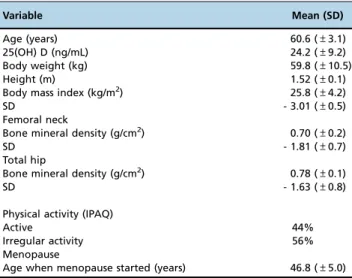
Table 1-Baseline characteristics of the study participants.
Variable Mean (SD)
Age (years) 60.6 (±3.1)
25(OH) D (ng/mL) 24.2 (±9.2)
Body weight (kg) 59.8 (±10.5)
Height (m) 1.52 (±0.1)
Body mass index (kg/m2) 25.8 (±4.2)
SD - 3.01 (±0.5)
Femoral neck
Bone mineral density (g/cm2) 0.70 (±0.2)
SD - 1.81 (±0.7)
Total hip
Bone mineral density (g/cm2) 0.78 (±0.1)
SD - 1.63 (±0.8)
Physical activity (IPAQ)
Active 44%
Irregular activity 56%
Menopause
Age when menopause started (years) 46.8 (±5.0)
25(OH) D: 25-hydroxyvitamin D; SD: standard deviation.
Table 2-Demographic and clinical characteristics of the study participants in each group (G).
Variable G1 (n=15) G2 (n=16) G3 (n=16) G4 (n=16)
25(OH)D (ng/mL) 14.0±3.3 19.5±2.3a 24.0±2.3b 36.7±7.7c
Age (years) 61.7±3.6 60.8±2.9 58.8±2.9 61.3±2.8
Body mass index (kg/m2) 25.1±4.6 26.2±3.5 25.6±3.7 26.2±5.2
Ethnicity
White 10 (66.7%) 14 (87.5%) 15 (93.7%) 11 (68.7%)
Black 4 (26.7%) 2 (12.5%) 1 (6.3%) 4 (25%)
Other 1 (6.6%) 0 (0%) 0 (0%) 1 (6.3%)
Menopause (years)* 16.2±7.3 14.0±5.8 13.1±5.2 12.7±3.9
Smoking 2 (13.3%) 3 (18.7%) 1 (6.2%) 3 (18.7%)
HRT 6 (40%) 11 (68.7%) 9 (56.2%) 10 (66.7%)
Calcium supplementation 8 (53.3%) 8 (50%) 15 (93.7%) 6 (37.5%)
Bone mineral density (g/cm2)
Vertebral column 0.72±0.07 0.75±0.05 0.74±0.06 0.72±0.09
Femur 0.77±0.06 0.76±0.09 0.76±0.07 0.81±0.12
Femoral neck 0.69±0.06 0.71±0.08 0.70±0.05 0.76±0.09
were configured to a frequency of 100 Hz with a fourth-order Butterworth filter and a cutoff frequency of 10 Hz. All subjects performed the test with standardized positioning in relation to the maximum width of the support base (smaller than hip width), with the arms down, against the body and the head looking straight at a target. The base of support was drawn on a piece of paper located at a fixed position on the force platform and corresponded to the anatomical points of the distal hallux phalanx, fifth metatarsal head, and lateral and medial malleolus for each foot. Three 60-second measure-ments were collected when the subjects’eyes were open (EO)
and when they were closed (EC) . The arithmetic mean of the results was calculated from the three tests conducted under each condition and was processed using Balance Clinics
software. The parameters used to measure the subjects’
stabi-lity when their eyes were open and closed were the mean square root of the displacement from the center of pressure (COP) in the mediolateral axis (XSD), the mean square root of the displacement from the COP in the anteroposterior axis (YSD), the mean velocity calculated from the total dis-placement of the COP in all directions (VAvg), and the total displacement area of the COP (Area95), as previously described (14,15).
Muscle force production was assessed using the Biodexs
Multi-joint System 3 (Biodex MedicalTM, Shirley, NY, USA). The isokinetic dynamometer was calibrated thirty minutes prior to each test. After a standardized warm-up, the subjects were positioned for the concentric evaluation of extension and flexion movements of the knee joint. The subjects remained seated, with the hips at 90o
of flexion and secured to the chair with belts. The test was started with the domi-nant limb. The limb was evaluated by aligning the lateral condyle of the femur with the mechanical axis of the dyna-mometer. All subjects performed four submaximal repeti-tions to familiarize themselves with the equipment, followed by a 60-second rest interval. Subsequently, two series of five maximal repetitions of knee extension and flexion were performed, starting with the dominant limb, with 60-second intervals between the series. Values from the second series were used to analyze the effects of motor learning on clinical isokinetic performance (16). Constant, standardized verbal encouragement was provided during the tests to promote maximum effort during contractions. The isokinetic variable was the maximum peak torque, which was corrected for body weight (PTQ/BW) and total work.
One milliliter of blood was collected from each subject and stored in a test tube to measure the serum 25(OH)D levels. The LIAISONs25 OH Vitamin D Total Assay kit (DiaSorin
Inc., MN, USA) was used with the direct competitive chemi-luminescent immunoassay for the quantitative determina-tion of the total serum 25(OH) D levels.
Statistical Analysis
All statistical analyses were performed using SigmaStatTM 3.5 for Windows (Systat Software Inc., San Jose, CA, USA) and SAS software version 9.3 (SAS Institute, Cary, NC). The data are reported as the means and standard deviations (SD) or 95% confidence intervals (Cis). Multiple linear regression was used to evaluate the association between the vitamin D (25(OH)D) levels and all balance, strength and functional outcomes, after controlling for age. The normality of the resi-duals was tested using the Shapiro-Wilk test, and homo-geneity of the variance of the residuals was tested with
standard regression analyses. The data were also analyzed after the subjects were divided into quartiles according to their 25(OH)D levels. The Kolmogorov-Smirnov test was applied to ensure a Gaussian distribution of the data. One-way analysis of variance (ANOVA) was used to indicate differences in the variables that followed the Gaussian distribution among the groups, and Bonferroni’s post hoc
analysis was used to identify significant differences between the groups, as indicated by the one-way ANOVA. The Kruskal-Wallis test (ANOVA on ranks) was used to indicate differences in the variables that did not follow a Gaussian distribution among the groups, and the Mann-Whitney post hoc test was used to identify significant differences between the groups, as indicated by the Kruskal-Wallis test. A mini-mum alpha level ofpp0.05 was used to determine statistical
significance.
’ RESULTS
The vitamin D levels ranged from 10.3 to 53.1 ng/mL (Table 1). Only 14 subjects (7.45%) were considered to have optimal vitamin D levels (430 ng/mL), whereas 12.77% had insufficient levels (21-29 ng/mL) and 79.79% exhibited a deficiency (o20 ng/mL).
As shown in the multiple linear regression analysis, the vitamin D (25(OH)D) status significantly predicted a normal-ized peak torque of the knee extensors (b=0.59;p=0.04) and TUGT (b=-0.07;po0.001) only after controlling for age. The
demographics and clinical characteristics of the study population that had been divided into quartiles according to their 25(OH)D levels are presented in Table 2. No signi-ficant differences in the demographic and clinical variables were observed between the groups, with the exception of the 25(OH)D level, which showed significant differences between the groups.
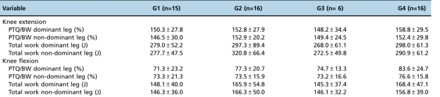
No significant between-group differences were observed in the dominant and non-dominant leg PTQ/BW and total work (Table 3). Likewise, no significant between-group dif-ferences were observed in XSD, YSD, VAvg and Area95 in the postural balance assessment (Table 4).
However, significant differences were observed in the TUGT, consistent with the results of the multiple linear regression analysis. Women with higher 25(OH)D levels (G4) showed better performance than those with lower 25(OH)D levels (G1). The women in groups G2 and G3 showed intermediate TUGT performance that was not significantly different from either G1 or G4 (Figure 1).
’ DISCUSSION
The principal finding of this study was that the serum 25(OH) vitamin D levels in postmenopausal women with osteoporosis were associated with knee extension strength and functional capacity. Vitamin D has been reported to influence neuromuscular transmission, cognition and exten-sor strength of the knee, important parameters for maintain-ing postural balance and functionality (6,17). Although the current findings did not support the hypothesis that the serum vitamin D levels contribute to balance, this study is one of the first to show an independent association between the serum vitamin D levels, strength and function within a sample of osteoporotic postmenopausal women.
and postural balance, and decreases the number of falls (8,18). Additionally, in as little as 3 months, vitamin D supplementa-tion may increase muscle strength in older women with osteo-porosis (19). Static and dynamic balance predict the risk of falls in older women, and the combination of vitamin D and
calcium may reduce the risk of falls by as much as 22-29% (12,18). Even a short period of supplementation with vitamin D and calcium improves postural balance in older women, particularly in the anterior-posterior direction, and prevent falls and non-vertebral fractures (20). Vitamin D supplementation Table 3-Isokinetic performance of the study participants in each group (G).
Variable G1 (n=15) G2 (n=16) G3 (n= 6) G4 (n=16)
Knee extension
PTQ/BW dominant leg (%) 150.3±27.8 152.8±27.9 148.2±34.4 158.8±29.5
PTQ/BW non-dominant leg (%) 146.5±30.0 152.9±20.2 149.4±24.5 152.4±29.8
Total work dominant leg (J) 279.0±52.2 297.3±89.4 268.0±61.1 298.0±61.3
Total work non-dominant leg (J) 277.7±47.5 320.8±66.4 272.5±49.8 290.9±61.2
Knee flexion
PTQ/BW dominant leg (%) 71.3±23.2 77.3±20.7 74.7±13.3 83.6±24.7
PTQ/BW non-dominant leg (%) 73.3±21.3 73.5±15.9 73.2±16.6 76.6±15.8
Total work dominant leg (J) 148.1±40.0 165.9±54.8 145.3±37.4 168.4±47.1
Total work non-dominant leg (J) 146.3±36.0 166.3±50.0 146.1±32.2 156.8±39.0
XSD: mean square root of the displacement from the COP in the mediolateral axis; YSD: mean square root of the displacement from the COP in the anteroposterior axis; VAvg: mean velocity calculated from the total displacement of the COP in all directions; Area95: total displacement area of the COP.
Table 4-Balance performance of the study participants in each group (G).
Variable G1 (n=15) G2 (n=16) G3 (n=16) G4 (n=16)
Eyes open
XSD (cm) 0.27±0.09 0.21±0.06 0.25±0.08 0.23±0.06
YSD (cm) 0.37±0.08 0.39±0.11 0.39±0.14 0.38±0.10
VAvg (cm/sec) 0.79±0.20 0.75±0.13 0.77±0.18 0.76±0.12
Area95 (cm2) 1.78±0.87 1.46±0.68 1.77±1.00 1.48±0.50
Eyes closed
XSD (cm) 0.27±0.11 0.22±0.07 0.29±0.15 0.22±0.06
YSD (cm) 0.38±0.12 0.41±0.10 0.45±0.15 0.41±0.16
VAvg (cm/sec) 1.10±0.31 0.98±0.23 1.10±0.37 1.03±0.20
Area95 (cm2) 20.3±1.28 1.63±0.70 2.49±1.64 1.71±0.86
XSD: mean square root of the displacement from the COP in the mediolateral axis; YSD: mean square root of the displacement from the COP in the anteroposterior axis; VAvg: mean velocity calculated from the total displacement of the COP in all directions; Area95: total displacement area of the COP.
in older people prevented a single fall in a group of 15 people treated with vitamin D because vitamin D supplementa-tion decreases the costs associated with falls (8) by preventing osteoporotic fractures and improving bone mineral density and muscle function (2).
In the present study, postural balance and vitamin D levels in osteoporotic women between the ages of 55 and 65 years were not associated, which has also been reported in another study (10). These results may have been influenced by the ages of the volunteers, even in a homogeneous sample regard-ing age. Some studies of patients who were older than those in our group found that postural instability is associated with low vitamin D concentrations (9) and the postural balance parameters can be improved with exercise intervention after three months (21). However, in another study, vitamin D supplementation did not improve physical function in older individuals (22).
Bischoff-Ferrari et al. (8) emphasized that high 25(OH)D concentrations (levels X40 ng/mL) are associated with better musculoskeletal function of the lower limbs and are recommended to maintain this functionality. However, in a cross-sectional study, impaired static balance was observed only in women with serum 25(OH)D levels of 25 nmol/L (23). An interventional study failed to show any significant improvement in the subjects’static balance after vitamin D
supplementation (24), and several studies failed to show any association between the 25(OH)D levels and quadricep strength (17,24). However, vitamin D supplementation at doses of 800 to 1,000 IU/d has a beneficial effect on balance and muscle strength (13); thus, the role of vitamin D as a contributing factor to functional capacity may well be mediated through the preservation of muscle function.
The association between vitamin D levels and performance on tests exploring mobility impairment has become a topic of much clinical discussion in the past two decades. The TUGT is a measure of functional mobility, muscle function, gait speed and balance (25). The concept of the TUGT is appealing because it describes a realistic mobility assessment that includes potential fall situations, such as getting into and out of a chair, walking, and turning around (25). Therefore, subjects must present muscle strength, particularly in the back and lower limbs, adequate extension and flexion of the hip and knee, postural control involving the interaction between the nervous and musculoskeletal systems to maintain balance, and cogni-tive skills to be able to complete the TUGT (26). Older subjects require more than 10 s to complete the TUGT and have an increasingly higher risk of falls (19).
The main components assessed by the TUGT are strength and functional capacity, which had important relationships with the 25(OH)D levels. These data corroborate the data reported by Toffanello et al. (27), who evaluated various functional tests, such as timed chair stands, gait speed, and a 6-minute walking distance test. Of all the performance measurements considered, the 6-minute walking distance test was the motor test that was most closely related to the vitamin D status in both sexes.
Vitamin D may indirectly contribute to the TUGT by regu-lating the musculoskeletal system, particularly muscle strength and neuromuscular coordination. In a study involving forty-two older women, the serum vitamin D concentrations were not significantly correlated with basic functional mobility, and higher serum vitamin D concentrations are not associated with a shorter time needed to complete the TUGT (26). How-ever, in a recent study of 540 elderly women, TUGT performance
was associated with age, exercise type and the serum 25(OH) D levels (28).
Although the causal effects of vitamin D on postural balance, muscle strength, and functional capacity are controversial, various studies have indicated an improvement in these parameters when subjects are provided vitamin D supple-ments, particularly in older individuals. Schacht and Ringe (19) conclude that daily treatment with 1 mcg of alfacalcidol is safe, increases muscle power, muscle function, and walking distance, decreases the fear of falls, and improves the per-formance in the TUGT and fall risk test. Even a single mega dose of vitamin D may be sufficient to increase the quality of life, decrease non-specific musculoskeletal pain, and prevent falls by improving functional mobility in older individuals (29).
Limitations
The literature on the influence of the vitamin D levels on muscle function, postural balance, gait, and functional independence is inconsistent. Regarding the tests conducted, the TUGT demands more action of the systems required for balance control. Therefore, vitamin D deficiency might accel-erate and aggravate the process of functional loss that occurs during ageing and vitamin D replacement is an important adjuvant factor.
Similar to all cross-sectional investigations, a limitation of this study is its inability to unravel the cause-effect relation-ship between exposure and the outcomes. Indeed, studies determining whether low vitamin D levels ‘‘cause’’ lower
knee extension strength and decrease functional mobility, or whether secondary conditions, such as general frailty syndrome, are drivers of poor nutrition habits, are interesting and complex. Nevertheless, based on these results, vitamin D levels are associated with knee extension strength and improved performance on the TUGT (dynamic equilibrium) in a group of postmenopausal women with osteoporosis. Another potential limitation is that we did not determine the vitamin D intake via diet or supplementation; therefore, we cannot determine whether differences in the vitamin D levels were due to exposure to the sun or oral intake. We did not measure other serum factors related to vitamin D metabo-lism, including calcium, parathyroid hormone, phosphorus, and sex hormone levels; thus we cannot determine whether these factors contributed to the‘‘vitamin D effect.’’
Clinical implications
A healthy 'musculoskeletal system' is required for indivi-duals to maintain independence as they age. Guided by the muscle, bones continuously adapt to their function. The deterioration of muscle power, function and balance con-tributes to the increased risk of falls and related fractures in older subjects with osteoporosis. Thus, obtaining a better under-standing of the importance of vitamin D in the musculoske-letal system and of its role in maintaining muscle strength, postural balance and functional capacity, is essential to plan preventive strategies to prevent falls in women with osteoporosis.
’ ACKNOWLEDGMENTS
The authors gratefully acknowledge the Fundac¸ão de Amparo à Pesquisa
do Estado de São Paulo for providingfinancial support (09/54568-2), and thefirst author acknowledges the Coordenac¸ão de Aperfeic¸oamento de
’ AUTHOR CONTRIBUTIONS
Brech GC was responsible for data collection, data analysis and prepara-tion of the manuscript. Ciolac EG was responsible for data analysis and preparation of the manuscript. Peterson MD edited the manuscript. D’Andrea Greve JM supervised the study and was responsible for manu-script editing.
’ REFERENCES
1. World Health Organization World Health Statistics – Large Gains in Life Expectancy. 2014 Accessed April 2015. Retrieved from: http://www. who.int/mediacentre/news/releases/2014/world-health-statistics-2014/en/. 2. Kuczyński M, Ostrowska B. Understanding falls in osteoporosis: the
visco-elastic modeling perspective. Gait Posture. 2006;23(1):51-8, http://dx.doi.org/ 10.1016/j.gaitpost.2004.11.018.
3. DATA SUS - Ministério da Saúde (2011) Quedas de idosos. http://portal. saude.gov.br/portal/saude/visualizar_texto.cfm?idtxt=33674&janela=1 Accessed 28 February 2011.
4. Pérez-López FR. Vitamin D and its implications for musculoskeletal health in women: an update. Maturitas. 2007;58(2):117-37, http://dx.doi. org/10.1016/j.maturitas.2007.05.002.
5. Shinchuk LM, Holick MF. Vitamin d and rehabilitation: improving func-tional outcomes. Nutr Clin Pract. 2007;22(3):297-304, http://dx.doi.org/ 10.1177/0115426507022003297.
6. Dukas L, Schacht E, Runge M. Independent from muscle power and balance performance, a creatinine clearance below 65 ml/min is a sig-nificant and independent risk factor for falls and fall-related fractures in elderly men and women diagnosed with osteoporosis. Osteoporos Int. 2010;21(7):1237-45, http://dx.doi.org/10.1007/s00198-009-1064-1. 7. Eriksen EF, Glerup H. Vitamin D deficiency and aging: implications
for general health and osteoporosis. Biogerontology. 2002;3(1-2):73-7, http://dx.doi.org/10.1023/A:1015263514765.
8. Bischoff-Ferrari HA, Dietrich T, Orav EJ, Hu FB, Zhang Y, Karlson EW, et al. Higher 25-hydroxyvitamin D concentrations are associated with better lower-extremity function in both active and inactive persons aged
4or =60 y. Am J Clin Nutr. 2004;80(3):752-8.
9. Boersma D, Demontiero O, Mohtasham Amiri Z, Hassan S, Suarez H, Geisinger D, et al. Vitamin D status in relation to postural stability in the elderly. J Nutr Health Aging. 2012;16(3):270-5, http://dx.doi.org/ 10.1007/s12603-011-0345-5.
10. Qutubuddin A, Cifu DX, Adler RA, Carne W, Gitchel G. A pilot study of vitamin D and balance characteristics in middle-aged, healthy indivi-duals. PM R. 2010;2(1):23-6, http://dx.doi.org/10.1016/j.pmrj.2009.10.007. 11. Mason C, Xiao L, Imayama I, Duggan CR, Foster-Schubert KE, Kong A,
et al. Influence of diet, exercise, and serum vitamin d on sarcopenia in postmenopausal women. Med Sci Sports Exerc. 2013;45(4):607-14, http://dx.doi.org/10.1249/MSS.0b013e31827aa3fa.
12. Bischoff-Ferrari HA, Conzelmann M, Stähelin HB, Dick W, Carpenter MG, Adkin AL, et al. Is fall prevention by vitamin D mediated by a change in postural or dynamic balance? Osteoporos Int. 2006;17(5):656-63, http://dx.doi.org/10.1007/s00198-005-0030-9.
13. Muir SW, Montero-Odasso M. Effect of vitamin D supplementation on muscle strength, gait and balance in older adults: a systematic review and meta-analysis. J Am Geriatr Soc. 2011;59(12):2291-300, http://dx.doi.org/ 10.1111/j.1532-5415.2011.03733.x.
14. Brech GC, Plapler PG, de Souza Meirelles E, Marcolino FM, Greve JM. Evaluation of the association between osteoporosis and postural balance in postmenopausal women. Gait Posture. 2013;38(2):321-5, http://dx.doi. org/10.1016/j.gaitpost.2012.12.012.
15. Brech GC, Fonseca ÂM, Bagnoli VR, Baracat EC, Greve JM. Ante-roposterior displacement behavior of the center of pressure, without visual reference, in postmenopausal women with and without lumbar osteoporosis. Clinics. 2013;68(10):1293-8, http://dx.doi.org/10.6061/ clinics/2013(10)01.
16. Brech GC, Ciolac EG, Secchi LL, Alonso AC, Greve JM. The effects of motor learning on clinical isokinetic performance of postmenopausal women. Maturitas. 2011;70(4):379-82, http://dx.doi.org/10.1016/j.maturitas. 2011.09.004.
17. Bischoff HA, Stahelin HB, Urscheler N, Ehrsam R, Vonthein R, Perrig-Chiello P, et al. Muscle strength in the elderly: its relation to vita-min D metabolites. Arch Phys Med Rehabil. 1999;80(1):54-8, http://dx. doi.org/10.1016/S0003-9993(99)90307-6.
18. Bischoff HA, Stähelin HB, Dick W, Akos R, Knecht M, Salis C, et al. Effects of vitamin D and calcium supplementation on falls: a randomized con-trolled trial. J Bone Miner Res. 2003;18(2):343-51, http://dx.doi.org/ 10.1359/jbmr.2003.18.2.343.
19. Schacht E, Ringe JD. Alfacalcidol improves muscle power, muscle func-tion and balance in elderly patients with reduced bone mass. Rheumatol Int. 2012;32(1):207-15, http://dx.doi.org/10.1007/s00296-010-1607-y. 20. Pfeifer M, Begerow B, Minne HW, Abrams C, Nachtigall D, Hansen C.
Effects of a short-term vitamin D and calcium supplementation on body sway and secondary hyperparathyroidism in elderly women. J Bone Miner Res. 2000;15(6):1113-8, http://dx.doi.org/10.1359/jbmr.2000.15.6.1113. 21. Swanenburg J, de Bruin ED, Stauffacher M, Mulder T, Uebelhart D. Effects of exercise and nutrition on postural balance and risk of falling in elderly people with decreased bone mineral density: randomized controlled trial pilot study. Clin Rehabil. 2007;21(6):523-34, http://dx.doi.org/10.1177/ 0269215507075206.
22. Latham NK, Anderson CS, Lee A, Bennett DA, Moseley A, Cameron ID, et al. A randomized, controlled trial of quadriceps resistance exercise and vitamin D in frail older people: the Frailty Interventions Trial in Elderly Subjects (FITNESS). J Am Geriatr Soc. 2003;51(3):291-9, http://dx.doi. org/10.1046/j.1532-5415.2003.51101.x.
23. Stewart JW, Alekel DL, Ritland LM, Van Loan M, Gertz E, Genschel U. Serum 25-hydroxyvitamin D is related to indicators of overall physical fitness in healthy postmenopausal women. Menopause. 2009;16(6):1093– 101, http://dx.doi.org/10.1097/gme.0b013e3181a8f7ed.
24. Annweiler C, Schott AM, Berrut G, Fantino B, Beauchet O. Vitamin D-related changes in physical performance: a systematic review. J Nutr Health Aging. 2009;13(10):893–8, http://dx.doi.org/10.1007/s12603-009-0248-x.
25. Podsiadlo D, Richardson S. The timed "Up & Go": a test of basic func-tional mobility for frail elderly persons. J Am Geriatr Soc. 1991;39(2):142-8, http://dx.doi.org/10.1111/j.1532-5415.1991.tb01616.x.
26. Laksmi PW, Setiati S, Oemardi M, Aries W, Siregar P. Correlation between vitamin D concentration and basic functional mobility in elderly women. Acta Med Indones. 2007;39(3):112-8.
27. Toffanello ED, Perissinotto E, Sergi G, Zambon S, Musacchio E, Maggi S, et al. Vitamin D and physical performance in elderly subjects: the Pro.V.A study. PLoS One. 2012;7(4):e34950, http://dx.doi.org/10.1371/journal. pone.0034950.
28. Nascimento NA, Moreira PF, Marin RV, Moreira LD, Castro ML, Santos CA, et al. Relation among 25(OH)D, Aquatic Exercises, and Multi-functional Fitness on Functional Performance of Elderly Women from the Community. J Nutr Health Aging. 2016;20(4):376-82, http://dx.doi.org/ 10.1007/s12603-015-0569-x.