Fisioter. Mov., Curitiba, v.30, Suppl 1, 2017 Licenciado sob uma Licença Creative Commons DOI: http://dx.doi.org/10.1590/1980-5918.030.S01.AO01
Effects of a therapeutic exercises program associated
with pompage technique on pain, balance and strength
in elderly women with knee osteoarthritis
Efeitos de um programa de exercícios terapêuticos
associado à técnica de pompage sobre dor, equilíbrio e
força muscular em idosas com osteoartrite de joelho
Ihana Thaís Guerra de Oliveira Gondim, Amanda Bruto da Costa Torres,
Amanda Telino Baudel de Lacerda, Danielle Queiroz Kühni Fernandes, Moisés Costa do Couto, Márcia Alessandra Carneiro Pedrosa*
Universidade Federal de Pernambuco (UFPE), Recife, PE, Brazil
[R]
Abstract
Introduction: Knee Osteoarthritis (OA) affects mainly elderly, being characterized by arthralgia, stiffness
and strength and balance deficits. Scientific evidence suggests beneficial effects of exercise therapy on these changes, but lacks detailed protocols. In addition, there were published studies on effects of therapeutic ex
-ercise associated with pompage that were not located. Objective: To investigate the effects of a therapeutic exercise program associated with pompage on pain, balance and muscle strength in elderly women with knee osteoarthritis. Methods: A randomized pilot clinical trial that included 22 women (60 - 80 years) di
-agnosed with knee OA, randomized and allocated by simple raffle into two groups: intervention and control. Intervention group (strengthening and balance exercises and knee pompage) performed two sessions per week for 12 weeks. The control group received educational lectures, they were evaluated before and after
* ITGOG: Doctoral student, e-mail: [email protected]
ABCT: undergrad, e-mail [email protected] ATBL: undergrad, e-mail: [email protected] DQKF: undergrad, e-mail: [email protected] MCC: MS, e-mail: [email protected]
12
12 weeks: arthralgia- subscale pain of the Western Ontario and McMaster Universities Osteoarthritis Index questionnaire (WOMAC); postural stability- postural balance protocol by Biodex Balance SD and muscle strength- concentric peak torque of the knee extensor by isokinetic dynamometer. The Independent Student t test was used for intergroup analysis. Results: The intervention group compared with the control showed better results for the outcomes pain, balance and muscle strength after 12 weeks. However, the difference was statistically significant (p < 0.05) for levels of antero-posterior oscillation (DM -0.28, 95% CI -0.54 a -0.02; p = 0.035) and global oscillation (DM -0.36, 95% CI -0.68 a -0.04; p = 0.028). Conclusion: The
inter-vention had a positive effect on postural balance in elderly women with knee osteoarthritis.
Keywords: Postural Balance. Physical Therapy Specialty. Osteoarthritis. Exercise Therapy. Muscle Strength.
Resumo
Introdução: Osteoartrite (OA) de joelhos acomete principalmente idosas, caracterizando-se por artralgia, rigidez, déficit de força e do equilíbrio. Evidências científicas apontam efeitos benéficos da terapia por exercícios sobre estas alterações, porém carecem de protocolos detalhados. Estudos sobre efeitos de exercícios terapêuticos associados à pompage também não foram localizados. Objetivo: Investigar efeitos de um programa de exercícios terapêuticos associados à pompage sobre dor, equilíbrio e força muscular em idosas com OA de joelhos. Métodos: Ensaio clínico randomizado piloto que incluiu 22 idosas (60 - 80 anos) com diagnóstico de OA de joelhos, sendo randomizadas por sorteio simples em 2 grupos: intervenção e controle. Grupo intervenção (exercícios de fortalecimento e equilíbrio associados à pompage) realizou 2 sessões semanais, por 12 semanas. Grupo controle recebeu palestras educativas. Foram avaliados antes e após 12 semanas: artralgia- subescala dor do questionário Western Ontario and McMaster Universities Osteoarthritis Index (WOMAC); equilíbrio postural- protocolo estabilidade postural do Biodex Balance SD e força muscular- pico de torque concêntrico dos extensores dos joelhos pelo dinamômetro isocinético. Utilizou-se o teste t Student independente para análise intergrupos. Resultados: Grupo intervenção comparado ao controle apresentou melhores resultados para os desfechos dor, equilíbrio e força muscular após as 12 semanas. Entretanto, a diferença foi estatisticamente significativa (p < 0.05) apenas para níveis de oscilação ântero-posterior (DM -0.28, 95% IC -0.54 a -0.02; p = 0.035) e global (DM -0.36, 95% IC -0.68 a -0.04; p = 0.028). Conclusão: A intervenção teve um efeito positivo sobre o equilíbrio postural em idosas com OA de joelhos.
Palavras-chave: Equilíbrio Postural. Fisioterapia. Osteoartrite. Terapia por Exercício. Força Muscular.
Introduction
Osteoarthritis (OA) is a condition due to cartilaginous degeneration and remodeling of the subchondral bone, being more common in women from 50 years of age (1, 2). When it affects the knee joint, it is characterized by stiffness, muscle weakness and arthralgia (3, 4).
Pain is a common symptom and, when severe and associated with movement, may lead to greater functional dependence (5). It can also alter the detection of information regarding position and joint movement, resulting in proprioceptive imprecision, with consequent alteration of postural control (6, 7).
A greater oscillation in the pressure center is observed in elderly women with knee OA, implying an increased risk of falls (8, 9). This is because the proprioceptive deficit changes the control of the
postural oscillation performed by the muscles, generating functional instability (9).
At the same time, knee OA is associated with sarcopenia (10, 11). This association causes changes in muscle properties, exacerbates failure of voluntary quadriceps muscle activation, and induces edema formation, synovial thickening, and atrophy of type II muscle fibers (12 - 15).
13 The knee pompage is performed in the direction of
articular decompression, which can combat stiffness and cartilaginous degeneration, delaying the evolution of OA. The maneuver used in clinical practice aims at reestablishing the hydric balance of cartilage and allowing functional recovery (17). Despite this, the effects of the association of pompage to a program of therapeutic exercises in knee OA are still unclear.
In this sense, the study intends to investigate the effects of a program of therapeutic exercises associated with the pompage technique on pain, balance and muscular strength in a group of elderly women with knee OA, suggesting a positive influence on these outcomes.
Methods
Randomized pilot clinical trial with blinding for the evaluator, that evaluating elderly women with diagnosis of knee OA recruited in the community in general and in the Nucleus of Attention to the Elderly of the Federal University of Pernambuco (UFPE) through posters and information leaflets about the research.
The study was conducted at the Physiotherapy Department of the UFPE, approved by the Research Ethics Committee of the institution (CAAE: 02993812.3.0000.5208) and registered at ClinicalTrials. gov (trial registration number: NCT 02560831). Participants signed the Informed Consent Term.
Inclusion criteria were: women aged 60 to 80 years, diagnosis of knee OA according to clinical and radiographic criteria of the American College of
Rheumatology (ACR) (18) and independent walking (without use of walking aid). Exclusion criteria were: unstable cardiovascular and / or respiratory diseases; cognitive disorder; knee and/or hips arthroplasty; have performed in the last six months lower limb surgery, corticosteroid injection in the knee and / or physiotherapy for OA; diseases contributing to balance deficit (ankylosing spondylitis, rheumatoid arthritis, uncontrolled diabetes mellitus, neurological diseases, vestibulopathies); fibromyalgia.
Procedures
Twenty-two women were randomized by a simple draw into two groups: FEP group (strengthening and balance exercises associated with knee pompage) and GC control (educational lectures), each with 11 participants.
FEP
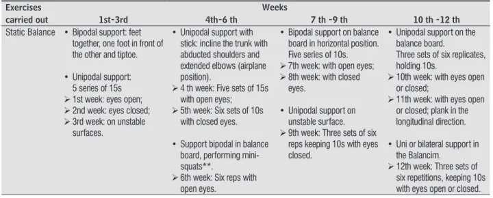
Static and dynamic balance and strength exercises for knee extensor and flexor muscles, associated with knee pompage. There were two weekly sessions (60 min) for 12 weeks, comprising: 10min of heating (up to 50% of maximal heart rate) and static stretching; 10min of balance training, 20min of muscle strengthening, 10min of knee pompage and 10min back to rest. Prescribed exercises were based on McKnight (19) and Chaipinyo and Karoonsupcharoen (20). The loads were progressive and adjusted according to the perceived exertion of less than 6 points on the Borg scale (21) (Table 1).
Table 1 - Progression of exercises at 12 weeks of treatment Exercises
carried out
Weeks
1st-3rd 4th-6 th 7 th -9 th 10 th -12 th
Static Balance • Bipodal support: feet together, one foot in front of the other and tiptoe.
• Unipodal support: 5 series of 15s Ø 1st week: eyes open; Ø 2nd week: eyes closed; Ø 3rd week: on unstable
surfaces.
• Unipodal support with stick: incline the trunk with abducted shoulders and extended elbows (airplane position).
Ø 4 th week: Five sets of 15s with open eyes;
Ø 5th week: Six sets of 10s with closed eyes.
• Support bipodal in balance board, performing mini-squats**.
Ø 6th week: Six reps with open eyes.
• Bipodal support on balance board in horizontal position. Five series of 10s. Ø 7th week: with open eyes; Ø 8th week: with closed
eyes.
• Unipodal support on unstable surface. Ø 9th week: Three sets of six
reps keeping 10s with eyes closed.
• Unipodal support on the balance board.
Three sets of six replicates, holding 10s.
Ø 10th week: with eyes open or closed;
Ø 11th week: with eyes open or closed; plank in the longitudinal direction.
• Uni or bilateral support in the Balancim.
Ø 12th week: Three sets of six repetitions, keeping 10s with eyes open or closed.
14
Table 1 - Progression of exercises at 12 weeks of treatment Exercises
carried out
Weeks
1st-3rd 4th-6 th 7 th -9 th 10 th -12 th
Dinamic Balance
• Circuit on flat surface Ø 1st week: the dynamic
balance was not worked on.
Ø 2nd week: walk forward (one foot in front of the other), sideways, following markings on the ground and zigzagging. Ø 3rd week: walk forward
(one foot in front of the other), sideways over 3 obstacles (canes, cones or PET bottles), zigzag or in eight shape.
• Unstable surface circuit. Ø 4 th week: on a mat, walk
forward (one foot in front of the other), sideways over five obstacles (sticks) and crossing one foot in front of the other. Ø 5th week: on the mat walk
forward (one foot in front of the other), overcome two obstacles (two elastic bands attached between two chairs) and work around two PET bottles, making a trajectory of eight.
Ø 6th week: keep your balance on two mats without removing your foot from the floor while trying to catch the ball thrown in several directions by the therapist and walk following markings on the mat.
• Circuit with obstacles. Ø 7th week: Hold a small ball
with elbows extended and walk one foot in front of the other on two mats, walk in an eight shape, bypassing two obstacles on the ground, and going up and down a cushion (repeat the circuit five times). Ø 8th week: hold a small ball
with elbows extended and walk with one foot in front of the other on two mats, walk sideways, on ground, surpassing four sticks and perform one-way support on a mat (repeat the circuit five times).
Ø 9th week: rise and fall of the balance board in the horizontal position (Ten repetitions).
• Circuit with obstacles. Ø 10th week: walk with one
foot in front of the other on two mats, sideways between four sticks, go up and down the balance board alternating the LL. Ø 11th week: walk with one
foot in front of the other on three mats, in zig-zag, with one foot in front of the other, tiptoe up and down and off the balance board alternating the LL. Ø 12th week: walk sideways
on the pillows in the parallel bar with one foot in front of the other and eyes closed and up and down the proprioceptive disk alternating LL.
Muscle Strengthening of Quadriceps
Ø 1st week: Knee flexion-extension in CKC (body weight resistance) and terminal extension of the knee (resistance of the leg weight).
Ø 2nd week: terminal extension* in CKC (elastic band resistance) and terminal extension in OKC (resistance of 1 kg) Ø 3rd week: terminal
extension* in OKC sitting on ball (resistance of 1kg) and up and down the 1st step of a ladder with one of the LL (3 sets of 6, 8 and 10 repetitions).
Ø 4th week: terminal extension* of the knee in OKC (resistance 1 kg, three sets of ten repetitions) and up and down the 1st step of a ladder with one of the LL (three sets of six, eight and ten repetitions). Ø 5th week: CKC, with the
elastic band passing under the foot and holding with both hands, perform mini-squats** yielding gradually to the elastic resistance in the flexion and surpassing it in the extension; Sit and lift from a chair without support from UL (three sets of six, eight and ten reps).
Ø 6th week: terminal extension* of knee in OKC sitting on the ball (resistance 2kg) and keep one of the lower limbs supported on the 1st step of the ladder, while lifting the body supporting the weight on this leg (three series of six, eight and ten repetitions).
Ø 7th week: mini-squats** with Swiss ball support (three sets of six, eight and ten repetitions) and sit and stand without support of the UL (three sets of six, eight and ten repetitions). Ø 8th week: knee extension
in OKC (2kg resistance, three sets of ten repetitions) and step up and down step alternating LL (ten repetitions). Ø 9th week: terminal extension* in OKC (resistance of 2kg, three series of eight, ten and 12 repetitions).
Ø 10th week: terminal extension* of the knee (resistance of 2kg, three series of ten,12 and 15 repetitions); Sit and stand up from the chair without support from UL (three sets of 10,12 and 15 repetitions). Ø 11th week: terminal
extension* of the knee, OKC (3kg resistance, three series of ten,12 and 15 repetitions); (three sets of ten repetitions) and mini-squatting** with Swiss ball support (three sets of ten,12 and 15 repetitions). Ø 12th week: knee extension
in OKC in bodybuilding machine*** (three sets of eight, ten and 12 repetitions (5kg resistance) and mini-squatting** with Swiss ball support (three sets of ten,12 and 15 repetitions).
15
Table 1 - Progression of exercises at 12 weeks of treatment Exercises
carried out
Weeks
1st-3rd 4th-6 th 7 th -9 th 10 th -12 th
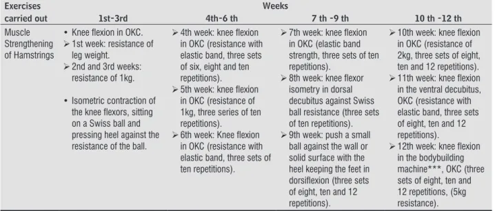
Muscle Strengthening of Hamstrings
• Knee flexion in OKC. Ø 1st week: resistance of
leg weight. Ø 2nd and 3rd weeks:
resistance of 1kg.
• Isometric contraction of the knee flexors, sitting on a Swiss ball and pressing heel against the resistance of the ball.
Ø 4th week: knee flexion in OKC (resistance with elastic band, three sets of six, eight and ten repetitions).
Ø 5th week: knee flexion in OKC (resistance of 1kg, three series of ten repetitions).
Ø 6th week: Knee flexion in OKC (resistance with elastic band, three sets of ten repetitions).
Ø 7th week: knee flexion in OKC (elastic band strength, three sets of ten repetitions).
Ø 8th week: knee flexor isometry in dorsal decubitus against Swiss ball resistance (three sets of ten repetitions). Ø 9th week: push a small
ball against the wall or solid surface with the heel keeping the feet in dorsiflexion (three sets of eight, ten and 12 repetitions).
Ø 10th week: knee flexion in OKC (resistance of 2kg, three sets of eight, ten and 12 repetitions). Ø 11th week: knee flexion
in the ventral decubitus, OKC (resistance with elastic band, three sets of eight, ten and 12 repetitions).
Ø 12th week: knee flexion in the bodybuilding machine***, OKC (three sets of eight, ten and 12 repetitions, (5kg resistance).
Note: LL = lower limbs; UL = upper limbs; OKC = Open Kinetic Chain; CKC = Closed Kinetic Chain; * Knee terminal extension = 15 to 0 degrees of flexion; ** Mini squat = zero to 45 degrees of flexion; *** Mega II Station for bodybuilding, Movement. (BRUDDEN Equipamentos Ltda, São Paulo, Brazil). Isotonic exercises in OKC for knee extensors were performed in the seated position and for the flexors in the standing position and ventral decubitus. Exercises for the knee extensors CKC were performed on foot.
Knee pompage was performed after exercises, with the patient lying in the supine position at the edge of the stretcher and the hanging leg placed between the legs of the therapist. Three decompression maneuvers of 15 to 20s each were performed by a slight retreat from the therapist's body, whose palms were placed laterally over the tibial tuberosity and the indicators folded under the knee, keeping it slightly flexed to relax the ligaments (17).
For the return to calm, patients relaxed to the sound of calm music, realizing respiratory cycles of slow and deep form.
GC
Educational lectures and group dynamics in four moments: after data collection and in the 4th, 8th and 12th weeks of treatment, according to Coimbra's precepts (16). The topics were: clarification on OA; effects of exercise therapy on quality of life; benefits of proper practice of activities; guidelines for ramps and stairs; ergonomics of domestic and professional work and living habits. GC received the same FEP treatment program after the study period.
Procedures related to the intervention and lectures were performed by physiotherapists and undergraduate students in Physical Therapy after the training period.
Instrumentation
Pain, postural balance, and muscle strength outcomes were collected by blind assessors before and after 12 weeks. An evaluation form has been completed for personal and clinical-functional information.
Visual Analog Scale (VAS)
For a better analysis of the concentric muscle torque peak of the knee extensors, the knee was more symptomatic, that is, the one that presented a higher level of pain for the patient, less symptomatic, using the VAS. This measurement consists of a 10 cm line, with anchors at its ends. In one of them it is marked "no pain" and the other "the worst pain". The magnitude of the pain is indicated by marking a line and a ruler is used for measurement. It is considered valid, reliable and responsive to evaluate pain in OA (22).
Western Ontario and McMaster Universities Osteoarthritis Index Questionaire (WOMAC)
Specifically for OA, it is composed of three domains: pain (5 questions), stiffness (2 questions) and functionality (17 questions), whose questions
16
are answered by the volunteer about their perception in the last 72h. In this study, the pain section score was used. The items were evaluated as "none", "low", "moderate", "intense" and "very intense" levels, being calculated using the Likert scale (23) of 5 points: 0, 25, 50, 75 and 100. Higher scores indicate a worse pain level (24).
Biodex Balance SD (BBSD) (Biodex Medical Systems, New York, USA)
It evaluates postural balance by quantifying body oscillation through the indexes of antero-posterior (API), mid-lateral (MLI) and global (OSI) stability on static or unstable surfaces (25). Studies have concluded that there is reliability in the stability and reproducibility indices of the equilibrium tests (26 - 29). The Postural Stability protocol (25) was used with bipodal evaluation, open eyes, eight resistance and three repetitions of 20s interspersed with rest. For positioning of the feet, the tendon line of the calcaneus and the middle toe were adopted as a reference. You should keep a cursor in the center of a target. A lower score, that is, smaller deviation of the cursor was desirable. Hand support or withdrawal of platform feet were not allowed. Before the test, the procedure was simulated.
Isokinetic Dynamometer HUMAC® NORM Testing & Rehabilitation System (CSMI Medical Solutions, Massachusetts, USA)
The concentric muscle torque peak of the knee extensors of both lower limbs was evaluated at a speed of 120° / s. Slower speeds (60 and 90° / s) are also recommended, but could contribute to increased pain during the test (30, 31). Participants sat in a chair with a 15° inclined backrest, without foot support, stabilized by a belt through the chest, pelvis and distal femur on the side tested (32). For measurement, they were instructed to perform knee extension and flexion at the maximum pain-free range. Five repetitions were performed, the
highest value being recorded as peak muscular torque. Prior to the test, five replicates were performed to familiarize the procedure. Data was corrected by gravity and recorded in Newton meters (Nm), adjusted for body weight (kg) and presented as percentage.
Statistical analysis
SPSS software version 16.0 was used. When the normal distribution was verified using the Shapiro Wilk test, we chose the independent Student t test for intergroup analysis and a p < 0.05. There was intention to treat analysis. The effect size was evaluated through mean difference (MD) and 95% confidence interval.
Results
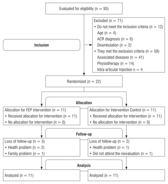
The elderly women in the study (Figure 1) had a mean age of 66 ± 5.42 years, mean height of 1.57 ± 0.06 m, average body weight of 72.5 ± 1.40 kg, and mean body mass index of 29.38 ± 4.54 kg/m². The characterization of the sample separated by group is shown in table 1, with no clinical and statistically significant difference between the groups at the beginning of the study.
According to table 2, the FEP group presented better results in pain outcomes, postural balance and muscle strength when compared to the control group.
Regarding pain, there was a reduction in pain in the FEP group when compared to control, however this result was not significant (Table 2).
The FEP group had lower levels of API, MLI and OSI when compared to the control group. However, there was only a significant difference for the API and OSI, evidenced by the comparison of mean differences between the FEP and control groups before and after twelve weeks of treatment.
17
Randomized (n = 22)
Inclusion
Allocation
Follow-up
Analysis
Excluded (n = 71)
• Do not meet the inclusion criteria (n = 12) Age (n = 4)
ACR diagnosis (n = 6) Deambulation (n = 2)
• They met the exclusion criteria (n = 59) Associated diseases (n = 41)
Physiotherapy (n = 14) Intra-articular Injection n = 4
Loss of follow-up (n = 2) • Health problem (n = 1)
• Did not attend the reevaluation (n = 1)
Analyzed (n = 11) Allocation for FEP Intervention (n = 11)
• Received allocation for intervention (n = 11) • No allocation for intervention (n = 0)
Loss of follow-up (n = 3) • Health problem (n = 2) • Family problem (n = 1)
Analyzed (n = 11)
Evaluated for eligibility (n = 93)
Allocation for Intervention Control (n = 11) • Received allocation for intervention (n = 11) • No allocation for intervention (n = 0)
Figure 1 - Sample constitution flowchart.
Table 2 - Characterization of the sample
Variables FEP (n = 11) GC (n = 11) P
Age (years) 65.09 (4.482) 66.91 (6.316) –
Weight (kg) 71.25 (9.87) 73.72 (17.71) –
Height (m) 1.55 (0.062) 1.59 (0.066) –
BMI (kg/m²) 29.67 (3.86) 29.08 (5.31) –
Pain 45.00 (17.46) 45.91 (19.98) 0.911
API 0.91 (0.37) 0.89 (0.51) 0.88
OSI 1.29 (0.51) 1.28 (0.61) 0.97
More symptomatic knee torque
peak (Nm/Kgx100) 61.68 (18.48) 66.04 (17.72) 0.579
Less symptomatic peak knee
torque (Nm/Kgx100) 76.91 (21.23) 71.31 (10.60) 0.443
18
Table 3 - Pain evaluation, postural balance and muscle strength after 12 weeks of treatment
FEP GC DM IC 95% P
Pain 25.81 (6.58) 33.52 (13.99) -7.70 -17.42 a 2.02 0.114
API 0.91 (0.26) 1.19 (0.32) -0.28 -0.54 a -0.02 0.035*
MLI 0.72 (0.25) 0.90 (0.30) -0.18 -0.43 a 0.06 0.142
OSI 1.29 (0.32) 1.65 (0.40) -0.36 -0.68 a -0.04 0.028*
More symptomatic knee
torque peak (Nm/Kgx100) 73.52 (23.70) 62.88 (18.84) 10.63 -8.40 a 29.67 0.258
Less symptomatic peak knee
torque (Nm/Kgx100) 80.02 (25.22) 66.08 (16.48) 13.93 -5.01 a 32.88 0.143
Note: Pain obtained by WOMAC. Assessment by BBSD- API = Antero-posterior stability index; MLI = Medium-lateral stability index; OSI = Overall stability index; Peak T knee = Peak of knee torque, Isokinetic Dynamometer - normalized by body weight (%). M1 = Mean (SD) at baseline; M2 = Mean (SD) after 12 weeks of intervention. DM = Difference of means; CI = confidence interval; * P<0.05 - T independent student test.
Discussion
The therapeutic exercise program associated to pompage promoted the reduction of the pain complaint, as well as the increase of the postural balance and of the knee extensor muscle strength in elderly women with knee OA. However, the improvement was statistically significant only for the postural balance outcome.
Systematic review by Fransen et al. (32) found high quality evidence for the reduction of OA pain after the practice of therapeutic exercises. In the present study, the reduction of pain intensity was not significant after the intervention, with a mean difference of ten points. The improvement in this outcome seems to be also associated with the gains on the extensor muscle strength (33), which in this study was also not significant.
Regarding the postural balance, the mean difference analysis showed a significant decrease in the API and OSI after treatment. The improvement of antero-posterior oscillation represents an important functional gain in this population, since according to Petrella et al. (8) it is exacerbated in elderly women with knee OA, resulting in an inability to maintain orthostatic posture. With regard to the significant improvement in the global oscillation, it is suggested that the OSI value has decreased as a function of the reduction of the API value, confirming Arnold and Schmitz (27).
The program of the present study did not address the strengthening of hip abductors, which may be one of the reasons why the decrease of the medium-lateral
oscillation (MLI) was not significant (8). Given that an increased MLI oscillation is related to the history of falls in the elderly (8), it is suggested that future studies should include strengthening of hip abductors in their programs.
In relation to quadriceps strength, the meta-analysis of Zacharias et al. (34) analyzed ten studies lasting between six and 12 weeks and found a high quality of evidence for improvement of muscle strength of knee extensors with low intensity resistance programs when compared to a control group. Despite the findings, the meta-analysis did not bring the detailed exercise protocols. This exercise program included isometric strengthening and, mainly, isotonic strengthening with resistance progression over 12 weeks. The increase in the concentric peak torque of knee extensors was not statistically significant, but the mean difference between the groups was greater than ten points for both the more symptomatic and less symptomatic knees.
Jan et al. (35), with a mean difference of approximately ten points after intervention, observed a statistically significant increase in peak torque of knee extensors in subjects with knee OA. However, they performed a program focused on low-intensity resistance exercises with three weekly sessions and duration of eight weeks, as well as a sample of 64 people. The present study was a pilot study. In addition, it covered not only muscle strengthening exercises, but also balance exercises and pompage.
19 action on pain may be associated to the effect of
the technique on joint decompression and combat cartilaginous degeneration, reestablishing the water balance and limiting cartilage dryness (17). According to Kul-Panza and Berker (36), the greater the degree of knee OA, the greater the difficulty in maintaining balance. Therefore, combating the evolution of cartilaginous degeneration through the pompage, we suggest the impact on postural balance. Despite this, the lack of evidence on the effects of pompage on the treatment of OA hinders a greater understanding about its therapeutic action.
The limitations of this study include the small sample size and the follow-up losses due to locomotion problems of the participants throughout the program. In spite of this, the relevance of presenting a program with a detailed description of the procedures used as: number and duration of sessions, type of exercise, repetitions, series and intensity of training is emphasized. In addition, the low cost and easy applicability of the program allow its implementation in the public health scenario.
Conclusion
Therapeutic exercise program associated with pompage promoted reduction of pain complaint, as well as increased postural balance and knee extensor muscle strength of elderly women with knee OA. However, the improvement was statistically significant only for the postural balance outcome. Despite the findings, this is a pilot study, and more evidence is needed. It is also suggested that future studies should analyze the minimal clinical difference detected by the elderly to know if the change was important for these patients.
Acknowledgements
To the Foundation of Assistance to the Research of the State of Pernambuco (FACEPE) and to the National Council of Scientific and Technological Development (CNPq) for the financial support.
References
1. Williams SB, Brand CA, Hill KD, Hunt SB, Moran H. Feasibility and outcomes of a home-based exercise program on improving balance and gait stability in women with lower-limb osteroarthritis or reumatoid arthritis: a pilot study. Arch Phys Med Rehabil. 2010;91(1):106-14.
2. Felson DT. An Update on the pathogenesis and epidemiology of osteoarthritis. Radiol Clin North Am. 2004;42(1):1-9, v.
3. Moreira C, Carvalho MAP. Reumatologia: Diagnóstico e Tratamento. 2nd ed. Rio de Janeiro: Medsi; 2001. 4. Moreira-Pfrimer LDF. Atividade física adaptada à
osteoartrite, fibromialgia e dor miofascial. São Paulo: Phorte; 2008.
5. Jinks C, Jordan K, Croft P. Osteoarthritis as a public health problem: the impact of developing knee pain on physical function in adults living in the community. Rheumatology (Oxford). 2007;46(5):877-81. 6. Sanchez-Ramirez DC, van der Leeden M, Knol DL,
van der Esch M, Roorda LD, Verschueren S, et al. Association of postural control with muscle strength, proprioception, self-reported knee instabilliy and activeIty limitations in patients with knee osteoarthritis. J Rehabil Med. 2013;45(2):192-7. 7. Bennell KL, Hinman RS, Metcalf BR, Crossley KM,
Buchbinder R, Smith M, et al. Relationship of knee joint proprioception to pain and disability in individuals with knee osteoarthritis. J Orthop Res. 2003;21(5):792-7.
8. Petrella M, Neves TM, Reis JG, Gomes MM, Oliveira RDR, Abreu DCC. Parâmetros do controle postural em mulheres idosas com ou sem histórico de quedas associadas ou não à osteoartrite de joelhos. Rev Bras Reumatol. 2012;52(4):512-7.
20
10. Walsh MC, Hunter GR, Livingstone MB. Sarcopenia in premenopausal and postmenopausal women with osteopenia, osteoporosis and normal bone mineral density. Osteoporos Int. 2006;17(1):61-7.
11. Scott D, Blizzard L, Fell J, Jones G. A prospective study of self-reported pain, radiographic osteoarthritis, sarcopenia progression and falls risk in community-dwelling older adults. Arthritis Care Res (Hoboken). 2012;64(1):30-7.
12. Bennell KL, Hunt MA, Wrigley TV, Lim BW, Hinman RS. Role of muscle in the genesis and management of knee osteoarthritis. Rheum Dis Clin North Am. 2008;34(3):731-54.
13. Lewek MD, Rudolph KS, Snyder-Mackler L. Quadriceps femoris muscle weakness and activation failure in patients with symptomatic knee osteoarthritis. J Orthop Res. 2004;22(1):110-5. 14. Meireles SM, Oliveira LM, Andrade MS, Silva AC,
Natour J. Isokinetic evaluation of the knee in patients with rheumatoid arthritis. Joint Bone Spine. 2002;69(6):566-73.
15. Stevens JE, Mizner RL, Snyder-Mackler L. Quadriceps
strength and volitional activation before and after
total knee arthroplasty for osteoarthritis. J Orthop Res. 2003;21(5):775-9.
16. Coimbra IB, Pastor EH, Greve JMD, Puccinelli MLC, Fuller R, Cavalcanti FS, et al. Osteoartrite (artrose): tratamento. Rev Bras de Reumatol. 2004;44(6):450- 3.
17. Bienfait M. Estudo e Tratamento do Esqueleto Fibroso: Fáscias e Pompages. São Paulo: Summus; 1995. 18. Altman R, Alarcón G, Appelrouth D, Bloch D,
Borenstein D, Brandt K, et al. The American College of Rheumatology criteria for the classification and reporting of osteoarthritis of the hip. Arthritis Rheum. 1991;34(5):505-14.
19. McKnight PE, Kasle S, Going S, Villanueva I, Cornett M, Farr J, et al. A comparison of strength training,
self-management, and the combination for early
osteoarthritis of the knee. Arthritis Care Res (Hoboken). 2010;62(1):45-53.
20. Chaipinyo K, Karoonsupcharoen O. No difference between based strength training and home-based balance training on pain in patients with knee osteoarthritis: a randomised trial. Aust J Physiother. 2009;55(1):25-30.
21. Borg GV, Noble B. Perceived exertion. In: Wilmore JH (ed). Exercise and Sport Sciences. Reviews. New York: Academic Press; 1974. p. 131-53.
22. Bellamy N. Osteoarthritis clinical trials: candidate variables and clinimetric properties. J. Rheumatol. 1997;24(4):768-78.
23. Likert R. A technique for the measurement of attitudes. Arch Psychol. 1932;22(140):5-55
24. Fernandes MI, Ferraz MB, Ciconelli RM. Tradução e validação do Questionário de Qualidade de Vida Específico para Osteoartrose (WOMAC) para a língua portuguesa. Rev Paulista Reumatol. 2003;10:25. 25. Biodex Medical Systems. Balance System SD: Manual
de instalação e procedimentos operacionais. New York; 2012 [cited 2015 June 14]. Available from: http://m.biodex.com/support/manuals.
26. Pincivero DM, Lephart SM, Henry TJ. Learning effects and reliability of the Biodex Stability System. J Athl Train. 1995;30:S35.
27. Arnold BL, Schmitz RJ. Examination of balance measures produced by the Biodex Stability System. J Athl Train. 1998;33(4):323-7.
28. Hinman MR. Factors affecting reliability of the Biodex Balance System: a summary of four studies. J Sport Rehab. 2000;9(3):240-52.
29. Hornyik ML, Harter RA. Reliability of limits of stability testing: a comparison of two dynamic postural stability evaluation devices. J Athl Train. 2001;36(2 Suppl):S78.
30. Perrin DH. Isokinetc exercise and assessment. Champaign, IL: Human Kinetics Publishers; 1993. 31. Almosnino S, Brandon SC, Sled EA. Does choice of
21 32. Fransen M, McConnell S, Harmer AR, Van der Esch M,
Simic M, Bennell KL. Exercise for osteoarthritis of the knee. Cochrane Database Syst Rev. 2015;1:CD004376. 33. Dubin, A. Managing Osteoarthritis and Other Chronic Musculoskeletal Pain Disorders. Med Clin North Am. 2016;100(1):143-50.
34. Zacharias A, Green RA, Semciw AI, Kingsley MI, Pizzari T. Efficacy of rehabilitation programs for improving muscle strength in people with hip or knee osteoarthritis: a systematic review with meta-analysis. Osteoarthritis Cartilage. 2014;22(11):1752-73. 35. Jan MH, Lin CH, Lin YF, Lin JJ, Lin DH. Effects
of weight-bearing versus nonweight-bearing exercise on function, walking speed, and position sense in participants with knee osteoarthritis: a randomized controlled trial. Arch Phys Med Rehabil. 2009;90(6):897-904.
36. Kul-Panza E, Berker N. Pedobarographic findings in patients with knee osteoarthritis. Am J Phys Med Rehabil. 2006;85(3): 228-33.
Received in 05/06/2016 Recebido em 06/05/2016