Protein Quality Control Disruption by PKC
b
II in Heart
Failure; Rescue by the Selective PKC
b
II Inhibitor,
b
IIV5-3
Julio C. B. Ferreira1,2, Berta Napchan Boer3, Max Grinberg3, Patricia Chakur Brum2, Daria Mochly-Rosen1*
1Department of Chemical and Systems Biology, Stanford University School of Medicine, Stanford, California, United States of America,2School of Physical Education and Sport, University of Sa˜o Paulo, Sa˜o Paulo, Brazil,3Heart Institute (InCor), University of Sa˜o Paulo, Medical School, Sa˜o Paulo, Brazil
Abstract
Myocardial remodeling and heart failure (HF) are common sequelae of many forms of cardiovascular disease and a leading cause of mortality worldwide. Accumulation of damaged cardiac proteins in heart failure has been described. However, how protein quality control (PQC) is regulated and its contribution to HF development are not known. Here, we describe a novel role for activated protein kinase C isoformbII (PKCbII) in disrupting PQC. We show that active PKCbII directly phosphorylated the proteasome and inhibited proteasomal activityin vitroand in cultured neonatal cardiomyocytes. Importantly, inhibition of PKCbII, using a selective PKCbII peptide inhibitor (bIIV5-3), improved proteasomal activity and conferred protection in cultured neonatal cardiomyocytes. We also show that sustained inhibition of PKCbII increased proteasomal activity, decreased accumulation of damaged and misfolded proteins and increased animal survival in two rat models of HF. Interestingly,bIIV5-3-mediated protection was blunted by sustained proteasomal inhibition in HF. Finally, increased cardiac PKCbII activity and accumulation of misfolded proteins associated with decreased proteasomal function were found also in remodeled and failing human hearts, indicating a potential clinical relevance of our findings. Together, our data highlights PKCbII as a novel inhibitor of proteasomal function. PQC disruption by increased PKCbII activityin vivoappears to contribute to the pathophysiology of heart failure, suggesting that PKCbII inhibition may benefit patients with heart failure. (218 words)
Citation:Ferreira JCB, Boer BN, Grinberg M, Brum PC, Mochly-Rosen D (2012) Protein Quality Control Disruption by PKCbII in Heart Failure; Rescue by the Selective PKCbII Inhibitor,bIIV5-3. PLoS ONE 7(3): e33175. doi:10.1371/journal.pone.0033175
Editor:Niels Olsen Saraiva Caˆmara, Universidade de Sao Paulo, Brazil
ReceivedOctober 31, 2011;AcceptedFebruary 5, 2012;PublishedMarch 30, 2012
Copyright:ß2012 Ferreira et al. This is an open-access article distributed under the terms of the Creative Commons Attribution License, which permits unrestricted use, distribution, and reproduction in any medium, provided the original author and source are credited.
Funding:This study was supported by National Institute of Health Grant HL076675 to DMR. JCBF held a post-doctoral fellowship from Fundac¸a˜o de Amparo a Pesquisa do Estado de Sa˜o Paulo (FAPESP 2009/03143-1) and and Coordenac¸a˜o de Aperfeic¸oamento de Pessoal de Ensino Superior – Programa de Doutorado no Paı´s com Esta´gio no Exterior (CAPES-PDEE, 2177-07-2) – Brasil. The funders had no role in study design, data collection and analysis, decision to publish, or preparation of the manuscript.
Competing Interests:Daria Mochly-Rosen is the founder of KAI Pharmaceuticals, Inc, a company that plans to bring PKC regulators to the clinic. However, none of the work described in this study is based on or supported by the company. The other authors have no disclosure. This does not alter the authors’ adherence to all the PLoS ONE policies on sharing data and materials.
* E-mail: [email protected]
Introduction
Maintenance of blood circulation during continual stress, such as hypertension or following cardiac ischemic events and infarction, contributes to cardiac wear and tear and results in accumulation of damaged cardiac proteins leading to cell death and further deterioration of cardiac functions. The cellular protein quality control (PQC) can detect, repair and dispose of cytotoxic damaged proteins using multiple control mechanisms, which include chaperone proteins, the ubiquitin-proteasome system (UPS) and autophagy [1]. The UPS is the primary effector of the PQC process, protecting long-lived cells, such as neurons and cardiomyocytes, from accumulation of aberrant and misfolded proteins [2]. The pathophysiological role of the PQC machinery in the heart emerged from studies showing accumulation of damaged proteins in humans and in animal models with cardiac diseases as well as cardiac mutations in PQC components [3,4]. There is also up-regulation of proteins involved in UPS and elevated levels of ubiquitinated proteins in hearts of human dilated cardiomyopathy [5]. Some studies found an overall decrease in proteasomal activity associated with and probably contributing to the increased steady
state level of ubiquitinated proteins and cell death [5,6]. However, others reported that several components of the ubiquitin-protein system and/or its overall activity are increased in experimental compensated cardiac hypertrophy and heart failure [7]. There-fore, it remains to be determined whether dysfunction of specific PQC components, such as the UPS, contribute to the develop-ment of end-stage heart failure and which signaling events regulate them.
Numerous studies have focused on identifying intracellular nodes where signals converge and serve as multi-effector brakes to suppress or reverse heart failure. We and others have identified PKCbII, which is over activated in failing hearts of humans [8] and in animal models [8,9,10,11], as a potential key player in heart failure. However, the molecular targets of PKCbII are still unknown.
models [12,13], disturbed cardiac PQC by decreasing proteasomal activity. Using different PKC-selective regulators [14], we then demonstrated here that the PKCbII-specific peptide inhibitor,
bIIV5-3, prevented the decline in PQC in cultured neonatal cardiac myocytes and that sustained PKCbII inhibition substan-tially increased survival and cardiac function in myocardial infarction-induced and hypertension-induced heart failure animal models in rats. The molecular bases of these events were also studied.
Results
PQC dysfunction parallels heart failure development in an animal model
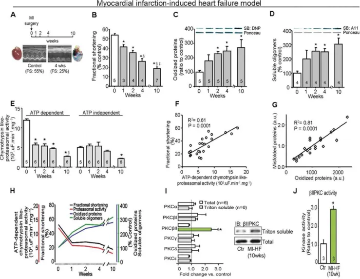
To investigate whether injury-induced progression to heart failure is associated with PQC dysfunction, we evaluated proteasomal activity and accumulation of damaged cardiac proteins in a rat model of myocardial infarction-induced heart failure (Fig. 1A). All measurements were performed in a region remote from the infarcted area in the left ventricle (non-infarcted zone). We found a progressive decline in proteasomal activity during 10 weeks following myocardial infarction that exhibited a tight correlation with the decline in cardiac function (R2= 0.61, p = 0.0001; Fig. 1B, E, F and H), reaching a deficit of 68% and 66%, respectively, when compared with sham-operated rats. The decreased proteasomal activity correlated with an increased accumulation of cardiac oxidized proteins and soluble oligomers of misfolded proteins in the failing hearts (R2= 0.81, p = 0.0001, Fig. 1C, D, G and H). Similar to results observed in human HF hearts [8,15], we found that, of the PKC isozymes present in rat heart, only PKCbII was activated in the myocardial infarction-induced failed hearts, as evidenced by its increased association with the cell particulate fraction (Fig. 1I); there was also a 3-fold increase in catalytic activity of PKCbII, as compared with that from control rat hearts (Fig. 1J).
PKCbII activation directly down-regulates proteasomal activity
We next determined whether PKCbII directly regulates proteasomal activity. Active PKCbII (but notaPKC or PKCbI) phosphorylated and decreased the proteolytic function of the purified 20S proteasome by 55%,in vitro(Fig. 2A, bottom and top panel, respectively). PKCbII activation-mediated proteasomal phosphorylation was essential to decreased proteasomal activity, since inactive PKCbII (added in the absence of its activators, PS/ DG/Ca+2) had no effect on proteasomal function (Fig. 2A). Further, treatment of cultured neonatal cardiomyocytes with 10 nM phorbol ester 12-myristate 13-acetate (PMA, an activator of most PKC isozymes) for 30 min induced oxidized protein accumulation, at least in part, by decreasing the 26S proteasomal ATP-dependent proteolytic activity (40% decrease) (Fig. 2B). Using selective peptide inhibitors for PKC isozymes, we found that only bIIV5-3, a PKCbII-specific peptide inhibitor [14,16,17], [and not selective inhibitors fora,bI or PKCe(aV5-3 andbIV5-3 and eV1-2, respectively), which are also present in cardiac myocytes; [18,19]] prevented this PMA-induced proteasomal inhibition and increased accumulation of damaged proteins (Fig. 2B). Confirming the pharmacological effect of bIIV5-3, PKCbknockdown using siRNA similarly abrogated PMA-induced proteasomal inhibition and oxidized protein accumulation in neonatal cardiomyocytes (Fig. 2C). In contrast, PKCaknockdown did not restore PMA-induced proteasomal activity unless cells
were pre-treated also withbIIV5-3 peptide (Fig. 2C). In addition,
bIIV5-3 treatment prevented damaged protein accumulation and diminished cell death in hydrogen peroxide-treated neonatal cardiomyocytes (Fig. S1) and epoxomicin (a selective proteasome inhibitor) abrogated the cytoprotective effect ofbIIV5-3.
Sustained PKCbII inhibition and cardiac PQC in the myocardial infarction-induced heart failure model
To evaluate the effect of PKCbII on cardiac PQC in heart failure, we further determined whether sustained administration of
bIIV5-3 in a myocardial infarction-induced heart failure model in rats (Fig. 3A) affected cardiac PQC, cardiac function and survival. PKCbII (but not PKCe, an abundant isozyme in the heart) co-immunoprecipitated with the proteasome and decreased its activity in these failing hearts (Fig. 3B and C). After the establishment of HF (4 weeks after myocardial infarction; MI), a subsequent six-week treatment with bIIV5-3 abolished the increased cardiac PKCbII translocation and activity (Fig. 3A), but not the activity of a, bI, d, c and PKCe (Fig. S2), and diminished the co-immunoprecipitation of PKCbII and the 20S proteasome as well as its phosphorylation (Fig. 3B). ThisbIIV5-3 treatment resulted also in a two-fold increase in both ATP-dependent (26S) and -inATP-dependent (20S) cardiac proteasomal activity back to control levels (Fig. 3C). There were no changes in the protein levels of cardiac proteasome subunits in failing hearts regardless of the treatment (Fig. 3D and Fig. S3). However, sustained PKCbII inhibition completely suppressed the accumu-lation of cardiac oxidized proteins, polyubiquitinated proteins and soluble oligomers of misfolded proteins in these rat samples (Fig. 3E–G). The increased abnormal protein accumulation in failed non-treated hearts was accompanied by an,50% increase
in the levels of the small chaperones,a-b-crystallin and HSP27, and a two-fold increase in caspase 3 activation, effects that were reversed by the sustained PKCbII inhibition (Fig. 3D and Fig. S3). In addition, chronic PKCbII inhibition abolished the HF-induced increase in the levels of well-known proteasome substrates, IkB and p53 (Fig. S3).
Sustained PKCbII inhibition and cardiac PQC in the hypertension-induced heart failure model
We next determined whether the role of PKCbII and PQC in heart failure is independent of the etiology of HF, using a hypertension-induced heart failure model in Dahl salt-sensitive rats (Fig. 5A). When placed on a high-salt diet from the age of 6 weeks, Dahl rats exhibit high blood pressure (230vs.160 mmHg), as previously reported [13]. These hypertensive rats develop compensatory left ventricular hypertrophy by the age of 11 weeks, and die from heart failure between 16 to 21 weeks [13,20]. Here we show that 17 week-old hypertensive Dahl salt-sensitive rats also exhibit decreased cardiac proteasomal ATP-dependent proteolytic activity and increased levels of oxidized proteins and soluble
misfolded protein oligomers (Fig. 5B–D). Similar to the effect of PKCbII inhibition in the myocardial infarction-induced HF model in rats, sustained treatment withbIIV5-3 (but not thebI inhibitor) between weeks 11–17, reduced PKCbII activity to basal levels (Fig. 5A), restored ATP-dependent proteasomal activity and decreased the levels of misfolded cardiac proteins to those seen in control animals (Fig. 5B–D). Importantly,bIIV5-3 (but not the
bI inhibitor) treatment prevented the decrease in fractional shortening in hypertensive rats (Fig. 5E). Further, while treated with the PKCbII inhibitor (between weeks 11–17), none of the hypertensive rats died, as compared with 60% death of hypertensive rats treated with vehicle control or the selective inhibitor of the alternatively spliced form,bIPKC (Fig. 5F).
Figure 1. Protein quality control disruption and PKCbII activation during progression of heart failure in a post-myocardial infarction (MI) model of HF in rats. A. MI-induced HF protocol (10 weeks follow up) and example of heart morphology and echo data before (left) and 4 weeks after myocardial infarction (right) in rats.B. Cardiac fractional shortening,C. Oxidized protein levels,D. Soluble oligomer level andE. Proteasomal activity during the progression of cardiac dysfunction induced by myocardial infarction. *, p,0.05 compared to week 0 (before surgery).
1, p,0.05 compared to week 1 after surgery.{, p,0.05 compared to week 2 after surgery.F. Concordance between fractional shortening and proteasomal activity, andG. Concordance between accumulation of misfolded cardiac proteins and oxidized proteins at weeks 0, 1, 2, 4 and 10 after myocardial infarction surgery in rats,H. Cardiac fractional shortening, proteasomal activity, oxidized and misfolded protein levels during the progression of cardiac dysfunction induced by myocardial infarction.I. The levels of total PKC isozymes (white bars) and translocated active PKCs (gray bars; Triton-soluble proteins or the particulate fraction/total fraction) at 10 weeks after myocardial infarction relative to control, age-matched rats (n = 6). Total proteins and Triton-soluble proteins of the particulate fraction were normalized against GAPDH and Gao, respectively. Representative blots are of PKCbII total level and translocation to particulate fraction. All biochemical analyses were performed in the ventricular remote area. Error bars indicate s.e.m. *, p,0.05 compared to control-TAT.J. PKCbII activity at 10 weeks after myocardial infarction relative to control age-matched rats (n = 3). *, p,0.05 compared to control.
doi:10.1371/journal.pone.0033175.g001
PQC in left ventricular remodeling and heart failure in humans
To determine the extent of PQC disruption in cardiac remodeling/failure, we used heart biopsies from seven patients with aortic stenosis-induced left ventricular remodeling who underwent aortic valve replacement. Heart biopsies from four patients with ischemic cardiomyopathy-induced HF and from autopsy specimens of 13 non-failing human hearts as controls were also examined (Table S1). Despite preserved systolic function, all patients with aortic stenosis displayed heart failure signs and symptoms, presenting functional class III–IV of the New York Heart Association [21]. Both ATPdependent (26S) and -independent (20S) proteasomal activities were lower by about 50% in aortic stenosis and ischemic failing hearts as compared with controls (Fig. 6A) and the levels of oxidized and poly-ubiquitinated cardiac proteins were two to three-fold higher in these patients as compared with control subjects (Fig. 6B–C). There was a negative correlation between proteasomal function and oxidized cardiac protein accumulation in failing human hearts (R2= 0.70, p = 0.001; Fig. 6D). There is an obvious caveat of using
autopsied hearts as a control. Nevertheless, when we examined PQC, an ATP-dependent function, we found a better PQC in the autopsied samples of control subjects relative to the biopsy samples from remodeled and failing human hearts, suggesting an impaired PQC in failing human hearts.
Because protein kinase C (PKC) isozymes have been implicated in HF [11,22] we determined whether specific PKC isozymes play a role in proteasomal activity and PQC in this disease. Similar to previous reports [15,23], both aortic stenosis and ischemic failing human hearts had a five-fold increase in total PKCbII levels, a two-fold increase in PKCaand no changes in the levels ofeand PKCbI relative to controls (Fig. 6E). Elevated PKCbII and PKCa
protein levels were accompanied by their activation in failing hearts, as evidenced by their increased association with the cell particulate fraction (Fig. 6F).
Discussion
In the present study, we showed that human hypertrophied and failing hearts of different etiologies, as well as myocardial
Figure 2. PKCbII activation inhibits proteasomal activity in vitro and disrupts protein quality control in cultured neonatal cardiomyocytes. A. Proteolytic activity (upper panel) and phosphorylation of purified proteasome 20S (middle and lower panels) by different recombinant PKC isozymes (n = 3 per group). Purified 20S proteasome (1 ug) was individually incubated with recombinant PKCa, PKCbI, PKCbII or PKCe(50 ng) at 37uC for 30 minutes. Proteasome phosphorylation was evaluated using serine/threonine phosphorylation antibody (1:1000) and [c32P] ATP incorporation. Histone phosphorylation was used to check the effectiveness of different PKC isozymes. Error bars indicate SEM. *, p,0.05 compared to other groups.B. Oxidized protein levels (upper panel) and ATP-dependent proteasomal activity (lower panel) in cultured neonatal cardiomyocytes. Cells were stimulated with PMA (a non-specific PKC activator) and the effect of PKC isozyme-specific peptide inhibitors (aV5-3, an
aPKC-specific inhibitor;bIV5-3, a bIPKC-specific inhibitor; bIIV5-3, abIIPKC-specific inhibitor [16]; and eV1-2, anePKC-specific inhibitor [46]; or epoxomicin were determined. Error bars indicate SEM. *, p,0.05 compared to control-TAT andbIIV5-3-treated groups. Representative blots of oxidized proteins and GAPDH. Oxidized proteins were normalized against GAPDH.C. Oxidized protein levels (upper panel) and ATP-dependent proteasomal activity (lower panel) in cultured neonatal cardiomyocytes. Cultured neonatal cardiomyocytes were PKCbor PKCadown-regulated (siRNA) plus/minusbIIV5-3 and challenged with phorbol ester (PMA). NC, negative control for siRNA. Representative blots of PKCbII and PKCashowed siRNA effectiveness. Oxidized proteins were normalized against GAPDH. Error bars indicate SEM. *, p,0.05 compared to control (non-treated cells), PKCbdown-regulated treated cells and PKCadown-regulated treated cells plusbIIV5-3.
infarction- and hypertensive-induced heart failure rat models displayed UPS dysfunction-mediated PQC disruption and elevat-ed PKCbII protein activity. We also demonstrated for the first time that PKCbII activation resulted in decreased proteasomal activity and accumulated damaged proteins. Moreover, improved proteasomal function by sustained inhibition of PKCbII, using the highly selective PKCbII inhibitor peptide, bIIV5-3 [16], signifi-cantly improved cardiac PQC, ventricular function and survival of myocardial infarction- and hypertensive-induced heart failure models in rats. Of interest, sustained proteasomal inhibition (by bortezomib treatment) abrogated PKCbII-mediated cardioprotec-tive effects and resulted in elevated mortality in the myocardial
infarction-induced rat heart failure model. Thus, PKCbII hyper-activation appears to contribute to UPS dysfunction-mediated PQC disruption and subsequent decreased cardiomyocyte viabil-ity, cardiac function and survival in HF (Fig. 7).
UPS-mediated PQC disruption has been involved in several chronic degenerative diseases, including neurodegenerative dis-eases, cancer and cardiac ischemia [24,25,26], where UPS malfunction culminates in accumulation of abnormal protein-mediated cellular dysfunction and apoptosis. These findings are extended to human heart failure, since we demonstrate here a
,50% decrease in proteasomal activity and an,3-fold increase in
both polyubiquitinated and oxidized proteins in human failing
Figure 3. Sustained PKCbII inhibition re-establishes protein quality control and improves cardiac function in myocardial infarction-induced model of heart failure. A. Schematic panel of PKCbII treatment in the post-MI heart failure model, representative blots of PKCbII total level and translocation to particulate fraction, and PKCbII activity from left ventricle tissue from 22-week-old myocardial infarction-induced heart failure (10 wks after MI surgery) TAT-treated,bIIV5-3-treated and control (sham) rats (n = 3 per group).B. 20S proteasome subunits (a5/7,b1,b5 and
b7) were precipitated from left ventricle tissue from 22-week-old myocardial infarction-induced heart failure (10 wks after MI surgery) TAT-treated,
bIIV5-3-treated and control (sham) rats (n = 3 per group), and then probed with PKCbII, PKCe and anti-serine and threonine phosphorylation antibodies. Equal sample loading was verified usinga5/7,b1,b5 andb7 proteasome subunits antibody.C. ATP-dependent and -independent cardiac proteasomal activity.D. Representative blots of proteasome 20S,a-b-crystallin, HSP27, caspase-3, cleaved caspase-3 and GAPDH in heart samples from 22 week-old rats (10 wks after MI surgery) (n = 6 per group). Data quantification and statistical details are in supplementary Fig. 4.E. Oxidized protein levels,F. polyubiquitinated protein levels andG. soluble oligomer accumulation in heart samples from control (sham, white bars), TAT-treated (gray bars) andbIIV5-3-treated (green bars) heart failure rats as determined by Western blot (E, F) and slot-blot analysis (G).H. Average fractional shortening data from each group at 16 weeks and 22 weeks. All biochemical analyses were performed in the ventricular remote area. Error bars indicate SEM. *, p,0.05 compared to control (sham) rats.1, p,0.05 compared tobIIV5-3-treated heart failure rats.
doi:10.1371/journal.pone.0033175.g003
hearts. Furthermore, decreased proteasomal activity was signifi-cantly correlated with increased accumulation of oxidized proteins in the failing human hearts (R2= 0.70, p = 0.0001, Fig. 6D). Thus, cardiac dysfunction, decreased UPS activity and PQC inadequacy seem to be common phenomena in developing heart failure, and the progressive PQC disruption paralleled cardiac function decline after myocardial infarction in rats (Fig. 1). Although the data using human-derived tissue samples were similar to those we obtained from the two animal models of HF, there is an inherent variability in disease type and comorbidity-associated factors. Further caveat is the use of pathological specimens from subjects who died of causes other than HF; the cause of death and the timing of sample collection relative to the time of death may introduce additional variable and therefore are not optimal controls. Despite these limitations, the similar findings in humans, in two animal models and in culture support our conclusion regarding the role of PKCbII in regulation of proteasomal function in failing hearts.
A number of studies have shown that activation of PKC contributes to a variety of heart diseases by targeting contractile myofilaments, mitochondrial proteins and transcriptional factors [26,27]. Different PKC isozymes have been implicated in HF [10]. Similar to previous findings from failed human hearts [15,23], we found that both aortic stenosis and ischemic-failing human hearts presented a significant increase in total PKCbII and PKCalevels accompanied by their activation. Similarly, we found that of the PKC isozymes expressed in heart, only PKCbII was activated in failing hearts from myocardial infarction- and hypertensive-induced HF rats. Further, we showed that PKCbII isozyme activation directly regulates proteasomal activity and PQC. PKCbII (but not aPKC or PKCbI) activation results in phosphorylation of the purified 20S proteasome in vitro with a reduction in its activity (Fig. 2A). The functional consequence of PKCbII-induced 20S proteasome phosphorylation was also demonstrated in isolated neonatal cardiomyocytes since both
bIIV5-3, a PKCbII-specific inhibitor (but not selective inhibitors fora,bI or PKCe) and PKCbknockdown using siRNA abrogated PMA-induced proteasomal dysfunction and the accumulation of damaged proteins (Fig. 2B–C). Since proteasomal activity is regulated by multiple factors, such as intracellular ATP levels [26] and post-translational modification of the proteasome [28,29,30,31], the in vitrofindings might not reflect proteasomal regulation in vivo. Thus, we next examined whether PKCbII activation disrupts cardiac PQC related to UPS dysfunction in myocardial infarction- and hypertensive-induced HF rats. Both HF animal models displayed accumulated misfolded proteins associated with proteasomal dysfunction. Indeed, PKCbII, which is over-activated in these failing rat hearts, co-immunoprecipitated with the 20S proteasome and was found to have decreased activity (Fig. 3B). Taken together, in vitro cell culture and in vivo data identify PKCbII as a key enzyme in down-regulating proteasomal activity, which resulted in disrupting cardiac PQC and worsening HF with increased mortality. Considering this scenario, the usage of selective inhibitors of PKCbII could provide a new pharma-cological tool against PQC disruption in heart failure.
The use of proteasome inhibitors for therapeutic purposes has been proposed based on the major role of proteasomes in degrading intracellular proteins involved in uncontrolled cell proliferation and growth [32]. In cardiac disease, both beneficial and detrimental effects were reported for pharmacologically-induced proteasome inhibition. While most studies on
pressure-overload hypertrophy have shown that systemic proteasome inhibition prevented or reversed concentric cardiac hypertrophy with no impact on cardiac function [33,34], cardiotoxic effects were attributed to proteasome inhibition in normal and ischemic hearts [26,35,36]. These findings raise important questions regarding the degree of proteasomal inhibition, which inhibitor should be used (reversible or irreversible) and the appropriate therapeutic time window (when and for how long) such an inhibitor should be used. The latter is of particular interest, since long-term use of proteasome inhibitors seem counterintuitive based on UPS dysfunction- mediated PQC disruption reported in chronic cardiac proteinopathies [37,38] and as reported here in human HF. Further, the chronic use of the proteasome inhibitor, bortezomib, for chemotherapy was reported to cause cardiac complications ranging from cardiotoxicity to HF in some cancer patients [39]. Relevant to these observations, we found that sustained bortezomib treatment resulted in 100% mortality in myocardial infarction-induced HF rats (fractional shortening below 25%) and blocked PKCbII-related cardioprotective effects. Since control animals treated with bortezomib did not die, these findings highlight the contribution of intact UPS function to cardiac integrity and strengthen our premise that the improve-ments in heart function/survival induced by PKCbII inhibition is mediated in part by protecting proteasomal function. It is also important to emphasize that most HF patients are elderly and that proteasome activity declines with ageing [40], which might suggest
Figure 5. PKCbII inhibition repairs protein quality control in hypertension-induced heart failure model in rats. A. Schematic panel of sustained PKCbI,bII oreinhibition in hypertension-induced model of heart failure in rats. Representative blots of PKCbII total level and translocation to particulate fraction.B. ATP-dependent and -independent cardiac proteasomal activity,C. Oxidized proteins (as determined by Western blot) and D. cardiac soluble oligomer accumulation (as determined by slot blot) in heart samples from 17 week-old normotensive rats (white bar), TAT-treated (gray bar),bIV5-3-treated (gray bar),bIIV5-3-treated (green bar) andeV1-2-treated (gray bar) hypertensive rats.E. Average fractional shortening data from each group at the age of 17 weeks-old.F.bIIV5-3 improved survival of rats with hypertension-induced heart failure. Error bars indicate SEM. *, p,0.05 compared to control (sham) rats.1, p,0.05 compared tobIIV5-3-treated heart failure rats. Data from Fig. b–d were analyzed by one-way analysis of variance (ANOVA) withpost-hoctesting by Tukey. Survival was analyzed by the standard Kaplan-Meier analysis with log-rank test. doi:10.1371/journal.pone.0033175.g005
that aged hearts are more susceptible to UPS dysfunction and PQC disruption.
Considering that proteasomal dysfunction is likely responsible for PQC disruption in HF, therapies that prevent or reverse selectively HF-induced proteasomal dysfunction may be of value for these patients. Our data in two models of HF in rats suggest
that sustained inhibition of PKCbII may provide such novel treatment for HF, since selective inhibition of PKCbII withb IIV5-3 appears safe, even after many weeks of treatment [9,11,17]. We showed in animal models that inhibition of PKCbII withbIIV5-3 treatment re-established cardiac PQC, and not only prevented further deterioration of cardiac function, but actually improved
Figure 6. Impaired protein quality control in left ventricular remodeling and heart failure in humans. A. ATPdependent and -independent proteasomal activity,B. oxidized protein levels (determined by Western blot) andC. polyubiquitinated protein levels (determined by Western blot) in biopsied hearts from aortic stenosis-induced left ventricular remodeling (LVR) patients (green bars), ischemic cardiomyopathy-induced heart failure patients (red bars) and autopsied non-failing human hearts (white bars).D. Negative correlation between proteasomal function and oxidized protein accumulation in failing (aortic stenosis-LVR and ischemic-HF) and non-failing heart samples.E. Total PKC levels in failing hearts compared to non-failing hearts andF. Representative blots of PKCbII and PKCaproteins in total and Triton-soluble fraction (particulate fraction) in biopsied hearts from aortic stenosis-induced left ventricular remodeling patients (n = 6, green bars) and ischemic cardiomyopathy-induced heart failure patients (n = 3, red bars) compared to autopsied non-failing human hearts (n = 6, trace). Total and Triton-soluble fractions were normalized against GAPDH and Gao, respectively. Error bars indicate SEM. *, p,0.05 compared to control (non-failing heart).1, p,0.05 compared to aortic
stenosis-LVR patients.
ventricular function (Fig. 3H) and prolonged the life span of two rat models of HF with etiologies most common to HF in humans (Fig. 4C and 5F). These results support a model in which the actions of PKCbII in hypertrophied and failing hearts involve primarily the inactivation of the proteasome. Further studies investigating both direct and indirect proteasomal regulation by PKCbII during heart failure progression are required. However, we cannot exclude the possibility that PKCbII exerts other effects that contribute to this pathology. Also, the contribution of other proteolytic systems such as autophagic/lysosomal pathways to cardiac protein quality control in HF should be considered.
Taken together, our data suggest that PKCbII-mediated impairment of cardiac PQC may be critical, at least in part, for the development of cardiac dysfunction in failing hearts. In addition, re-establishment of proteasomal function and PQC with
bIIV5-3 treatment suggests specific PKCbII inhibition may be a valuable therapeutic approach for patients with HF.
Methods
Ethics statement
The animal protocols were approved by the Stanford University Institutional Animal Care and Use Committee (Protocol ID: 14746) and by the Ethical Committee of the School of Physical Education and Sport of the University of Sa˜o Paulo, Brazil (Protocol ID: 2009/13). Human biopsies were taken according to the procedure approved by the Human Ethical Committee in Brazil (Protocol ID: CAPP2409/04/029) and USA (IRB number: 350, protocol ID: 96726). Written informed consent was also
obtained from all patients undergoing aortic valve replacement surgery.
Myocardial infarction-induced heart failure model
Myocardial infarction was induced by ligation of the left anterior descending coronary artery (LAD) in Wistar normoten-sive rats at 12 weeks of age, as described [41]. After the LAD surgery, animals were followed up to 10 weeks to establish the time window of proteasome activity and protein quality control. In addition, to determine the effect of sustained PKCbII inhibition on PQC during heart failure, another group of animals with fractional shortening ,25% (MI-HF) was treated between the ages of 16 and 22 weeks with TAT47–57-bIIV5-3 (3 mg/kg/day) or
with equimolar concentration of TAT47–57-carrier peptide, using
Alzet osmotic pumps, which were replaced every two weeks. The sham-infarcted group was subjected to TAT treatment as a negative control. Echocardiography (Acuson Sequoia, 14-MHz) to evaluate fractional shortening was performed 10 weeks after LAD surgery as well as before and after sustained PKCbII inhibition. In addition, MI-HF rats were treated between the ages of 16 and 22 weeks with a specific proteasome inhibitor (bortezomib, 0.2 mg/ kg, thrice weekly) either alone or together with bIIV5-3. The bortezomib dose was previously shown to produce blood concentrations of bortezomib comparable with those seen in humans [36,42]. All the biochemical analyses were performed in the ventricular remote area.
Hypertension-induced model of heart failure
Male Dahl rats were fed with an 8% NaCl-containing diet (high salt diet) or with a 0.3% NaCl low salt diet from the age of 6 weeks onward, as described [13]. All peptides were delivered using osmotic pumps implanted subcutaneously and replaced every two weeks, Dahl rats were treated between the ages of 11 and 17 weeks with the selective PKCbI inhibitor peptide, TAT47–57-bIV5-3
(3 mg/kg/day); the selective PKCbII inhibitor peptide, TAT47–57 -bIIV5-3 (3 mg/kg/day); the selective PKCe inhibitor peptide, TAT47–57-eV1-2 (3 mg/kg/day); or an equimolar concentration
of TAT47–57carrier peptide alone (1.6 mg/kg/day), as a control. Cell culture
Cardiac myocytes were isolated from 1-day-old Sprague-Dawley rat litters, as described [43]. Short interfering RNA was transfected into cardiac myocytes as described [43].
Proteasome phosphorylation assay
1 ug of purified 20S proteasome (PW8720, Enzo Lif Sci, PA) was incubated with 50 ng of recombinant PKCa, PKCbI, PKCbII or PKCe(Cell Signaling, MA) in assay buffer (25 mM Tris-HCl, pH 7.5, 1 mM CaCl2, 20 mM MgCl2, 1 mM DTT,
25 nM ATP) at 37uC for 30 minutes. Proteasome phosphorylation was evaluated using serine/threonine phosphorylation antibody (1:1000) and [c32
P] ATP incorporation, as described [44]. Histone phosphorylation was used to check the effectiveness of different PKC isozymes.
Kinase assay
The kinase assay was performed as described [44].
Proteasome activity
ATP-dependent (26S) and -independent (20S) chymotrypsin-like activity of the proteasome was assayed in the total lysate from heart or isolated cardiomyocyte using the fluorogenic peptide Suc-Leu-Leu-Val-Tyr-7-amido-4-methylcoumarin (LLVY-AMC,
Figure 7. Scheme depicting a possible mechanism of PKCb II-mediated PQC disruption during heart failure establishment. doi:10.1371/journal.pone.0033175.g007
25mM) in a microtiter plate (FlexStation II384, Molecular Device Inc, CA), in Tris-HCl buffer (25 mmol/L, pH 7.5). Kinetic analyses were carried out using 50mg of protein for 30 min at
37uC in the absence and presence of 25mmol/L ATP plus 5.0 mmol/L MgCl2, with the difference attributed to
ATP-dependent proteasomal activity. Excitation/emission wavelengths were 350/440 nm. Data were normalized by proteasomal activity in the presence of 2mmol/L of epoxomicin (a selective proteasome inhibitor). The purified 20S proteasome activity (Fig. 2A) was carried out after finishing thein vitroproteasome phosphorylation assay (see method for proteasome phosphorylation assay).
Cellular oxidized proteins
Protein oxidation was determined as previously described [45]. We evaluated oxidatively modified proteins using an Oxyblot kit (S7150, Millipore, MA). Samples were normalized by GAPDH and expressed as percent control.
Statistics
Data are expressed as mean 6 s.e.m. One-way analysis of variance (ANOVA) with post-hoc testing by Tukey was used to analyze data from Fig. 1b–e,i, 2a–c, 3a,c,e–g, 5b–e and 6a–c,e. Two-way repeated measures analysis of variance (ANOVA) with
post-hoctesting by Tukey was used to analyze data from Fig. 3h. Student’st-test (one-tailed distribution/two-sample equal variance) was used to analyze data from Fig. 1j and 3e. Linear regression analysis and correlation test by Pearson’s method were used to assess concordance of Fig. 1f,g and 6d. Survival was analyzed by the standard Kaplan-Meier analysis with log-rank test. A value of
P,0.05 was considered significant.
Reagents, peptide synthesis, electron microscopy, tissue fractionation, immunoprecipitation, immunoblot assays and human samples
Detailed methods can be found in the Supporting Information S1.
Supporting Information
Figure S1 PKCbII inhibition improved ATP-dependent proteasomal activity and decreased hydrogen peroxide-induced accumulation of oxidized proteins and cell death in cultured neonatal cardiomyocytes. a. Schematic panel of PKCbII inhibition and hydrogen peroxide challenge in cultured neonatal cardiomyocytes. b. Cultured neonatal cardio-myocytes were pre-treated with TAT or bIIV5-3 plus/minus epoxomicin and challenged with hydrogen peroxide for 5 min. Proteasomal activity, oxidized proteins accumulation and cell death were evaluated after 24 hrs. Cell death was evaluated by lactate dehydrogenase (LDH) release assay in the medium after 24 hrs. Epoxomicin abrogated the bIIV5-3 cytoprotective effect. Error bars indicate SEM. *, p,0.05 compared to control (non-treated cells).1, p,0.05 compared tobIIV5-3-treated cells. Data
were analyzed by one-way analysis of variance (ANOVA) with
post-hoctesting by Tukey. (DOC)
Figure S2 Sustained treatment with bIIV5-3 decreased PKCbII translocation to active fraction in myocardial infarction-induced heart failure rats.The ratio of translo-cation of PKC to active fraction (Triton-soluble proteins or the particulate fraction/total fraction) in 22-week old rats (10 wks after MI surgery) (n = 6 per group). Total and Triton-soluble fractions were normalized against GAPDH and Gao, respectively. Error bars indicate SEM. *, p,0.05 compared to control (sham rats, trace).1, p,0.05 compared tobIIV5-3-treated heart failure rats.
Total PKC levels and translocations were analyzed by one-way analysis of variance (ANOVA) withpost-hoctesting by Tukey. (DOC)
Figure S3 Sustained bIIV5-3 treatment decreased a-b -crystallin, HSP27, cleaved caspase-3, p53 and IkB protein levels in myocardial infarction-induced heart failure. a. Representative blots of 20S proteasome subunits (a5/ 7, b1, b5 and b7), a-b-crystallin, HSP27, caspase-3, cleaved caspase-3, p53, IkB and GAPDH protein levels in heart samples from 22 week-old rats (10 wks after MI surgery) (n = 6 per group).
b. Cardiac proteasome 20S proteasome subunits (a5/7, b1, b5 and b7), c. a-b-crystallin, d. HSP27, e. HSP 90, f. Ratio of cardiac cleaved caspase-3/caspase-3, g. p53 and h. IkB were measured in left ventricle tissue from 22 week-old myocardial infarction-induced heart failure (10 wks after MI surgery) TAT-treated (gray bar),bIIV5-3-treated (green bar) and control (sham, white bar) rats. Data were normalized against GAPDH. Error bars indicate SEM. *, p,0.05 compared to control (sham).1, p,0.05
compared to bIIV5-3-treated heart failure rats. Data were analyzed by one-way analysis of variance (ANOVA) withpost-hoc
testing by Tukey. (DOC)
Table S1 Individual characteristics of left ventricular remodeling and heart failure patients.
(DOC)
Supporting Information S1 Reagents, peptide synthesis, electron microscopy, tissue fractionation, immunoprecipitation, immunoblot assays and human samples.
(DOC)
Acknowledgments
The authors are grateful to Katt C. Mattos and Marcele Coelho for technical assistance.
Author Contributions
Performed the experiments: JCBF. Wrote the paper: JCBF PCB DMR. Contributed to study design: JCBF. Collaborated on experiments shown in Figure 4: BNB MG. Collaborated on experiments shown in Figure 2: PCB. Directed and designed the study: DMR.
References
1. Wang X, Robbins J (2006) Heart failure and protein quality control. Circ Res 99: 1315–1328.
2. Patterson C, Ike C, Willis PWt, Stouffer GA, Willis MS (2007) The bitter end: the ubiquitin-proteasome system and cardiac dysfunction. Circulation 115: 1456–1463. 3. Liu J, Chen Q, Huang W, Horak KM, Zheng H, et al. (2006) Impairment of the ubiquitin-proteasome system in desminopathy mouse hearts. FASEB J 20: 362–364.
4. Weekes J, Morrison K, Mullen A, Wait R, Barton P, et al. (2003) Hyperubiquitination of proteins in dilated cardiomyopathy. Proteomics 3: 208–216.
5. Weekes J, Wheeler CH, Yan JX, Weil J, Eschenhagen T, et al. (1999) Bovine dilated cardiomyopathy: proteomic analysis of an animal model of human dilated cardiomyopathy. Electrophoresis 20: 898–906.
6. Fu HY, Minamino T, Tsukamoto O, Sawada T, Asai M, et al. (2008) Overexpression of endoplasmic reticulum-resident chaperone attenuates car-diomyocyte death induced by proteasome inhibition. Cardiovasc Res 79: 600–610.
8. Bowman JC, Steinberg SF, Jiang T, Geenen DL, Fishman GI, et al. (1997) Expression of protein kinase C beta in the heart causes hypertrophy in adult mice and sudden death in neonates. J Clin Invest 100: 2189–2195.
9. Ferreira JC, Koyanagi T, Palaniyandi SS, Fajardo G, Churchill EN, et al. (2011) Pharmacological inhibition of betaIIPKC is cardioprotective in late-stage hypertrophy. J Mol Cell Cardiol 51: 980–987.
10. Ferreira JCB, Brum PC, Mochly-Rosen D (2011) betaIIPKC and epsilonPKC isozymes as potential pharmacological targets in cardiac hypertrophy and heart failure. J Mol Cell Cardiol 51: 479–484.
11. Palaniyandi SS, Ferreira JCB, Brum PC, Mochly-Rosen D (2011) PKCbetaII inhibition attenuates myocardial infarction induced heart failure and is associated with a reduction of fibrosis and pro-inflammatory responses. J Cell Mol Med 15: 1769–1777.
12. Ferreira JC, Moreira JB, Campos JC, Pereira MG, Mattos KC, et al. (2011) Angiotensin receptor blockade improves the net balance of cardiac Ca(2+) handling-related proteins in sympathetic hyperactivity-induced heart failure. Life Sci 88: 578–585.
13. Inagaki K, Iwanaga Y, Sarai N, Onozawa Y, Takenaka H, et al. (2002) Tissue angiotensin II during progression or ventricular hypertrophy to heart failure in hypertensive rats; differential effects on PKC epsilon and PKC beta. J Mol Cell Cardiol 34: 1377–1385.
14. Souroujon MC, Mochly-Rosen D (1998) Peptide modulators of protein-protein interactions in intracellular signaling. Nat Biotechnol 16: 919–924.
15. Simonis G, Briem SK, Schoen SP, Bock M, Marquetant R, et al. (2007) Protein kinase C in the human heart: differential regulation of the isoforms in aortic stenosis or dilated cardiomyopathy. Mol Cell Biochem 305: 103–111. 16. Stebbins EG, Mochly-Rosen D (2001) Binding specificity for RACK1 resides in
the V5 region of beta II protein kinase C. J Biol Chem 276: 29644–29650. 17. Kim J, Choi YL, Vallentin A, Hunrichs BS, Hellerstein MK, et al. (2008)
Centrosomal PKCbetaII and pericentrin are critical for human prostate cancer growth and angiogenesis. Cancer Res 68: 6831–6839.
18. Inagaki K, Chen L, Ikeno F, Lee FH, Imahashi K, et al. (2003) Inhibition of delta-protein kinase C protects against reperfusion injury of the ischemic heart in vivo. Circulation 108: 2304–2307.
19. Johnson JA, Mochly-Rosen D (1995) Inhibition of the spontaneous rate of contraction of neonatal cardiac myocytes by protein kinase C isozymes. A putative role for the epsilon isozyme. Circ Res 76: 654–663.
20. Iwanaga Y, Kihara Y, Hasegawa K, Inagaki K, Yoneda T, et al. (1998) Cardiac endothelin-1 plays a critical role in the functional deterioration of left ventricles during the transition from compensatory hypertrophy to congestive heart failure in salt-sensitive hypertensive rats. Circulation 98: 2065–2073.
21. Paulus WJ, van Ballegoij JJ (2010) Treatment of heart failure with normal ejection fraction: an inconvenient truth! J Am Coll Cardiol 55: 526–537. 22. Wakasaki H, Koya D, Schoen FJ, Jirousek MR, Ways DK, et al. (1997)
Targeted overexpression of protein kinase C beta2 isoform in myocardium causes cardiomyopathy. Proc Natl Acad Sci U S A 94: 9320–9325. 23. Bowling N, Walsh RA, Song G, Estridge T, Sandusky GE, et al. (1999)
Increased protein kinase C activity and expression of Ca2+-sensitive isoforms in the failing human heart. Circulation 99: 384–391.
24. Bence NF, Sampat RM, Kopito RR (2001) Impairment of the ubiquitin-proteasome system by protein aggregation. Science 292: 1552–1555. 25. Sohn D, Totzke G, Schulze-Osthoff K, Janicke RU (2006) Friend or foe? The
proteasome in combined cancer therapy. Cell Cycle 5: 841–845.
26. Churchill EN, Ferreira JC, Brum PC, Szweda LI, Mochly-Rosen D (2010) Ischaemic preconditioning improves proteasomal activity and increases the degradation of deltaPKC during reperfusion. Cardiovasc Res 85: 385–394. 27. Palaniyandi SS, Sun L, Ferreira JC, Mochly-Rosen D (2009) Protein kinase C in
heart failure: a therapeutic target? Cardiovasc Res 82: 229–239.
28. Lu H, Zong C, Wang Y, Young GW, Deng N, et al. (2008) Revealing the dynamics of the 20 S proteasome phosphoproteome: a combined CID and electron transfer dissociation approach. Mol Cell Proteomics 7: 2073–2089. 29. Asai M, Tsukamoto O, Minamino T, Asanuma H, Fujita M, et al. (2009) PKA
rapidly enhances proteasome assembly and activity in in vivo canine hearts. J Mol Cell Cardiol 46: 452–462.
30. Zong C, Gomes AV, Drews O, Li X, Young GW, et al. (2006) Regulation of murine cardiac 20S proteasomes: role of associating partners. Circ Res 99: 372–380.
31. Gomes AV, Young GW, Wang Y, Zong C, Eghbali M, et al. (2009) Contrasting proteome biology and functional heterogeneity of the 20 S proteasome complexes in mammalian tissues. Mol Cell Proteomics 8: 302–315. 32. Willis MS, Schisler JC, Portbury AL, Patterson C (2009) Build it up-Tear it
down: protein quality control in the cardiac sarcomere. Cardiovasc Res 81: 439–448.
33. Hedhli N, Lizano P, Hong C, Fritzky LF, Dhar SK, et al. (2008) Proteasome inhibition decreases cardiac remodeling after initiation of pressure overload. Am J Physiol Heart Circ Physiol 295: H1385–1393.
34. Meiners S, Dreger H, Fechner M, Bieler S, Rother W, et al. (2008) Suppression of cardiomyocyte hypertrophy by inhibition of the ubiquitin-proteasome system. Hypertension 51: 302–308.
35. Farout L, Mary J, Vinh J, Szweda LI, Friguet B (2006) Inactivation of the proteasome by 4-hydroxy-2-nonenal is site specific and dependant on 20S proteasome subtypes. Arch Biochem Biophys 453: 135–142.
36. Nowis D, Maczewski M, Mackiewicz U, Kujawa M, Ratajska A, et al. (2010) Cardiotoxicity of the anticancer therapeutic agent bortezomib. Am J Pathol 176: 2658–2668.
37. Chen Q, Liu JB, Horak KM, Zheng H, Kumarapeli AR, et al. (2005) Intrasarcoplasmic amyloidosis impairs proteolytic function of proteasomes in cardiomyocytes by compromising substrate uptake. Circ Res 97: 1018–1026. 38. Su H, Wang X (2010) The ubiquitin-proteasome system in cardiac
proteino-pathy: a quality control perspective. Cardiovasc Res 85: 253–262.
39. Orciuolo E, Buda G, Cecconi N, Galimberti S, Versari D, et al. (2007) Unexpected cardiotoxicity in haematological bortezomib treated patients. British Journal of Haematology 138: 396–403.
40. Bulteau AL, Szweda LI, Friguet B (2002) Age-dependent declines in proteasome activity in the heart. Arch Biochem Biophys 397: 298–304.
41. Johns TN, Olson BJ (1954) Experimental myocardial infarction. I. A method of coronary occlusion in small animals. Ann Surg 140: 675–682.
42. Hemeryck A, Geerts R, Monbaliu J, Hassler S, Verhaeghe T, et al. (2007) Tissue distribution and depletion kinetics of bortezomib and bortezomib-related radioactivity in male rats after single and repeated intravenous injection of 14 C-bortezomib. Cancer Chemother Pharmacol 60: 777–787.
43. Vallentin A, Mochly-Rosen D (2007) RBCK1, a protein kinase CbetaI (PKCbetaI)-interacting protein, regulates PKCbeta-dependent function. J Biol Chem 282: 1650–1657.
44. Disatnik MH, Boutet SC, Lee CH, Mochly-Rosen D, Rando TA (2002) Sequential activation of individual PKC isozymes in integrin-mediated muscle cell spreading: a role for MARCKS in an integrin signaling pathway. J Cell Sci 115: 2151–2163.
45. Antony JM, van Marle G, Opii W, Butterfield DA, Mallet F, et al. (2004) Human endogenous retrovirus glycoprotein-mediated induction of redox reactants causes oligodendrocyte death and demyelination. Nat Neurosci 7: 1088–1095.
46. Gray MO, Karliner JS, Mochly-Rosen D (1997) A selective epsilon-protein kinase C antagonist inhibits protection of cardiac myocytes from hypoxia-induced cell death. J Biol Chem 272: 30945–30951.