(Annals of the Brazilian Academy of Sciences) ISSN 0001-3765
www.scielo.br/aabc
Role of renin-angiotensin system in development of heart failure
induced by myocardial infarction in rats
DANIEL C. TRINDADE1,3, RAQUEL C. TRINDADE1, MICHELLE P. MARASSI2, ORNÉLIA P.P.R. MARTINS1, RICARDO H. COSTA-E-SOUSA2,3, ELISABETE C. MATTOS4, ALCIDES MARINHO-JR.1,3,
LUÍS C. REIS3 and EMERSON L. OLIVARES2,3
1Universidade de Barra Mansa, Rua Vereador Pinho de Carvalho, 267, 27330-550 Barra Mansa, RJ, Brasil 2Instituto de Biofísica Carlos Chagas Filho, CCS, UFRJ, Av. Brigadeiro Trompowsky, s/n
Ilha do Fundão, 21949-900 Rio de Janeiro, RJ, Brasil
3Departamento de Ciências Fisiológicas, Instituto de Biologia, Universidade Federal Rural do Rio de Janeiro
BR 465, Km 7, 23890-000 Seropédica, RJ, Brasil
4Ecodata Exames Médicos LTDA, 22020-120 Rio de Janeiro, RJ, Brasil
Manuscript received on December 27, 2005; accepted for publication on July 19, 2006; presented byLUCIAMENDONÇAPREVIATO
ABSTRACT
We investigated the morphologic and functional changes of infarcted rat hearts under a paradigm of angiotensin-converting enzyme inhibition. Myocardial infarction was induced by left coronary artery ligation and a control group (SHAM) underwent sham-operation. Infarcted rats received normal drinking water with (CAP group) or without (INF group) captopril. Functional assessment was performed by electro (ECG) and echocardiogram (ECHO) just before and 21 days after surgery. The ECG of INF and CAP showed similar values and resembled healed infarct after surgery. The most outstanding differences between INF and CAP were the prevention of the increase of P-wave and attenuation both in rightward deviation of the QRS axis and Q-wave amplitude in CAP compared with INF. The ECHO showed that captopril treatment improved the diastolic filling more than systolic performance. Cardiac dilatation and left congestive heart failure were observed only in INF. Both infarcted groups showed a scar tissue in the left ventricular wall, but the INF showed a higher scar area than CAP (49.7±5.24 vs. 22.33±6.19 respectively). These data suggest that the
renin-angiotensin system induces morphologic and functional changes in post-infarcted rat hearts and which can be assessed by non-invasive exams.
Key words:myocardial infarction, electrocardiogram, echocardiogram, renin-angiotensin system, captopril.
INTRODUCTION
Myocardial infarction (MI) often evokes left ventricu-lar dilatation associated with hypertrophy and fibroses of non-infarcted myocardium. Compensatory mecha-nisms, that help the compromised tissue in the early steps of the ischemic insult, are detrimental in the chronic stage of this disease (Francis 1985). Because one of these compensatory mechanisms, the circulating and lo-cal renin-angiotensin system, has long been considered
Correspondence to: Prof. Emerson Lopes Olivares E-mail: [email protected]
inhibited the normalization of maximum coronary flow without affecting the hypertrophic response. The authors suggested that this negative result could be attributed to the inhibition of endothelial proliferation and vascu-lar growth after infarction. Schoemaker et al. (1991) also suggested a decrease in cardiac performance after early (0-3 weeks after MI) and an increase after late (3-5 weeks after MI) iACE. In the other hand, Kalkman et al. (1999), working with isolated rat heart, demonstrated that early iACE (1-3 weeks after MI) prevented the hypertrophy, but not adaptive vascular growth, increasing perfusion on viable myocardium. However, the same group did not demonstrate any improvement of cardiac function adopting this protocol (Schoemaker et al. 1991).
Despite of the beginning and period of treatment, the beneficial effects of the ACE inhibitors on cardiac function are still not clear. Although this pharmacolog-ical approach is effective in decreasing afterload in re-sponse to vasodilatation, many studies, as the Litwin et al. (1996), showed only a slight improvement in systolic function.
Our group already described significant changes in electro and echocardiogram (Santos and Masuda 1991, Olivares et al. 2004) of healed infarcted rat hearts. But, a serial functional assessment based on non-invasive exam as a tool to investigate the role of renin-angiotensin sys-tem in myocardial infarction is still poor in literature.
Thus, the aim of this study was to report the electro/ echocardiographic and morphologic changes of healed infarcted rat hearts under a paradigm of iACE since early stage of myocardial infarction.
MATERIALS AND METHODS
This investigation conforms to the Guide for the Care and Use of Laboratory Animals published by the US National Institutes of Health (NIH Publication No. 85-23, revised 1996) and was approved by the institutional animal welfare committee.
ANIMALS ANDGENERALPROCEDURES
Wistar rats (200 to 250 g) were obtained from Univer-sidade Federal do Rio de Janeiro (UFRuralRJ/Brazil). Animals were housed at controlled temperature (23◦C) with daily exposure to a 12 h light-dark cycle and free access to water and standard rat chow.
MYOCARDIALINFARCTION AND CAPTOPRIL-TREATMENT
Left ventricular myocardial infarction (MI) was induced following the procedure described by Johns and Olson (1954). Briefly, the rats were anesthetized with Halo-thane PA (Merck, Deutschland) and a 2 cm incision was made on the left side of the thorax, parallel to the ster-num. The fifth and sixth ribs were separated, exposing the heart, and the left anterior descending coronary artery (LAD) was permanently occluded as it passes under-neath the left atrial appendage by ligation with a 6-0 silk thread. The chest was then closed with continuous silk stitch and the rats allowed recovering.
The infarcted rats were randomized to receive cap-topril (Extracto Vital, 2 g/L in drinking water), as de-scribed by Pfeffer et al. (1987), soon after surgery and supplied continuously until the end of the experiment (28 days after surgery).
FUNCTIONALSTUDY
Electrocardiography
The electrocardiogram (ECG) was performed as de-scribed before (Olivares et al. 2004). Rats were anes-thetized with Ketamine (50 mg/kg, Dopalen) and Xi-lazyne (5 mg/kg, Amasedan) administered intraperi-toneally and placed in the supine position for ECG re-cording (Cardimax FX-2111-Fukuda Denshi) in clas-sic configuration: six limb leads (L1, L2, L3, aVR, aVL, aVF) and two chest leads (Vantrecorded half-way from the sternal manubrium and the xyphoid process and Vlat, recorded at the mid-axillary line, at the same height of the first chest lead). In order to obtain reproducible ECG recordings in the rat, the anesthetized animals were fixed on a board with their anterior paws in orthogonal direction with respect to the body and posterior limbs were kept free. Additionally, all animals were carefully examined at the time ofpost mortemstudy in order to
The ECG was calibrated for 50 mm/s with a sensi-tivity of the 2 cm = 1 mV. The parameters studied were as previously described (Olivares et al. 2004): heart rate, presence of a P wave larger than 0.1 mV in L1, L2 or L3, PR interval, QRS duration, QT interval, frontal QRS axis (ÂQRS), QRS amplitude index (I-QRS, the sum of QRS complex voltage in L1, L2 and L3) and presence of Q wave in L1, L2, L3, aVF, Vantand Vlat. In all infarcted animals (n = 15) the presence of an ÂQRS larger than 90◦(right deviation) was used as evidence of an exten-sive infarction as demonstrated bypost mortem
patho-logical analysis that confirmed the presence of trans-mural scar tissue (Santos and Masuda 1991, Olivares et al. 2004).
Echocardiography
The echocardiogram (ECHO) was performed as de-scribed before (Olivares et al. 2004). In this study we used an echocardiographic color-system (Megas/ Esaote) equipped with a 10 MHz electronic-phased-array trans-ducer. Under Ketamine and Xylazyne anesthesia, the chests of the animals were shaved and they were main-tained either in left lateral decubitus or supine position. All ECO analysis was performed blind by the same echocardiographist and included morphologic and func-tional parameters. Images were obtained from the left paraesternal and apical windows. Short-axis 2-dimen-sional views of the left ventricle (LV) were taken at the level of the papillary muscles to obtain the M-mode recordings. Anterior and posterior diastolic and end-systolic wall thickness, LV, left atrium and aorta internal dimensions (LA/Ao) ratio, and relative wall thickness (RWT, 2X posterior end-diastolic wall thickness/LV in-ternal end-diastolic dimension, LVEDd) were measured following the American Society of Echocardiography (ASE) leading-edge method. The systolic function was expressed by the ejection fraction (EF%), calculated by Simpson’s method after left ventricular (LV) volume cal-culation: systolic and diastolic LV long axis were mea-sured on the long-axis view and systolic and diastolic LV short axis, traced at papillary muscles, were measured on transversal view. The pulsed-wave Doppler spectra of mitral inflow were recorded from the apical four-chamber view with the guidance of the color Doppler. All Doppler spectra (mitral flow velocity pattern: peak early
dias-tolic filling velocity, E velocity; peak filling velocity at atrial contraction, A velocity; and their ratio: E/A) were recorded and morphologic parameter values were mea-sured during the echocardiographic exam.
Post-mortemSTUDY
The rats were anesthetized with ether, killed by cervical dislocation 28 days post-surgery. The pleural effusion was collected by needle aspiration and its volume was measured by gravimetric method (Prunier et al. 2002). This procedure was performed blind by the same inves-tigator and the data were showed as the number of ani-mals with pleural effusion volume>0.1 ml. Thereafter, the heart, lung and liver were removed and their weights measured and corrected by the body weight of the animal and expressed as heart, lung and liver index respectively.
Histopatology
EXPERIMENTALPROTOCOL
Twenty-five normal rats underwent functional assess-ment (ECG and ECHO) just before the surgical proce-dures. Thereafter, one group was infarcted (n = 15) and other underwent sham-operation (n = 10). The sham group was subject to the same surgical procedure as the infarcted group but the LAD was not ligated.
The infarcted group (n = 15) was randomized to re-ceive normal drinking water with (CAP group, n = 9) or without (INF group, n = 6) captopril as described be-fore. The second functional assessment was performed 21 days after surgery and thepost-mortemstudy in the
end of the study (28 days after surgery) in all groups.
STATISTICALANALYSIS
All values are expressed as mean±SEM. Differences be-tween electrocardiographic and echocardiographic mea-surements before and after surgery, and between groups at the same period of observation were evaluated by paired and unpaired Studentttest respectively. Values
were considered different when P<0.05 after Bonfer-roni’s correction. When frequency data were compared, the non-parametric Fischer exact test with the 0.05 level of probability was used.
RESULTS
FUNCTIONALSTUDY
Electrocardiography
The ECG data from SHAM, INF and CAP performed before surgery did not significantly differ between the three groups as showed in Table I. Otherwise, the INF showed significant differences after surgery that resem-bled a large myocardial infarction. The main changes were: the presence of P-wave>0.1 mV in 50% of the animals, significant decrease of I-QRS, rightward of the frontal QRS axis (ÂQRS), and presence of the Q-wave in L1 in all 6 animals compared with dada obtained before surgery.
The ECG tracings from 9 captopril-treated animals (CAP) showed improvement in some parameters. The most outstanding one were: the prevention of P-wave
> 0.1 mV in 100% of the animals and attenuation of rightward deviation of the QRS compared with non-treated infarcted rats. Although captopril treatment has
not been efficient to avoid the Q-wave in L1, the Q-wave amplitude was significantly decreased in CAP compared with INF (Table I). The other ECG parameters have not shown any differences between infarcted groups.
Echocardiography
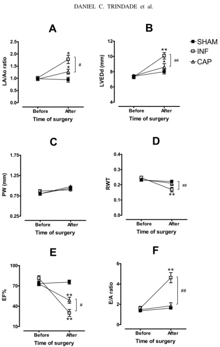
Heart rate was constantly monitored during echocardio-graphy and was not statistically different between all groups, ranging from 250 to 300 beats per minute under anesthesia in both periods of observation. The groups have shown similar echocardiographic values before surgery (Figure 1). However, the INF presented clear sings of severe heart failure 21 days after myocardial infarction. The LA/Ao ratio, LVEDd and E/A ratio (re-strictive mitral flow) were increased, and the RWT (rel-ative wall thickness) and EF% (ejection fraction) were decreased compared with the same group before surgery and SHAM 21 after surgery. No major changes were detected in other ECO parameters.
Despite of significant increase in LA/Ao ratio (Fig-ure 1A) and LVDd (Fig(Fig-ure 1B) showed by CAP, the PW (Figure 1C) and RWT (Figure 1D) did not change 21 days after infarction. Actually, the captopril-treated group exhibited an attenuation of the left atrial and ven-tricular dilatation compared with INF after infarction.
Regarding systolic function, the ejection fraction of the captopril-treated rats decreased significantly after infarction, but was still higher than infarcted rats with-out captopril-treatment (Figure 1E).
The data obtained from Doppler analysis was the most outstanding result found in CAP. This group showed a normal E/A velocity ratio (E/A ratio<3.0) compared with INF, which exhibited a significantly increase in this parameter 21 days after surgery (Figure 1F).
Post-mortemstudy
The hearts from the all groups were carefully examined
post mortemand none were found to present either
TABLE I
Electrocardiographic parameters recorded from SHAM, INF and CAP just before and 21 days after surgery. Data are means±SEM. n = number of rats, I-QRS = QRS amplitude index, ÂQRS = frontal QRS axis.
*P<0.05and **P<0.01vs. before surgery.#P<0.05vs. INF after surgery.
GROUP TIME P A R A M E T E R S
Heart rate P>0.1 mV QRS-index ÂQRS Q-wave in Q-amplitude (L1)
(BPM) (n) (mV) (degrees) L1 (n) (mV)
SHAM before 277±8.49 0 0.99±0.03 69.82±3.47 0 0
(n = 10) after 261±6.08 0 1.17±0.05 51.31±21.61 3 0.02±0.01
INF before 287±8.66 0 1.08±0.1 69.90±4.40 0 0
(n = 6) after 293±11.6 3∗ 0.57±0.06∗∗ 153.3±6.01∗∗ 6∗∗ 0.19±0.03∗∗
CAP before 258±10.3 0 1.11±0.06 67.75±2.10 0 0
(n = 9) after 288±8.56 0 0.70±0.11∗∗ 124.5±7.89∗∗# 9∗∗ 0.11±0.03∗∗#
pleural effusion was observed in 100% of the infarcted non-treated rats (INF) during necropsy procedure. In contrast, the captopril-treated rats (CAP) exhibited a de-crease in both heart and lung index compared with INF and pleural effusion was absent 28 days after captopril treatment. No changes were observed in liver index.
Histopathology
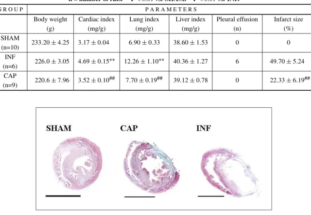
A gross examination showed a large transmural postin-farct scar in all hearts from INF 28 days after surgery. As showed in Table II and Figure 2, infarct size was decreased in captopril-treated group compared with INF. Figure 2 shows representative cross section of heart slices at papillary level obtained from SHAM, INF and CAP 28 days after surgery.
DISCUSSION
The local and/or circulating renin-angiotensin system (RAS) has long been considered responsible for car-diac remodeling and ventricular dysfunction. In the present report we evaluated the role of RAS on cardiac remodeling and function of rats with myocardial infarc-tion through the early angiotensin converting enzyme (ACE) inhibition. The functional data presented here strengthen the evidences that RAS plays an important role on healing process. The ECG and ECHO parame-ters of captopril-treated animals improved significantly when compared to non-treated animals, suggesting a di-rectly and/or indidi-rectly involvement of the RAS in the
chain of events that ultimately led to the heart failure. According with functional data, the histological exami-nation confirmed that the block of RAS was effective to decrease the scar tissue formation and left ventricular dilatation, the so-called “cardiac remodeling”. Whereas some studies as like Lapointe et al. (2002) supported these findings, several ones showed no marked effects on fibrosis after early treatment with ACE inhibitors com-pared with non-treated animals (Nelissen-Vrancken et al 1998, Shao et al 1999, Zornoff et al 2000). We hy-pothesized that controversial results could be explained by differences in selected ACE inhibitor, i.e., pharma-cological properties as tissue distribution (Keilani et al. 1995), which could be exploited to inhibit some local renin-angiotensin system while leaving others relatively intact, dose and route of drug administration and dif-ferences in infarct size induced by the surgery proce-dure. Although several works have assessed the effect of ACE inhibitors on infarcted rat hearts, these contro-versial data show that the real role of renin angiotensin system in heart function and cardiac remodeling in this model is far from clear. The congestive heart failure was also prevented as showed in post-mortem study:
A
Before After 0.0 0.5 1.0 1.5 2.0 2.5*
*
#Time of surgery
L A /A o r a ti o
B
Before After 4 6 8 10 12**
##*
SHAM INF CAPTime of surgery
L V E D d ( m m )
C
Before After 0.25 0.75 1.25 1.75Time of surgery
P W ( m m )
D
Before After 0.0 0.1 0.2 0.3 0.4**
##Time of surgery
R W T
E
Before After 10 40 70 100**
#**
Time of surgery
EF %
F
Before After 0 2 4 6 ##**
Time of surgery
E /A r a ti o
Fig. 1 – Echocardiographic parameters recorded from SHAM, INF and CAP just before and 21 days after surgery. Left atrium-to-aorta diameter (LA/Ao) ratio (A), left ventricular end-diastolic diameter (LVEDd, B), posterior wall thickness in diastole (PW, C), relative wall thickness (RWT, D), ejection fraction (EF%, E) and E/A velocity ratio (E/A ratio, F). Data are means±SEM. *P<0.05 and **P<0.01 vs. before and#P<0.05 and ##P<0.01 CAP vs. INF.
infarcted rats as described herein and in the other study (Santos and Masuda 1991), was not observed in 50% of the treated rats. In a previous study, using the same model of myocardial infarction in 84 rats, the ÂQRS was a sensible ECG parameter altered in the infarct group
TABLE II
Post-mortem study performed in SHAM, INF and CAP 28 days after surgery. Data are means±SEM. n = number of rats.∗∗P<0.01vs. SHAM.##P<0.01vs. INF.
G R O U P P A R A M E T E R S
Body weight Cardiac index Lung index Liver index Pleural effusion Infarct size
(g) (mg/g) (mg/g) (mg/g) (n) (%)
SHAM
233.20±4.25 3.17±0.04 6.90±0.33 38.60±1.53 0 0
(n=10) INF
226.0±3.05 4.69±0.15∗∗ 12.26±1.10∗∗ 40.36±1.27 6 49.70±5.24
(n=6) CAP
220.6±7.96 3.52±0.10## 7.70±0.19## 39.12±0.78 0 22.33±6.19##
(n=9)
Fig. 2 – Representative cross section of heart slices at papillary level obtained from SHAM, CAP and INF 28 days after surgery. Note that captopril-treated rat showed a smaller scar tissue and left ventricular dilatation than non-treated rat. Bar = 0.5 cm. Tricromo gomori staining.
in L1 was another sensible ECG parameter. There was a correlation between Q-wave measured one day post-surgery and infarct size suggesting that ECG recorded at this time could estimate infarct size. For this reason, we excluded from the present study two animals (one from INF and one from CAP) which showed small (or not showed) Q-wave in L1 one day after infarction. We thought to minimize or abolish the most limitation of in-farct model induced by coronary ligation, i.e., the large range of infarct size. Therefore, the fact those captopril-treated rats have presented smaller infarct size than non-treated rats could be attributed to effect of captopril and not to heterogeneity from the model. Regarding the main electrocardiographic signs of the large myocardial in-farction in rats, the Q-wave in L1 and rightward devia-tion of the frontal QRS-axis (Santos and Masuda 1991, Olivares et al. 2004), the treatment with captopril
atten-uated both the amplitude of Q-wave in L1 and ÂQRS, even with the presence of Q-wave in 100% of the treated rats. Whether these findings are explained by the small infarct size presented by this group, future studies need to be done. Anyway, the ECG was considered sensi-ble to detect the real contribution of the RAS in de-velopment of the heart failure induced by myocardial infarction in rats.
pre-sented here suggest that RAS was more relevant in a chain of events that led to cardiac remodeling and conse-quently to diastolic dysfunction than affect directly the systolic performance. This late may result from diverse other mechanism as the loss of cardiac tissue and the increase of the neurohumoral drive involved in the car-diac dysfunction (Francis 1985).
These data suggest that the renin-angiotensin sys-tem induces morphologic and functional changes in post-infarcted rat hearts and which can be assessed by non-invasive exams.
ACKNOWLEDGMENTS
The authors are grateful to Prof. Sylvia E.C. Cabezas by the essential technical support and animal care. This study was partially supported by Conselho Nacional de Desenvolvimento Científico e Tecnológico (CNPq).
RESUMO
Nós investigamos as alterações funcionais e morfológicas em corações de ratos infartados, sob o paradigma de inibição da enzima conversora de angiotensina. O infarto do miocárdio foi produzido pela ligadura da artéria coronária esquerda e um grupo falso-operado serviu de controle para o experimento. Os ratos infartados receberam água normal com (grupo CAP) ou sem (grupo INF) captopril. A avaliação funcional foi feita através de eletro (ECG) e ecocardiografia (ECO) momentos antes e 21 dias depois da cirurgia. O ECG dos grupos INF e CAP foram similares e compatíveis com infarto cicatrizado após a cirurgia. As principais diferenças entre os grupos INF e CAP foram: a prevenção do aumento da onda P e a aten-uação tanto do desvio do eixo de despolarização ventricular como da amplitude da onda Q no CAP comparado com o INF. O ECO revelou que o tratamento com captopril foi mais efe-tivo em melhorar o enchimento diastólico do que aumentar a função sistólica. A dilatação e a falência cardíaca congestiva foram observadas apenas no INF. Ambos os grupos infartados exibiram um tecido cicatricial no ventrículo esquerdo, mas no INF esta se mostrou maior do que no CAP (49.7±5.24 vs.
22.33±6.19 respectivamente). Estes dados sugerem que o
sistema renina angiotensina produz alterações morfológicas e funcionais em corações de ratos infartados e que estas podem ser detectadas por exames não invasivos.
Palavras-chave: infarto do miocárdio, eletrocardiograma,
ecocardiograma, sistema renina-angiotensina, captopril.
REFERENCES
FRANCIS GS. 1985. Neurohumoral mechanism evolved in
congestive heart failure. Am J Cardiol 55: 15–21. GODYRJ. 1986. Conceptual and therapeutic approaches to
inhibition of the renin-angiotensin system in chronic heart failure. J Cardiovasc Pharmacol 8: 58–65.
GOTTLIEBSS, DICKSTEINK, FLECKE, KOSTISJ, LEVINE
TB, LEJEMTELT ANDDEKCOK M. 1993. Hemody-namic and neurohormonal effects of the angiotensin II antagonist losartan in patients with congestive heart fail-ure. Circulation 88: 1602–1609.
JOHNSTNPANDOLSONJB. 1954. Experimental
myocar-dial infarction: a method of coronary occlusion in small animals. Ann Surg 140: 675–682.
KALKMANEAJ, VANHARENP, SAXENAPRANDSCHOE
-MAKER, RG. 1999. Early captopril prevents myocardial infarction-induced hypertrophy but not angiogenesis. Eur J Pharmacol 369: 339–348.
KEILANIT, SCHLUETERWANDBATLED. 1995. Selected
aspects of ACE inhibitor therapy for patients with renal disease: impact on proteinuria, lipids and potassium. J Clin Pharmacol 35: 87–97.
KOBERLARS ET AL. 1995. A clinical trial of the angiotensin-converting-enzyme inhibitor trandolapril in patients with left ventricular dysfunction after myocardial infarction. N Engl J Med 333: 1670–1676.
LAPOINTEN, BLAIS-JRC, ADAMA, PARKER T, SIROIS
MG, GOSSELIN H, CLEMENT R AND ROULEAU JL.
2002. Comparison of the effects of an angiotensin con-verting enzyme inhibitor and a vasopeptidase inhibitor after myocardial infarction in rat. Am Coll cardiol 39: 1692–1698.
LITWIN SE, KATZSE, MORGANJP ANDDOUGLASPS. 1996. Long-term captopril treatment improves diastolic filling more than systolic performance in rats with large myocardial infarction. J Am Coll Cardiol 28: 773–781. NELISSEN-VRANCKENHJMG, KUIZINGAMC, DAEMEN
MJAPANDSMITSJFM. 1998. Early Captopril inibis
DNA synthesis in endothelial cells and normalization of maximal coronary flow in infarcted rat hearts. Cardiovasc Res 40: 156–164.
OLIVARES EL ET AL. 2004. Bone marrow stromal cells improve cardiac performance in healed infarcted rat hearts. Am J Physiol Heart Cir Physiol 287: 464–470. PFEFFER JM, PFEFFER MA AND BRAUNWALD E. 1987.
long-term captopril therapy in rats myocardial infarction and heart failure. Circulation 75: 1149–1155.
PFEFFERMA, PFEFFERJM, STEUNBERGCANDFINN P.
1985. Survival after an experimental myocardial infarc-tion: beneficial effects of long-term therapy with capto-pril. Circulation 72: 406–412.
PRUNIER F, GAERTNER R, LOUEDEC L, MICHEL JB, MERCADIER JJ AND ESCOUBET B. 2002. Doppler echocardiographic estimation of left ventricular end-diastolic pressure after MI in rats. Am J Physiol Heart Circ Physiol 283: 346–352.
SANTOSPEBANDMASUDAMO. 1991. The
electrocardio-gram of rats with an old extensive myocardial infarction. Brazilian J Med Biol Res 24: 1173–1177.
SCHOEMAKER RG, DEBETS JJM, STRUYKER-BOUDIER
HAJ ANDSMITSJFM. 1991. Delayed but not imme-diate captopril therapy improves cardiac function in con-scious rats, following myocardial infarction. J Mol Cell Cardiol 23: 187–197.
SHAO Q, REN B, ZARAIN-HERZBERG A, GANGULYPK ANDDHALLANS. 1999. captopril treatment improves
the sarcoplasmatic reticular Ca2+ transport in heart fail-ure due to myocardial infarction. J Mol Cell Cardiol 31: 1663–1672.
SIGURDSSONA, HELDPANDSWEDBERGK. 1993. Short-and long-term neurohumoral activation following acute myocardial infarction. Am Heart J 126: 1068–1076. SPADAROJ, FISHBEINMC, HAREC, PFEFFER MAAND
MAROKO PR. 1980. Characterization of myocardial
infarcts in the rat. Arch Pathol Lab Med 104: 179–183. ZORNOFF LAM, MATSUBARA BB, MATSUBARA LS,