Necrotic Hepatocyte Death through IRGM1-Mediated
Lysosomal Membrane Disruption
Chih-Peng Chang1,3, Ming-Chen Yang2, Huan-Yao Lei1,2,3*
1Department of Microbiology and Immunology, College of Medicine, National Cheng Kung University, Tainan, Taiwan,2Institute of Basic Medical Sciences, College of Medicine, National Cheng Kung University, Tainan, Taiwan,3Infectious Disease and Signaling Research Center, College of Medicine, National Cheng Kung University, Tainan, Taiwan
Abstract
Interferon-gamma (IFN-c), a potent Th1 cytokine with multiple biological functions, can induce autophagy to enhance the clearance of the invading microorganism or cause cell death. We have reported that Concanavalin A (Con A) can cause autophagic cell death in hepatocytes and induce both T cell-dependent and -independent acute hepatitis in immunocompetent and immunodeficient mice, respectively. Although IFN-cis known to enhance liver injury in Con A-induced hepatitis, its role in autophagy-related hepatocyte death is not clear. In this study we report that IFN-ccan enhance Con A-induced autophagic flux and cell death in hepatoma cell lines. A necrotic cell death with increased lysosomal membrane permeabilization (LMP) is observed in Con A-treated hepatoma cells in the presence of IFN-c. Cathepsin B and L were released from lysosomes to cause cell death. Furthermore, IFN-c induces immunity related GTPase family M member 1(IRGM1) translocation to lysosomes and prolongs its activity in Con A-treated hepatoma cells. Knockdown of IRGM1 inhibits the IFN-c/Con A-induced LMP change and cell death. Furthermore, IFN-c2/2mice are
resistant to Con A-induced autophagy-associated necrotic hepatocyte death. We conclude that IFN-cenhances Con A-induced autophagic flux and causes an IRGM1-dependent lysosome-mediated necrotic cell death in hepatocytes.
Citation:Chang C-P, Yang M-C, Lei H-Y (2011) Concanavalin A/IFN-Gamma Triggers Autophagy-Related Necrotic Hepatocyte Death through IRGM1-Mediated Lysosomal Membrane Disruption. PLoS ONE 6(12): e28323. doi:10.1371/journal.pone.0028323
Editor:Rafael Linden, Universidade Federal do Rio de Janeiro, Brazil
ReceivedJune 6, 2011;AcceptedNovember 5, 2011;PublishedDecember 5, 2011
Copyright:ß2011 Chang et al. This is an open-access article distributed under the terms of the Creative Commons Attribution License, which permits unrestricted use, distribution, and reproduction in any medium, provided the original author and source are credited.
Funding:This work was supported by grant NSC-98-2320-B-006-031-MY2 from the National Science Council, Taiwan (http://web1.nsc.gov.tw/). The funders had no role in study design, data collection and analysis, decision to publish, or preparation of the manuscript.
Competing Interests:The authors have declared that no competing interests exist. * E-mail: hylei@mail.ncku.edu.tw
Introduction
Programmed cell death has been classified as type I apoptosis and type II autophagy-associated cell death [1]. Autophagy is an evolutionarily conserved lysosomal pathway to generate energy by digesting cytoplasmic proteins and organelles. Although autophagy allows cells to survive from starvation or stress, an extended or massive autophagy process turns to kill cells [2,3,4]. Necrosis is usually considered as a non-programmed cell death without features of apoptosis and autophagy [5,6]. However, necrosis can also be regulated and controlled by cellular proteins, such as lysosomal protease cathepsins [7]. Programmed cell death can switch to necrosis by inhibition of specific proteins of apoptosis or autophagy [6]. Cross-regulation between these different types of cell death remains unclear. Lysosomes are intracellular organelles which contain various hydrolytic enzymes to control turnover of cellular macromolecules and eliminate intracellular pathogens [8]. However, they can also be responsible for mediating programmed cell death or necrosis [9]. Lysosome membrane permeablization (LMP) changes will initiate a lysosome-mediated cell death. When the lysosome membrane is damaged through LMP, cathepsins or other hydrolytic enzymes will be released from lysosomes to the cytosol and cause cell death [10]. Autophagy is a lysosome-dependent pathway and can cause a type II programmed cell death. Accumulated lysosomes or autophagolyso-somes are increased in autophagy-associated cell death. However, the role of LMP on autophagy-associated cell death is poorly understood.
Interferon-gamma (IFN-c), a major Th1 cytokine, is an important mediator for immune-mediated hepatitis [11,12]. A p53-dependent cell cycle arrest and apoptosis caused by IFN-cin hepatocytes is suggested [13]. However, IFN-ccan also regulate the autophagic response to eliminate intracellular pathogen infection in macrophages or cause cell death of epithelial cells [14,15]. The immunity related GTPase family proteins (IRG), particularly IRGM1, have been considered as key mediators for IFN-c-induced autophagy [16]. We have reported that Concanavalin A (Con A) can induce both T cell-dependent and T cell-independent acute hepatitis [17]. A Bcl-2/adenovirus E1B 19 kDa-interacting protein 3 (BNIP3)-related mitochondria autophagy was involved in the hepatocyte death [18]. In this study, we further report that IFN-c can enhance Con A-induced autophagic flux and hepatocyte death. The enhanced cell death by IFN-c/Con A represents a lysosomal proteases, cathepsin B- and cathepsin L-dependent necrosis. IRGM1 participates in this IFN-c-stimulated LMP-associated necrosis in Con A-treated hepatocytes.
Results
IFN-cenhances autophagic flux and causes necrotic type cell death in Con A-treated hepatocytes
autophagic pathway in hepatocytes [18]. The double membrane autophagosomes can be found in Con A-treated ML-14acells but
reduced in the presence of autophagy inhibitor, 3-methylade-nine, from electron microscopy (Figure S1). This pathway is further extended to autophagic flux and is enhanced by IFN-c. The BNIP3 was induced by Con A or IFN-c alone, and enhanced while co-treatment in ML-14a cells. However, only
slightly increase of BNIP3 expression was detected at 12 hours post treatment of IFN-c/Con A in HepG2 cells, which might be due to the high level of basal BNIP3 expression in HepG2 cells. The LC3-II conversions were both slightly increased in IFN-c/ Con A-treated ML-14aor HepG2 cells (Figure 1A). We next used
GFP-LC3 processing to follow autophagic flux, and an increase of free GFP from GFP-LC3 processing was found in IFN-c/Con A-treated ML-14aor HepG2 cells (Figure 1B). This suggests that
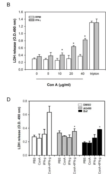
IFN-cenhances the autophagic flux on Con A-treated hepatoma cells. We next determined the effect of IFN-con Con A-induced autophagic cell death. Con A dose-dependently induced HepG2 and ML-14a cell death, the addition of IFN-c significantly
enhanced the Con A-induced cell death, especially at the low dose of 5–10mg/ml of Con A (Figure 1C). The increase of cell death by IFN-con Con A-induced cell death was diminished by 3-methyadenine (autophagic inhibitor) or LC3 siRNA, but not by Z-Vad-fmk (apoptosis inhibitor), suggesting that this enhance-ment in cell death is not due to apoptosis (Figure 1D and 1E). To further confirm this phenomenon, we monitored the fragmented DNA from apoptotic cells by propidium iodide (PI) staining. The apoptotic inducer, puromycin, induced fragmented DNA with increase of sub G1 population in ML-14aells. Instead, no such
increased-sub G1 population was detected in Con A-, IFN-c- or IFN-c/Con A- treated ML-14a cells, suggesting that these cells
were not died by apoptosis (Figure S2). These data indicate that IFN-c enhances Con A-induced autophagic cell death in hepatocytes. Con A alone-induced cell death can be easily detected by propidium iodide or eosin Y exclusion assay (data not shown), but not by LDH release, a feature of necrotic cell death with detection in the culture supernatant. However, early fusion of autophagosomes and lysosomes by IFN-cin Con
A-Figure 1. IFN-cenhances autophagic flux and cell death in Con A -treated hepatoma cell lines. A. Con A/IFN-cinduces autophagy in hepatoma cells. ML-14aand HepG2 cells were treated with PBS, Con A, IFN-cor IFN-c/Con A and then collected cell lysates at indicated time. The expressions of phospho-STAT1, STAT1, BNIP3, LC3-I and LC3-II were determined by Western blot.B. Generation of free GFP from pGFP-LC3-expressed hepatoma cells by ConA/IFN-ctreatment. pGFP-LC3-transfected ML-14aor HepG2 cells were treated with Con A and IFN-cfor 24 hours. The cell lysates were extracted to determine expression of GFP-LC3 and free GFP by Western blot.C, IFN-cenhances Con A-induced cell death in hepatoma cells. HepG2 and ML-14acells were treated with various concentrations of Con A in the presence or absence of IFN-cfor 24 hours. Cell death was determined by propidium iodide staining and analyzed by flow cytometry.D and E. IFN-cenhances autophagy-dependent cell death in Con A-treated hepatoma cells. ML-14acells were pre-incubated with 3-methyladenine or Z-Vad-fmk (D), or transfected with control or LC3 siRNA (E). Then these cells were co-treated with Con A and IFN-cfor 24 hours. The knock down effect of LC3 was monitored by Western blot. The cell death of all treatments was determined by propidium iodide staining. All results are representative of two to three experiments. * shows statistically significant differences relative to PBS group. (p,0.05).
treated ML-14acells suggested that lysosome-mediated necrosis
might be induced (Figure 2A). As results showed in Fig. 2B, we can detect the LDH release in Con A and IFN-c treated hepatoma cells. Another marker of necrosis, the release of nuclear protein, HMGB1, was also found to be released from Con A -treated hepatoma cells, and IFN-ccould enhanced this release (Figure 2C). Furthermore, IFN-ccan increase Annexin V/PI double positive cell population, which suggests as necrotic cells, of Con A-treated ML-14a cells, and pretreatment of
3-methyladenin will reduce this enhancement (Figure S3). AG490 (Jak2 inhibitor), or bafilomycin A1 (autophagic flux inhibitor) both inhibited the IFN-c-triggered LDH release in Con A-treated hepatoma cells (Figure 2D). These results suggest that IFN-ccan enhance Con A-induced autophagic flux and cause a necrotic type cell death.
IFN-ccauses a necrotic cell death with increase of lysosomal membrane permeabilization (LMP) in Con A-treated hepatocyte
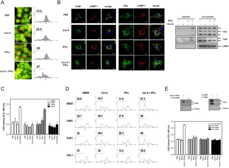
We next determined how IFN-c enhances Con A-induced necrotic type cell death. The LMP was determined by acridine orange (AO), a lysosomotropic fluorochrome used to measure cellular increase of the LMP. AO-stained normal cells showed red punctate on microscopic observation and intact red fluorescence on flow cytometry. The Con A or IFN-ctreatment alone have the same pattern with that of normal cells. However, IFN-cplus Con A caused a dramatic loss of the punctate staining and red fluorescence on AO-stained hepatoma cells compared with the Con A or IFN-ctreatment groups (Figure 3A). We also found that the lysosomal proteases, cathepsin B and L, were released into the cytoplasm. They are normally localized in LAMP1-positive
Figure 2. IFN-ccauses necrotic type cell death in Con A-treated hepatoma cell lines. A. The colocalization of punctate LC3 and LAMP-1 in ML-14acells. ML-14acells were treated with PBS, Con A, IFN-cor IFN-c/Con A for 6 hours. All cells were fixed and stained with anti-LC3 or LAMP1 antibodies. The colocalization of punctate LC3 and LAMP 1 was assayed using confocal microscopy. Cell nucleus was stained with Hoechst (blue).
lysosomes of ML-14acells. After treatment with IFN-cand Con A,
increases of LAMP1-negative cathepsin B or L were found in the cytosol, indicating that cathepsin B and L were released following the IFN-cplus Con A-induced increment of LMP (Figure 3B). We further determined whether cathepsin can mediate the IFN-c-triggered necrosis of Con A-treated hepatoma cells. Using several specific inhibitors to block the IFN-cplus Con A-induced LDH release and LMP increment, we found that inhibitors of cathepsin B (Z-Phe-Phe-CH2F) and L (CA-074Me), but not an inhibitor of
cathepsin D (pepstatin A), can inhibit the IFN-c-induced LDH release and increase of LMP in Con A-treated hepatoma cells (Figure 3C and 3D). To further verify the role of cathepsin B, L and autophagy in the IFN-c-triggered necrotic cell death, the siRNA of cathepsin B, L and LC3 were used. The IFN-c/Con A induced LDH release was inhibited by cathepsin B, L and LC3 siRNA treatment (Figure 3E). These results suggest that IFN-c-induced LMP causes cathepsin B and L-dependent necrotic cell type death in Con A-treated hepatoma cells.
IRGM1 mediates IFN-c-enhanced LMP-associated lysosomal cell death in Con A-treated hepatocytes
The immunity-related GTPase, IRGM1, is essential for IFN-c-dependent anti-microbial function and autophagy induction [16]. Since IFN-ccauses an autophagy-associated necrotic cell death in Con A-treated hepatoma cells, we further studied the role of IRGM1 on this phenomenon. To our surprise, Con A, not IFN-c, enhanced a temporary increase of IRGM1 at 3 hours post treatment in ML14acells. However, the increased level of IRGM1
could be sustained to 12 hours in IFN-c/Con A-treated cells, suggesting the IRGM1 expression were enhanced by IFN-c (Figure 4A). With confocal microscopy observation, we found that IRGM1 was co-localized with mitochondria-related protein, Tom20, in IFN-cor IFN-c/Con A-treated ML-14acells, but not
in PBS or Con A-treated cells. In addition to mitochondria translocation, IRGM1 was found to co-localize with LAMP1 only in IFN-c/Con A-treated cells, but not in IFN-c-treated cells
Figure 3. IFN-cinduces a necrotic type cell death with increase of LMP in Con A-treated hepatoma cells. A. IFN-ccauses increase of LMP in Con A-treated hepatocytes. ML-14acells were stained with acridine orange and treated with Con A and IFN-c. The red fluoresce of acridine orange was observed in florescence microscope or analyzed by flow cytometry.B. IFN-cinduces cathepsin-B/L release in Con A-treated hepatoma cells. After treatment of Con A and IFN-cfor 12 hours, ML-14acells were fixed and stained with anti-cathepsin B, anti-cathepsin-L or anti-LAMP1 antibody or extracted the cytosolic proteins by digitonin as described in Materials and Methods. The distribution of cathepsinB/L was analyzed using a confocal fluorescence microscope or immunoblotting. Cell nucleus was stained with Hoechst (blue).C–E. Cathepsin B/L mediates IFN-c-induced LDH release and increase of LMP in Con A-treated hepatoma cells. ML-14acells were pretreated with DMSO or different cathepsin inhibitors (C,D) or transfected with siRNA of control, LC3, cathepsin B or cathepsin L (E), and then treated with Con A and IFN-cfor another 24 hours. The release of LDH and cellular LMP in all treatments were both determined as described above. The knock down effect of cathepsin B and L were monitored by Western blot. All results are representative of two to three experiments.* shows statistically significant differences relative to DMSO group. (p,0.05).
(Figure 4B). These data suggest that IFN-c/Con A enhance not only the IRGM1 expression, but also translocation of IRGM1 to lysosome. Furthermore, the IRGM1 expression was knocked down by siRNA to further investigate its role in IFN-c/Con A-induced LMP-associated necrosis (Figure 4C).We found that the IFN-c/Con A-caused increase of LMP in hepatoma cells was significantly inhibited by IRGM1 siRNA, suggesting that IRGM1 is involved in IFN-c/Con A-caused LMP change (Figure 4D). Because the activity of cathepsin B and L is crucial for IFN-c/Con A-caused LMP change and necrotic cell death as shown above, and might be regulated by IRGM1. Indeed, the cytosolic cathepsin B/L activity as well as the LDH release were also abolished by IRGM1 siRNA in IFN-c/Con A-treated hepatoma cells (Figure 4E and 4F). These results suggest that IRGM1 regulates the LMP change that releases the cathepsin B/L to
causes lysosomal cell death in IFN-c/Con A-treated hepatoma cells.
IFN-c2/2mice are resistant to Con A-induced autophagy-associated necrotic type cell death
To further evaluate this phenomenonin vivo, we compared the Con A-induced hepatitis in IFN-c2/2mice with that in wild type mice. In wild type mice, an intravenous injection of 40 mg/kg of Con A increased the serum ALT, and necrosis in the liver was observed by histological staining at 6 hours post injection, and the mice finally die within 10 h. In contrast, the IFN-c2/2mice were resistant to Con A-induced hepatitis, with less mortality, no increase of serum ALT, and no necrosis in the liver (Figure 5A– 5C). We also found that the LC3-II conversion was induced in Con A-treated wild type mice, but not in IFN-c2/2 mice Figure 4. IRGM1 mediates IFN-c-enhanced LMP-associated necrosis-like cell death in Con A-treated hepatoma cells. A. IRGM1 expression in IFN-c/Con A-treated hepatoma cells. ML-14acells were treated with PBS, Con A, IFN-cor IFN-c/Con A and collected cell lysates at indicated time. The expression of IRGM1 was determined by Western blot.B. IRGM1 translocates to mitochondria and lysosomes. After treatment of Con A or IFN-cfor 12 hours, ML-14acells were fixed and stained with anti-IRGM1, Tom20, and LAMP-1 antibodies. The co-localization of IRGM1 and Tom20 or LAMP-1 was analyzed using a confocal fluorescence microscope. Cell nucleus was stained with Hoechst (blue).C. Knock down of IRGM1 by siRNA. ML-14acells were transduced with siRNA of control or IRGM1 as described in Materials and Methods. The expression of IRGM1 was determined by Western blot.D. Knock down of IRGM1 attenuates IFN-c-enhanced LMP in Con A-treated hepatoma cells. ML-14acells were transduced with siRNA of control or IRGM1 and treated with Con A and IFN-c. The cells were then stained with acridine orange to monitor the LMP.E. IRGM1 mediates IFN-c -induced increase of cytosolic cathepsin B/L activities in Con A-treated hepatoma cells. After knock down of IRGM1 by siRNA, ML-14acells were treated with Con A and IFN-cand the cytosolic fraction was collected. The activities of cytosolic cathepsin B/L were then determined.F. IRGM1 mediates
IFN-c-enhanced LDH release from Con A-treated hepatoma cells. ML-14acells were pretreated with siRNAs or bafilomycine A1 and then incubated with Con A and IFN-c. The activity of LDH in cell culture supernatants was determined. All results are representative of two to three experiments.* shows statistically significant differences relative to control group. (p,0.05).
(Figure 5D). The serum LDH and HMGB1 were also significantly inhibited in IFN-c2/2 mice comparing with wild type mice at 6 hours post Con A injection (Figure 5E). This suggests that IFN-c can enhance the Con A-induced necrotic type cell deathin vivo.
Discussion
Autophagy is a cell survival system to protect cells from damage by protein aggregates or injured organelles, but it can also lead to cell death with characteristics of apoptosis or necrosis under different conditions. We have previously reported that autophagic induction resulted in hepatocyte death with Con A-induced acute hepatitis. In this study we further showed that IFN-c, an important mediator for immune-mediated liver injury, enhances the autophagic flux and triggers a lysosomal cell death in Con A-treated hepatocytes. This autophagy-dependent necrotic cell death is mediated by IFN-c-related IRGM1-induced LMP to release lysosomal proteases, cathepsin B/L.
IFN-c can induce hepatocytes to undergo cell arrest and apoptosis after in vitro culture for 48 to 72 hours, where an IRF-1-dependent caspase-3 and p53 expression mediates this IFN-c-caused hepatocyte apoptosis [13]. IFN-chas also been shown to
enhance apoptosis with other cytokines or death-related receptors [19,20,21]. In our study of the hepatoma ML-14a cell line, we
found neither any caspase-3 activation, nor any characteristics of apoptosis within 24 hours post IFN-ctreatment. Tagawa Y et al. have reported that IFN-c would enhance Con A-induced hepatocyte apoptosis on Con A-induced acute hepatitis in mice [22]. In our earlier study, however, we reported that autophagy occurred before the Con A-induced apoptosis in mice [17]. The dose used can explain this discrepancy, as a higher dose (40 mg/ kg) of Con A is required to induce autophagy in mice. The LC3-II conversion was found in wild type mice, but not in IFN-c2/2 mice (Figure 5D). The autophagy-related cell death has the characteristics of LDH and HMGB1 release into the circulation. Necrosis was found in the liver, with no immune cell infiltrations, at 6 h post injection (Figure 5B). Therefore, the effect of IFN-con Con A-treated hepatocytes must be direct, and is not mediated by the enhancement of immune cells. Our in vitro study demonstrates that IFN-ccan enhance the Con A-stimulated autophagic flux to cause lysosomal cell death.
A massive breakdown of lysosomes could cause cell death by necrosis, which is related to increase of cytosolic acidification [10]. IFN-ccan regulate lysosome activities by the alteration of vacuolar Figure 5. IFN-c2/2mice are resistant to Con A-induced autophagy-associated necrotic type cell death. A. Serum level of ALT on Con A-treated mice. Wide type or IFN-c2/2mice received intravenous injection of Con A and the serum were collected at indicated time. The serum level of ALT was determined as described in Materials and Methods. (n = 5)B. Histological staining of mice liver tissue. The liver tissues of wild type or IFN-c2/2 mice were collected to stain with haematoxylin and eosin at indicated time post Con A injection. The arrow head indicates necrotic hepatocytes.C.
IFN-c2/2mice are resistant to Con A-induced mortality. Wild type or IFN-c2/2mice were injected intravenously with Con A and the survival was monitored. (n = 7)D. IFN-c2/2mice show reduction of Con A-induced autophagy. The liver extracts of wild type or IFN-c2/2mice were collected to detect the LC3-II conversion by immune blotting at 3 hours post Con A injection.E. Serum level of LDH and HMGB1 on Con A-treated mice. Sera of Con A-treated wild type or IFN-c2/2mice were collected at indicated time. The level of LDH or HMGB1 was determined as described above. (n = 5) All results are representative of two to three experiments. * shows statistically significant differences relative to control group. (p,0.05).
pH or the activation of cathepsin expression to cause cell death [23,24]. Here we found no significant increase in hepatocytes’ acidification and protein level of cathepsin B/L upon IFN-c stimulation. Instead, small increase in cell acidification can be detected at 6 hour post Con A treatment, which might lead to activate cathepsins in lysosome (Figure S4.) However, this increase in acidification is not enhanced by IFN-c, suggesting that other factors are involved in IFN-c/Con A-caused necrosis. In this study we further demonstrate that IFN-cenhances the autophagic flux induced by Con A to cause LMP-associated necrotic cell death in hepatocytes. In addition, the interferon-induced GTPase, IRGM1, plays a crucial role to mediate this autophagy-associated necrosis. IRGM1 is reported to recruit to pathogen-containing phagosomes and autophagosomes, and to facilitate the acidification of theses vacuoles and their fusion with lysosomes [16,25]. However, we did not observe IRGM1 in autophagosomes in IFN-c/Con A-treated hepatocytes (data not shown). Instead, IFN-c/Con A treatment enhances or sustains the IRGM1 expression and causes its redistribution to mitochondria and lysosomes, resulted in lysosome membrane disruption. The redistribution of IRGM1 to lysosomes has also been reported by Zhao et al. in IFN-c-induced mouse embryonic fibroblasts [26]. How IRGM1 regulates lysosomal membrane permeabilization remains further investigation. Hu-man IRGM1 recently been reported to regulate IFN-c-induced autophagy by binding mitochondrial lipid to control mitochon-drial fission [27]. We have reported that accumulation of Con A on mitochondria after endocytosis causes mitochondrial damage and autophagy of damaged mitochondria [18]. In this study, we further showed that co-localized of damaged mitochondria with IRGM1 in IFN-c-treated murine hepatocytes, and its transloca-tion to lysosome. Both Con A and IRGM1 binding to mitochondria might increase mitochondria membrane damage and enhance autophagolysosome process. Here we also found that Con A can up-regulate IRGM1 expression in hepatocytes (Figure 4A). Since BNIP3 is an important mediator to Con A-induced autophagy, the interaction of IRGM1 and BNIP3 in regulating autophagy process is worth to further study. The lysosome-mediated cell death induced by IFN-c/Con A in hepatocytes causes HMGB1 release. HMGB1 can act as an inflammatory mediator to aggravate to inflammation-related disease [28]. The autophagy-regulated HMGB1 release in tumor cells, either in cell culture or in the serum of Con A-injected wild type mice, is consistent with the observation of Thurhurn J et al. [29].
Cell death with apoptotic or autophagic features can lead to different immune responses [30]. Clearance of apoptotic cells is considered to induce an anti-inflammatory reaction by produc-tion of transforming growth factor-b(TGF-b), prostaglandin E2, and platelet activating factor (PAF) [31]. Apoptotic cell death induced by most chemotherapeutic drugs and radiation in cancer therapy creates an immunosuppressive environment around the tumor site, which may promote further tumor growth. Cell death with autophagy stimulates a pro-inflammatory response that subsequently activates the immune system and facilitates efficient antigen cross-priming to CD8+
T cells [32,33]. We have reported that Con A with both autophagic induction and immuno-modulation can generate a CD8+
T cell-dependent anti-hepatoma effect in mice [34]. These findings therefore support the use of induction of autophagy as a new target for tumor therapy. The release of HMGB1 post IFN-c/Con A treatment by lysosomal cell death will provide a link to further anti-hepatoma CD8+
cytotoxic T cell activation. This is worthy of further investigation.
Materials and Methods
Reagents and antibodies
Con A, pepstatin A, 3-methyladenine (3-MA) and E64D were purchased from Sigma-Aldrich (St. Louis, MO, USA). The cell-permeable cathepsin L inhibitor, Z-Phe-Phe-CH2F, and cathepsin
B inhibitor, CA-074Me, were purchased from Calbiochem (La Jolla, California, USA). Control, cathepsin B and IRGM1 siRNA were purchased from Sigma-Aldrich. Microtubule-associated proteins light chain 3 (LC3) and cathepsin L siRNA were purchased from Santa Cruz (Santa Cruz, CA, USA). The antisera used were: anti-signal transducer and activator of transcription 1 (STAT1) and phospho-STAT1 (Tyr 701) from Cell Signaling (Beverly, MA), LC3 from MBL (Nagoya, Japan), anti-mitochondrial preprotein translocases of the outer membrane 20 (Tom20) and anti-green fluorescent protein (GFP) from Santa Cruz, anti-Bcl-2/adenovirus E1B 19 kDa-interacting protein 3 (BNIP3) from Sigma-Aldrich, anti-cathepsin B and anti-cathepsin L from R&D system (Minneapolis, MN, USA), anti-lysosomal-associated membrane protein 1 (LAMP 1) from ebioscience (San Diego, CA, USA), immunity-related GTPase (IRGM), anti-high mobility group box 1 protein (HMGB1) and anti-b-actin from Abcam (Cambridge, UK). The recombinant human and murine interferon gamma were purchased from Peprotech (Rocky Hill, NJ, USA).
Cell culture
The murine hepatoma cell line, ML-14acell, was adapted from
ML-1 cells in BALB/c mice as previously described [18]. HepG2 cells were obtained from the Cell Collection and Research Center (CCRC, Hsin-Chu, Taiwan). In inhibition experiments, the cells were pre-treated with several inhibitors, including pepstatin A (10mg/ml), E64D (10mg/ml), 3-MA (2 mM), Z-Vad-fmk (100mM), bafilomycin A1 (2 nM), AG490 (25mM), Z-Phe-Phe-CH2F (2 nM), and CA-074Me (50mM), 1 h before addition of
Con A (20mg/ml) or IFN-c (500 U/ml). Cell death was determined by propidium iodide exclusion staining and analyzed by flow cytometry. To detect the release of lactate dehydrogenase (LDH) from cells, the cell culture supernatants were collected for a LDH activity assay according to the guidelines of the detection assay kit (Roche Diagnostics, Mannheim, Germany). To monitor the status of lysosomal stability, ML-14a cells were stained with
1mg/ml acridine orange at 37uC for 30 minutes. The red acridine orange fluorescence was visualized by a fluorescent microscope or analyzed by flow cytometry. ML-14acells were planted on a
cover-slide and transfected with pGFP-LC3 or siRNAs using Lipofecta-mine 2000 (Invitrogen Corp., Carlsbad, CA). Cells were fixed and stained with primary antibody against LC3, IRGM1, Tom20, LAMP-1, cathepsin B or cathepsin L followed by secondary antibody conjugated with Alexa Fluor 488 or 594. The cellular distribution of proteins was observed under a confocal fluores-cence microscope (Olympus FV 1000, Japan).
Mice
injected intravenously with Con A at a dose of 40 mg/kg body weight, the serum being collected at various time points post injection. The serum alanine aminotransferase (ALT) was determined by using the Hitachi type 717 automatic analyzer (Hitachi, Tokyo).
Western blot analysis
Cell lysates were prepared by extracting proteins with lysis buffer (Cell Signaling, Beverly, MA). Proteins were separated by SDS-PAGE and transferred to PVDF membranes. The mem-branes were blocked and incubated with primary antibodies. After incubation with peroxidase-conjugated secondary antibodies, the blots were visualized by enhancing chemiluminescence reagents (PerkinElimer Life Sciences, Boston, MA).
Analysis of cytosolic cathepsin B/L activity
To measure the cytosolic cathepsin B/L activity, the cytosolic fraction was prepared as previously described [35]. In brief, the treated or non-treated cells were harvested and washed with phosphate buffered saline (PBS) twice. The digitonin-based extraction buffer (50mg/ml digitonin, 250 mM sucrose, 20 mM HEPES, 10 mM KCl, 1.5 mM MgCl2, 1 mM EDTA, 1 mM
EGTA, pH 7.5) was added to cells and put on ice for 30 minutes. The cytosolic fraction was then collected to measure cathepsin B/ L activity by InnoZymeTM Cathepsin L Fluorogenic Activity Kit (Calbiochem) or to detect cathepsin B/L protein expression by Western blotting as described above.
Supporting Information
Figure S1 Electron micrographs of Con A-treated ML-14a cells. ML-14acells were pretreated with 3-methyladenin
(3-MA) for 30 minutes and incubated with Con A for another 12 hours. A. The electron micrographs (610000) of PBS-, Con A-,
3-MA- and Con A/3-MA-treated ML-14acells are presented. B.
The selected-areas of electron micrographs (660000) from Con A-treated ML-14a cells are presented. M: mitochondria; AP:
autophagosome. (TIF)
Figure S2 Analysis of apoptotic cells with sub G1 population in IFN-c/Con A-treated ML-14acells.ML-14a
cells were treated with PBS, Con A, IFN-c, IFN-c/Con A or puromycin for 24 hours. The cells were fixed with ethanol and stained with PI to analyze the sub G1 population by flow cytometry.
(TIF)
Figure S3 Autophagy contributes to IFN-c/Con
A-induced necrotic cell death. ML-14a cells were pretreated
with DMSO or 3-methyladenin for 30 minutes and incubated with PBS, Con A, IFN-c, or IFN-c/Con A for another 24 hours. The cells were harvested and stained with Annexin V-FITC/PI. The cell population was analyzed by flow cytometry.
(TIF)
Figure S4 Acidification and total cathepsin B/L protein expression in IFN-c/Con A-treated hepatocytes. A.
ML-14acells were pretreated with DMSO or bafilomycin A1 (Baf) and
incubated with PBS, Con A, IFN-c, or IFN-c/Con A for another 6 hours. These cells were stained with AO for 30 minutes and analyzed the acidification by flow cytometry.B. ML-14acells were
with PBS, Con A, IFN-c, or IFN-c/Con A for 12 hours and extracted the total cell lysates. The expression of cathepsin B and L were determined by Western blot.
(TIF)
Acknowledgments
We thank Dr. Yu-Jung Cheng and Yu-Ping Lin for their help in experiments.
Author Contributions
Conceived and designed the experiments: C-PC H-YL. Performed the experiments: C-PC M-CY. Analyzed the data: C-PC. Contributed reagents/materials/analysis tools: H-YL. Wrote the paper: C-PC.
References
1. Hotchkiss RS, Strasser A, McDunn JE, Swanson PE (2009) Cell death. N Engl J Med 361: 1570–1583.
2. Wang CW, Klionsky DJ (2003) The molecular mechanism of autophagy. Mol Med 9: 65–76.
3. Yorimitsu T, Klionsky DJ (2005) Autophagy: molecular machinery for self-eating. Cell Death Differ 12(Suppl 2): 1542–1552.
4. Kondo Y, Kanzawa T, Sawaya R, Kondo S (2005) The role of autophagy in cancer development and response to therapy. Nat Rev Cancer 5: 726–734. 5. Festjens N, Vanden Berghe T, Vandenabeele P (2006) Necrosis, a
well-orchestrated form of cell demise: signalling cascades, important mediators and concomitant immune response. Biochim Biophys Acta 1757: 1371–1387. 6. Golstein P, Kroemer G (2007) Cell death by necrosis: towards a molecular
definition. Trends Biochem Sci 32: 37–43.
7. Conus S, Simon HU (2008) Cathepsins: key modulators of cell death and inflammatory responses. Biochem Pharmacol 76: 1374–1382.
8. Luzio JP, Pryor PR, Bright NA (2007) Lysosomes: fusion and function. Nat Rev Mol Cell Biol 8: 622–632.
9. Turk B, Turk V (2009) Lysosomes as ‘‘suicide bags’’ in cell death: myth or reality? J Biol Chem 284: 21783–21787.
10. Boya P, Kroemer G (2008) Lysosomal membrane permeabilization in cell death. Oncogene 27: 6434–6451.
11. Mizuhara H, Uno M, Seki N, Yamashita M, Yamaoka M, et al. (1996) Critical involvement of interferon gamma in the pathogenesis of T-cell activation-associated hepatitis and regulatory mechanisms of interleukin-6 for the manifestations of hepatitis. Hepatology 23: 1608–1615.
12. Gilles PN, Guerrette DL, Ulevitch RJ, Schreiber RD, Chisari FV (1992) HBsAg retention sensitizes the hepatocyte to injury by physiological concentrations of interferon-gamma. Hepatology 16: 655–663.
13. Kano A, Haruyama T, Akaike T, Watanabe Y (1999) IRF-1 is an essential mediator in IFN-gamma-induced cell cycle arrest and apoptosis of primary cultured hepatocytes. Biochem Biophys Res Commun 257: 672–677. 14. Gutierrez MG, Master SS, Singh SB, Taylor GA, Colombo MI, et al. (2004)
Autophagy is a defense mechanism inhibiting BCG and Mycobacterium tuberculosis survival in infected macrophages. Cell 119: 753–766.
15. Pyo JO, Jang MH, Kwon YK, Lee HJ, Jun JI, et al. (2005) Essential roles of Atg5 and FADD in autophagic cell death: dissection of autophagic cell death into vacuole formation and cell death. J Biol Chem 280: 20722–20729.
16. Singh SB, Davis AS, Taylor GA, Deretic V (2006) Human IRGM in-duces autophagy to eliminate intracellular mycobacteria. Science 313: 1438– 1441.
17. Chang CP, Lei HY (2008) Autophagy induction in T cell-independent acute hepatitis induced by concanavalin A in SCID/NOD mice. Int J Immunopathol Pharmacol 21: 817–826.
18. Chang CP, Yang MC, Liu HS, Lin YS, Lei HY (2007) Concanavalin A induces autophagy in hepatoma cells and has a therapeutic effect in a murine in situ hepatoma model. Hepatology 45: 286–296.
19. Suk K, Kim S, Kim YH, Kim KA, Chang I, et al. (2001) IFN-gamma/TNF-alpha synergism as the final effector in autoimmune diabetes: a key role for STAT1/IFN regulatory factor-1 pathway in pancreatic beta cell death. J Immunol 166: 4481–4489.
20. Giammarioli AM, Vona R, Gambardella L, Ascione B, Maselli A, et al. (2009) Interferon-gamma bolsters CD95/Fas-mediated apoptosis of astroglioma cells. FEBS J 276: 5920–5935.
22. Tagawa Y, Sekikawa K, Iwakura Y (1997) Suppression of concanavalin A-induced hepatitis in IFN-gamma(2/2) mice, but not in TNF-alpha(2/2) mice: role for IFN-gamma in activating apoptosis of hepatocytes. J Immunol 159: 1418–1428.
23. Khalkhali-Ellis Z, Abbott DE, Bailey CM, Goossens W, Margaryan NV, et al. (2008) IFN-gamma regulation of vacuolar pH, cathepsin D processing and autophagy in mammary epithelial cells. J Cell Biochem 105: 208–218. 24. Li JH, Pober JS (2005) The cathepsin B death pathway contributes to TNF plus
IFN-gamma-mediated human endothelial injury. J Immunol 175: 1858–1866. 25. MacMicking JD, Taylor GA, McKinney JD (2003) Immune control of
tuberculosis by IFN-gamma-inducible LRG-47. Science 302: 654–659. 26. Zhao YO, Konen-Waisman S, Taylor GA, Martens S, Howard JC (2010)
Localisation and mislocalisation of the interferon-inducible immunity-related GTPase, Irgm1 (LRG-47) in mouse cells. PLoS One 5: e8648.
27. Singh SB, Ornatowski W, Vergne I, Naylor J, Delgado M, et al. (2010) Human IRGM regulates autophagy and cell-autonomous immunity functions through mitochondria. Nat Cell Biol 12: 1154–1165.
28. Ulloa L, Messmer D (2006) High-mobility group box 1 (HMGB1) protein: friend and foe. Cytokine Growth Factor Rev 17: 189–201.
29. Thorburn J, Horita H, Redzic J, Hansen K, Frankel AE, et al. (2009) Autophagy regulates selective HMGB1 release in tumor cells that are destined to die. Cell Death Differ 16: 175–183.
30. Krysko DV, Vandenabeele P (2008) From regulation of dying cell engulfment to development of anti-cancer therapy. Cell Death Differ 15: 29–38.
31. Fadok VA, Bratton DL, Konowal A, Freed PW, Westcott JY, et al. (1998) Macrophages that have ingested apoptotic cells in vitro inhibit proinflammatory cytokine production through autocrine/paracrine mechanisms involving TGF-beta, PGE2, and PAF. J Clin Invest 101: 890–898.
32. Petrovski G, Zahuczky G, Majai G, Fesus L (2007) Phagocytosis of cells dying through autophagy evokes a pro-inflammatory response in macrophages. Autophagy 3: 509–511.
33. Uhl M, Kepp O, Jusforgues-Saklani H, Vicencio JM, Kroemer G, et al. (2009) Autophagy within the antigen donor cell facilitates efficient antigen cross-priming of virus-specific CD8+T cells. Cell Death Differ 16: 991–1005. 34. Lei HY, Chang CP (2009) Lectin of Concanavalin A as an anti-hepatoma
therapeutic agent. J Biomed Sci 16: 10.