43 Radiol Bras. 2013 Jan/Fev;46(1):43–50
Congenital upper urinary tract abnormalities: new images
of the same diseases
*
Anomalias congênitas do trato urinário superior: novas imagens das mesmas doenças
Carol Pontes de Miranda Maranhão1, Christiana Maia Nobre Rocha de Miranda2, Carla Jotta Justo dos Santos1, Lucas de Pádua Gomes de Farias3, Igor Gomes Padilha3
Congenital upper urinary tract abnormalities imply a variable clinical spectrum of morphofunctional changes ranging from asymptomatic conditions to renal failure and incompatibility with life. Computed tomography, which has overcome excretory urography imaging, has been playing a key role in the diagnosis of congenital anomalies, serving as a better guidance in the therapeutic and surgical decision-making process, besides acting as an essential tool in the identification of associated complications and aiding in the performance of minimally invasive surgery techniques.
Keywords: Congenital abnormalities; Urinary tract; Multislice computed tomography.
As anomalias congênitas do trato urinário superior implicam modificações morfofuncionais com espectro clínico variá-vel, desde manifestações assintomáticas até falência renal e incompatibilidade com a vida. A tomografia computado-rizada, além de ter superado o método de imagem da urografia excretora, tem desempenhado papel fundamental no diagnóstico das anomalias congênitas, orientando melhor nas decisões terapêuticas clínicas e cirúrgicas, além de atuar como ferramenta essencial na identificação de complicações associadas e no melhor desempenho de técnicas ope-ratórias menos invasivas.
Unitermos: Anomalias congênitas; Trato urinário; Tomografia computadorizada multidetectores. Abstract
Resumo
* Study developed at Clínica de Medicina Nuclear e Radiolo-gia de Maceió (MedRadiUS), Maceió, AL, Brazil.
1. Titular Members of Colégio Brasileiro de Radiologia e Diag-nóstico por Imagem (CBR), Physicians at Clínica de Medicina Nuclear e Radiologia de Maceió (MedRadiUS), Maceió, AL, Bra-zil.
2. PhD, Physician at Clínica de Medicina Nuclear e Radiolo-gia de Maceió (MedRadiUS), Professor of Radiology and Imaging Diagnosis at Universidade Federal de Alagoas (UFAL), Maceió, AL, Brazil.
3. Sixth-year Students at School of Medicine, Universidade Federal de Alagoas (UFAL), Maceió, AL, Brazil.
Mailing Address: Dra. Christiana Maia Nobre Rocha de Mi-randa. Rua Hugo Corrêa Paes, 104, Farol. Maceió, AL, Brazil, 57050-730. E-mail: maiachristiana@globo.com.
Received June 16, 2012. Accepted after revision October 8, 2012.
Maranhão CPM, Miranda CMNR, Santos CJJ, Farias LPG, Padilha IG. Congenital upper urinary tract abnormalities: new images of the same diseases. Radiol Bras. 2013 Jan/Fev;46(1):43–50.
asis has also been recently reported(8,11,12).
The only potential limitation of CT – its limited accuracy in the evaluation of the mucosal surface of the renal collecting sys-tem and of the ureters –, has been overcome with recent developments of multidetector computed tomography (MDCT), allowing the acquisition of increasingly thinner slices over a short period of time, which implies the utilization of a higher radiation dose. The multiplanar reconstructions and post-processing images of the MDCT ap-paratuses allow for a more accurate diag-nosis(6,13).
The present article is aimed at demon-strating by means of MDCT, the main con-genital renal abnormalities which can be detected in patients from both sexes and varied age groups by means of the conven-tional anatomic planes and volume render-ing techniques (VRT).
SIZE ABNORMALITIES
Abnormalities in size, shape and posi-tioning of the kidneys occur at the early stages of development and result from the Congenital upper urinary tract
abnor-malities, including the milder forms, are not rare(5). Some abnormality of kidneys
and in the ureters occur in 3% to 4% of the newborns, with abnormalities in the kid-neys´ position and shape being the most common ones(1). Most of such disorders are
only clinically followed-up, hence the ne-cessity of a correct diagnosis of the mor-phological change as well as a correct evaluation of possible complications.
Formerly, plain abdominal radiography and excretory urography (EU) were the methods of choice for imaging diagnosis of kidneys and urinary tract conditions. How-ever, the introduction of computed tomog-raphy (CT) and magnetic resonance imag-ing (MRI) had a considerable influence on the utilization of imaging methods in the diagnosis and treatment of such conditions. Over the past decade, CT has overcome EU in the evaluation of the genitourinary tract(6–8).
CT is reportedly superior to EU and ul-trasonography (US) in the detection and characterization of renal masses(9,10), and its
superiority in detecting urinary tract
lithi-INTRODUCTION
Embryologically, the urinary and geni-tal systems are closely related to each other, as the nephrogenic cord and the gonadal ridge develop from a longitudinal elevation of the mesoderm at each side of the dorsal aorta(1). About 10% of the individuals are
born with potentially significant urinary system malformations(2), and structural and
incorrect union between metanephric blast-emas(1).
In cases of hypoplastic kidneys, there is a developmental failure, and in spite of small in size and number, the components of the calyceal system present a normal functioning and keep a relationship with the volume of the parenchyma, character-istics that must be differentiated from ac-quired atrophic kidney, which is small and contracted. The presence of renal hypopla-sia has been associated with infections and arterial hypertension(14,15).
Hyperplasia, another size abnormality, is associated with contralateral agenesis or hypoplasia, and is more appropriately named compensatory hypertrophy(16,17).
SHAPE ABNORMALITIES
As the kidneys migrate into the renal fossa, they cross the umbilical arteries. Any change in the position of such arteries may cause the fusion of nephrogenic blast-emas(18), that may be either partial, like in
cases of horseshoe kidneys and crossed fused renal ectopia, or total, like in the case of pancake kidneys(19,20).
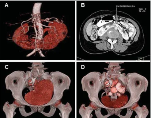
Horseshoe kidney (Figures 1A and 1B) is the most common and most frequently found renal abnormality among men. Re-nal fusion occurs at variable degrees, in most cases between the lower poles of the kidneys, which are closer to the midline than in normal kidneys. The isthmus, most commonly located in front of the aorta or of the inferior vena cava, connects the two renal masses, and may contain functioning parenchyma or fibrous tissue, and for such reason a functional evaluation utilizing a radionuclide, may be necessary before any interventional approach. The isthmus itself poses some difficulty for the renal rotation, as well as its ascent due to the inferior mesenteric artery. Its blood supply may be varied and usually the collecting system is anteriorized(1,18–21).
Most patients are asymptomatic and the finding is incidentally observed in the course of imaging procedures. In symptom-atic cases, generally hydronephrosis (Fig-ure 2), infection or development of calculi are reported. Such abnormality has been associated with a higher predisposition to development of malignant neoplasms, such
Figure 1. Renal shape abnormalities. MDCT image with VRT (A) and axial view (B) of a “horseshoe” kid-ney showing the connection between the inferior renal poles. Note the contrast uptake by the isthmus and its anterior position in relation to the aorta artery, next to the common iliac arteries bifurcation. MDCT images with VRT (C, D) of a “pancake” kidney.
Figure 2. MDCT images of a “horseshoe” kidney. Coronal image (A) and VRT (B) identifying the renal connection by the inferior poles and presence of hydronephrosis at left.
as Wilms’ tumor, as well as systemic mal-formations, as in the case of Turner’s syn-drome(1,18,20).
In cases of abnormality characterizing pancake kidneys (Figures 1C and 1D), the kidneys form a single medial mass located in the pelvic cavity or at the level of the aor-tic bifurcation, with a flat, lobulated and non-reniform appearance, with anteriorized collecting system and short ureters
drain-ing into independent orifices or into a single ureter(20). Its blood irrigation, also
anomalous, is a risk factor for the renal vascular length, from the event of a simple gestation to pelvic traumas(19).
dif-ferential diagnoses simulating pelvic mass which cannot be unequivocally removed or injured(19).
POSITION ABNORMALITIES
The ectopic kidney (Figures 3 and 4) is the result of failure in its migration from the pelvic cavity towards the renal fossa(15) and
is frequently related to poor rotation(14,18).
A slightly higher prevalence of such abnor-mality in the left side is observed, and 10% of cases may be bilateral(18).
Cranial ectopia is usually intrathoracic, and caudal ectopia can be classified into abdominal, iliac (lumbar) and pelvic
(sac-ral), the latter being most frequently found(15), besides the association with
geni-tal malformations(14).
In cases of congenital renal ectopia, the lower positioning of the kidney position is associated with a shorter ureter, renal ves-sels with ectopic origin (adjacent blood vessels, sometimes irrigated by multiple vessels) and some degree of collecting sys-tem malformation(1,18), increasing the
sus-ceptibility to reflux, infection, lithiasis and obstructive conditions(2). It should neither
be confused with abnormally mobile kid-ney nor with ptotic kidkid-ney(18).
In simple ectopia (Figure 3), the kidney is at the same side in which it originated.
The pelvic location, the most common one, is associated with the absence of its ha-bitual morphology as the kidney is fre-quently malrotated and has its image super-imposed over the pelvic bones, which makes its identification difficult(18).
Crossed renal ectopia (Figure 4) occurs when one of the kidneys is contralateral in relation to the site of insertion of its ureter into the urinary bladder. Almost always, the ectopic kidney has a shorter ureter, and, therefore, a lower position as compared with the normal kidney, which may present varied degrees of ptosis and malrotation. The normal kidney may remain separated from the ectopic kidney or form a single mass with the ectopic one(14,15). Crossed
renal ectopia with fusion (85% of the cases) can be identified in several presentations, with the most common one being the fu-sion of the upper pole of the ectopic kid-ney with the inferior pole of the other(18,20).
ROTATION ABNORMALITIES
The kidney migration from the pelvic cavity into its definitive location at the lum-bar site, occurs simultaneously with its ro-tation in the longitudinal plane. Each kid-ney undergoes a medial rotation of approxi-mately 90° as it migrates cephalically. Thus, the hila are oriented towards the midline, aligned and anteromedially ori-ented towards each other(1,18). It is
imptant to establish a correct diagnosis in
or-Figure 4. MDCT images of crossed renal ectopia. Oblique image (A) and VRT (B,C) demonstrating renal duplicity at right without fusion of renal masses. Note that the ureter of the upper kidney has its pathway at right, while the ureter of the lower (ectopic) kidney crosses to the contralateral side and inserts into the urinary bladder.
der to rule out other pathological condi-tions which may cause similar distortion in the kidneys(18).
Renal malrotation (Figure 5) is com-monly associated with an ectopic kidney or fusion, besides the possibility of partial obstruction of the pelvis and the kidneys’ ureters. Both kidneys may be affected, and incomplete rotation or nonrotation at all are
more frequently observed as compared with other subtypes(18). Rarely, there is a
hyper-rotation, placing the renal hilum to-wards the dorsum(15).
NUMBER ABNORMALITIES
Renal agenesis, with a probable multi-factorial etiology, is defined as the absence
of renal tissue secondary to embryogenesis failure, occurring either unilateral or bilat-erally(1). Supernumerary kidney is
ex-tremely rare, is separated from the normal kidney and has its own blood supply(14).
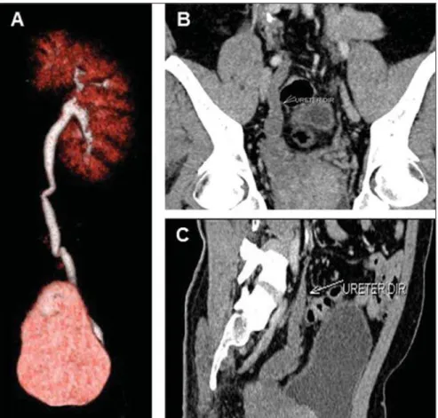
Unilateral agenesis (Figure 6) is a rela-tively common abnormality, occurring ap-proximately once in every 1,000 new-borns(1). The prognosis is good when the
Figure 6. Unilateral renal agenesis. MDCT image VRT (A) showing a solitary left kidney. Note, at coronal (B) and sagittal (C) images, that the right ureter is rudimentary and ectopically drains into the right semi-nal vesicle.
Figure 5. Renal rotation abnormalities. Diagram illustrating a primitive fetal kidney (A); normal kidney in adult individual (B); incomplete rotation (C); hyper-rotation (D); exaggerated hyper rotation (E) and reversed rotation (F). (Adapted from Prando et al.(5)). Axial MDCT image (G) demonstrating renal hyperrotation at left.
condition is not associated with other sys-temic abnormalities and is related to a con-tralateral, usually hypertrophic kidney, as a compensatory effect(1,2). Agenesis of the
ipsilateral adrenal gland is found in 10% of the cases(22) and the renal artery and vein
do not develop. The corresponding ureter is absent in most cases, sometimes corre-sponding to a fibrotic cord which may end ectopically, for example, in the contralat-eral seminal vesicle(15). One should suspect
of unilateral renal agenesis in children with only one umbilical artery(1).
Bilateral agenesis occurs once in every 3,000 newborns, is incompatible with life, and is generally found in stillborns(1,2).
Such children present a characteristic facial appearance(1,23) and frequently there are
as-sociation with other congenital disorders, like in the case of Potter’s syndrome(2,24).
Fetal urine is not produced, resulting in severe oligohydramnios(24).
LOBAR ANATOMY ABNORMALITIES
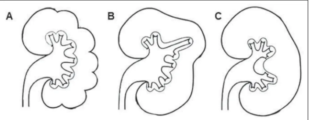
frequent anatomic variations of the renal parenchyma and may simulate renal tu-mors, with an otherwise healthy paren-chyma(21,23) (Figure 7).
Renal contour lobulations (Figure 7A) are found in approximately 5% of adults submitted to kidney imaging studies(23).
Such abnormality corresponds to persis-tence of well defined cortical sulci on the renal surface which are found in the fetal kidney and usually disappear during the childhood as a consequence of growth and increase in the number of nephrons(1). It
may also be confused with renal scars(23,25).
On the other hand, dromedary hump kidney (Figure 7B) is characterized by a
change in the shape and contour of the posterolateral aspect of the left kidney, as a consequence of a focal prominence of the renal parenchyma, probably due to impres-sion of the spleen in the course of fetal life(23,25).
Another benign condition which may simulate neoplasia is hypertrophied col-umn of Bertin (Figures 7C and 8), which correspond to columns of renal cortical tis-sue located between the pyramids, and re-sulting from fusion of two or more renal lobes. Such columns may be thicker, hyper-trophic and deep, protruding in the renal sinus and manifesting as a regular and well defined cortical nodule located at the
junc-tion between the upper and medial renal thirds(23,26).
Hypertrophied column of Bertin pre-sents suggestive, but nonspecific signs at EU and US, so CT(25), whose findings are
well characterized, should be performed. Such CT findings are isodense in relation to the cortical parenchyma and the postcontrast uptake is uniform.
ABNORMALITIES OF THE CALYCES AND PAPILLAE
The pyelocalyceal diverticulum is a cys-tic cavity covered by urothelium, located inside the renal parenchyma, which may be either acquired or congenital (most com-mon) and single or multiple(2,15). Such
ab-normalities may be divided into two types, as follows: 1) the most frequent one is rep-resented by minor lesions affecting the minor calyces, and are located close to the region of the upper renal pole (Figure9); 2) the less frequent one, centrally located in the kidneys, is related to the renal pelvis or major calyces(27,28).
Minor diverticula are typically asymp-tomatic, and are incidentally found at im-aging studies. The major ones are generally symptomatic and urinary stasis favors the
Figure 8. Hypertrophied column of Bertin. Axial (A), coronal (B) and sagittal (C) MDCT images demonstrating septal hypertrophy. Figure 7. Lobar anatomy abnormalities. Schematic illustration of persistent fetal lobulations (A);
development of urinary infection and for-mation of calculi(2,27). While the incidence
of pyelocalyceal diverticulum is low, the frequency of associated calculi formation is high(29).
ABNORMALITIES OF THE RENAL PELVIS AND OF THE URETER
The renal collecting system is a frequent site of anatomic variations with respect to size, shape, degree of ramification and de-gree of rotation in relation to the renal hilum. Ureteropelvic junction (UPJ) stenosis (Figure 10) is the most common abnormal-ity in the childhood and is more frequent in male children, normally being diagnosed at the first year of life, sometimes remain-ing undetected until adulthood, and in such
age range it is more frequently found in women(1,22). Such abnormality is
character-ized by narrowing of the UPJ, generally at left, and may be caused either by an intrin-sic muscle lesion or by a functional discon-tinuity in this segment, impairing the ap-propriate emptying of the renal pelvis, lead-ing to hydronephrosis(15,22).
The stenosis may also be determined by a pyeloureteral mucous fold with valvular behavior, or by extrinsic compression by an aberrant vessel which compresses the in-fundibulum of the renal pelvis, impairing its emptying(15). It is one of the main causes
of urinary tract dilation (approximately 35% to 40% of the cases) and its origin is still to be completely understood(18).
However, in most of cases, renal pelvis and ureter abnormalities present as
duplic-ity of the collecting system, a common cause of dimension asymmetry between the kidneys during the childhood, which occurs in 1% to 2% of the population, most fre-quently among female individuals(15,22,30).
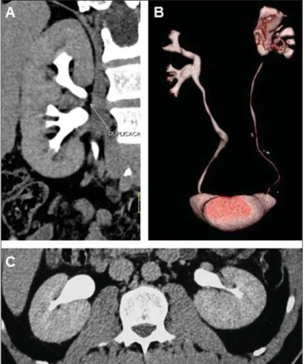
Such duplicity may be complete or incom-plete (Figure 11), with higher prevalence for the unilateral presentation, and is fre-quently associated with various complica-tions(22,30). The kidney with duplicated
col-lecting system is larger, particularly along its longitudinal axis, and likewise the vol-ume of the parenchyma.
In cases of complete duplicity (Figures 11A and 11B), there are two collecting sys-tems for a single kidney, and two ureters at the same side, draining into separate ori-fices. According to the Weigert-Meyer rule (Figure 11B), the ureter which drains the upper part goes over the urinary bladder wall to insert itself inferiorly and medially in relation to the normal insertion site. Fre-quently, such an insertion is defective, as-sociated with ureteroceles and, when ec-topic, it may drain into the posterior uretra, vagina or the vulva. The ureter of the lower segment inserts close to the normal site and is subject to vesicoureteral reflux because of the distortion it undergoes as it crosses the urinary bladder wall associated with ureteroceles(15,22). At radiography, the
com-plete dilation is seen as the characteristic and well known dropping lily sign(21).
Complications from complete duplica-tion include infecduplica-tions, vesicoureteral reflux and UPJ obstruction. Reflux in the collect-ing system of the lower segment may cause scars and deformities in that segment(22). Figure 9. Pyelocalyceal diverticulum. VRT (A) and coronal (B) images of left kidney demonstrating
ca-lyceal diverticulum in the caca-lyceal group of the right kidney.
In incomplete duplicity (Figure 11C) there are two collecting systems and two ureters that fuse together at any level be-tween the kidney and the bladder (normally in the lower third of that pathway), origi-nating a single ureter which drains nor-mally into the vesical base. In cases where the junction is at a level above the vesical dome, the ureter presents a “Y” configura-tion, and in cases where the junction occurs at the level of the intramural segment of the ureters, the ureter presents a “V” configu-ration(15,22). There may be uretero-ureteral
reflux because of the ureteral peristalsis asynchrony before the confluence.
Pelvic anomalies are other abnormali-ties resulting from the division of the col-lecting system (Figure 12). In cases of bifidus renal pelvis (Figures 12A and 12B) only the renal pelvis is divided and there is only one UPJ. This is a relatively common anomaly, which occurs in up to 10% of the population and there is no association with other anomalies(22). The position of the
re-nal pelvis is also quite variable. Te pelvis is classified as intrarenal when there is abundant renal tissue around it. On the other hand, the pelvis is extrarenal (Figures 12C and 12 D) (more common), when it is actually outside the hilum which is occu-pied only by the calyceal infundibula. In general, it is associated with other
anoma-Figure 11. Duplicity of collecting system. MDCT – coronal (A) and VRT (B) images showing complete duplicity at left. Observe the Weigert-Meyer rule at the posterior view on (B). The image with VRT (C) shows incomplete duplicity at left, where the fusion of the ureters in the inferior medial third of the ureteral pathway is observed. Note the subtle pyelocalyceal dilation as well as dilation of the two ureters above the junction.
lies such as malrotation or position defects, with the possibility of stasis and predispo-sition to infections(22).
CONCLUSION
Many of the morphological renal changes can be evaluated by means of US and EU, but CT, with its more modern tech-nological resources, has contributed over the past years for a better characterization of morphological changes. Computed to-mography is essential in the diagnosis of congenital abnormalities, offering a better guidance in the clinical and surgical/thera-peutic decisions making process, addition-ally to its role as a relevant tool for identi-fying associated complications. Also, the several resources of this imaging method allow for renal evaluation with respect to size, position and shape.
The present article demonstrates how the new images from the same congenital renal abnormalities have contributed for a more accurate diagnosis and better evalu-ation of associated complicevalu-ations in such patients.
REFERENCES
1. Moore KL, Persaud TVN. Embriologia clínica. 6ª ed. Rio de Janeiro, RJ: Guanabara Koogan; 2000.
2. Kumar V, Abbas AK, Fausto N. Robbins e Cotran – Patologia: bases patológicas das doenças. 7ª ed. Rio de Janeiro, RJ: Elsevier; 2005.
3. Reidy KJ, Rosenblum ND. Cell and molecular biology of kidney development. Semin Nephrol. 2009;29:321–37.
4. Sanna-Cherchi S, Ravani P, Corbani V, et al. Re-nal outcome in patients with congenital anoma-lies of the kidney and urinary tract. Kidney Int. 2009;76:528–33.
5. Prando A, Prando D, Caserta NMG, et al. Urolo-gia: diagnóstico por imagem. São Paulo, SP: Sar-vier; 1997.
6. Caoili EM, Cohan RH, Korobkin M, et al. Urinary tract abnormalities: initial experience with multi-detector row CT urography. Radiology. 2002;222: 353–60.
7. Nikken JJ, Krestin GP. MRI of the kidney – state of the art. Eur Radiol. 2007;17:2780–93. 8. Galvão Filho MM, D’Ippolito G, Hartmann LG,
et al. O valor da tomografia computadorizada he-licoidal sem contraste na avaliação de pacientes com dor no flanco. Radiol Bras. 2001;34:129–34. 9. Warshauer DM, McCarthy SM, Street L, et al. Detection of renal masses: sensitivities and speci-ficities of excretory urography/linear tomography, US, and CT. Radiology. 1988;169:363–5. 10. Jamis-Dow CA, Choyke PL, Jennings SB, et al.
Small (< or = 3-cm) renal masses: detection with CT versus US and pathologic correlation. Radi-ology. 1996;198:785–8.
11. Smith RC, Verga M, McCarthy S, et al. Diagno-sis of acute flank pain: value of unenhanced CT. AJR Am J Roentgenol. 1996;166:97–101. 12. Levine JA, Neitlich J, Verga M, et al. Ureteral
calculi in patients with flank pain: correlation of plain radiography with unenhanced helical CT. Radiology. 1997;204:27–31.
13. Kawashima A, Sandler CM, Ernst RD, et al. CT evaluation of renovascular disease. Radiographics. 2000;20:1321–40.
14. Barbaric ZL. Principles of genitourinary radiol-ogy. 2nd ed. New York, NY: Thieme; 1994. 15. Kim SH. Radiology illustrated – uroradiology.
2nd ed. Berlin: Springer-Verlag; 2012. 16. Hartshorne N, Shepard T, Barr M Jr.
Compensa-tory renal growth in human fetuses with unilat-eral renal agenesis. Teratology. 1991;44:7–10. 17. Cho JY, Moon MH, Lee YH, et al. Measurement
of compensatory hyperplasia of the contralateral
kidney: usefulness for differential diagnosis of fetal unilateral empty renal fossa. Ultrasound Obstet Gynecol. 2009;34:515–20.
18. Fotter R. Pediatric uroradiology. 2nd ed. Berlin: Springer-Verlag; 2008.
19. Gun S, Ciantelli GL, Takahashi MAU, et al. Fu-são renal completa em criança com infecção re-corrente do trato urinário. Radiol Bras. 2012;45: 233–4.
20. Türkvatan A, Ölçer T, Cumhur T. Multidetector CT urography of renal fusion anomalies. Diagn Interv Radiol. 2009;15:127–34.
21. Dyer RB, Chen MY, Zagoria RJ. Classic signs in uroradiology. Radiographics. 2004;24 Suppl 1: S247–80.
22. Brant WE, Helms CA. Fundamentals of diagnos-tic radiology. 3rd ed. Philadelphia, PA: Lippincott Williams & Wilkins; 2007.
23. Quaia E. Radiological imaging of the kidney. 1st ed. Berlin: Springer-Verlag; 2011.
24. Zhou Q, Cardoza JD, Barth R. Prenatal sonography of congenital renal malformations. AJR Am J Roentgenol. 1999;173:1371–6.
25. Bhatt S, MacLennan G, Dogra V. Renal pseudo-tumors. AJR Am J Roentgenol. 2007;188:1380– 7.
26. Lafortune M, Constantin A, Breton G, et al. Sonography of the hypertrophied column of Bertin. AJR Am J Roentgenol. 1986;146:53–6. 27. Rathaus V, Konen O, Werner M, et al. Pyelocalyceal
diverticulum: the imaging spectrum with empha-sis on the ultrasound features. Br J Radiol. 2001;74:595–601.
28. Abad PG, González IF, Peso AC, et al. Percuta-neous treatment of stone-containing calyceal di-verticulum. Arch Esp Urol. 2009;62:42–8. 29. Stunell H, McNeill G, Browne RF, et al. The
im-aging appearances of calyceal diverticula compli-cated by uroliathasis. Br J Radiol. 2010;83:888– 94.


