Angle Class I malocclusion treated with lower incisor extraction
» The author reports no commercial, proprietary or financial interest in the products or companies described in this article.
» The patient displayed in this article previously approved the use of her facial and intraoral photographs.
Contact address: Vanessa Leal Tavares Barbosa
Rua José Alexandre Buaiz, nº 160, salas 904/906 – Enseada do Suá / Brazil CEP: 29.050-545 - Vitória/ES - E-mail: [email protected] Vanessa Leal Tavares Barbosa1
In planning orthodontic cases that include extractions as an alternative to solve the problem of negative space dis-crepancy, the critical decision is to determine which teeth will be extracted. Several aspects must be considered, such as periodontal health, orthodontic mechanics, functional and esthetic alterations, and treatment stability. De-spite controversies, extraction of teeth to solve dental crowding is a therapy that has been used for decades. Pre-molar extractions are the most common, but there are situations in which atypical extractions facilitate mechanics, preserve periodontal health and favor maintenance of the facial proile, which tends to unfavorably change due to facial changes with age. The extraction of a lower incisor, in selected cases, is an efective approach, and literature describes greater post-treatment stability when compared with premolar extractions. This article reports the clini-cal case of a patient with Angle Class I malocclusion and upper and lower anterior crowding, a balanced face and harmonious facial proile. The presence of gingival and bone recession limited large orthodontic movements. The molars and premolars were well occluded, and the discrepancy was mainly concentrated in the anterior region of the lower dental arch. The extraction of a lower incisor in the most ectopic position and with compromised periodon-tium, associated with interproximal stripping in the upper and lower arches, was the alternative of choice for this treatment, which restored function, providing improved periodontal health, maintained facial esthetics and allowed inishing with a stable and balanced occlusion. This case was presented to the Brazilian Board of Orthodontics and Dentofacial Orthopedics (BBO), as part of the requirements for obtaining the BBO Diplomate title.
Keywords:Crowding. Lower incisor extraction. Gingival recession.
1Specialist in Orthodontics and Dentofacial Orthopedics, State University of Rio de
Janeiro (UERJ). Diplomate of the Brazilian Board of Orthodontics and Dentofacial Orthopedics (BBO).
* Clinical case, category 2, accepted by the Brazilian Board of Orthodontics and Dentofacial Orthopedics, BBO.
How to cite this article: Barbosa VLT. Angle Class I malocclusion treated with lower incisor extraction. Dental Press J Orthod. 2013 May-June;18(3):150-8.
No planejamento ortodôntico de casos que incluem extrações como alternativa para solucionar o problema de discre-pância de espaço negativa, a decisão crítica é determinar quais dentes serão extraídos. Devemos considerar vários as-pectos, como a saúde periodontal, mecânica ortodôntica, alterações funcionais e estéticas, e estabilidade do tratamento. Apesar das controvérsias, a extração de dentes para solucionar apinhamentos dentários é uma terapêutica que tem sido utilizada há décadas. As extrações de pré-molares são as mais comuns, mas há ocasiões em que extrações atípicas facilitam a mecânica, preservam a saúde periodontal e favorecem a manutenção do peril, que tende a se alterar desfa-voravelmente devido às modiicações faciais decorrentes da idade. A extração de um incisivo inferior, em casos bem selecionados, é uma abordagem eiciente; e a literatura descreve maior estabilidade pós-tratamento, quando comparada com a opção de extração de pré-molares. O presente artigo relata um caso clínico de uma paciente com má oclusão de Classe I de Angle e apinhamento anterior superior e inferior, face equilibrada e peril harmonioso. A presença de recessões gengivais e ósseas limitava grandes movimentações ortodônticas. Os molares e pré-molares estavam bem relacionados, e a discrepância concentrava-se principalmente na região anterior da arcada dentária inferior. A extração de um incisivo inferior em posição mais ectópica e com periodonto comprometido, associada a desgastes interproxi-mais nas arcadas superior e inferior, foi a alternativa de escolha para o tratamento, que restabeleceu a função, propor-cionando melhoria da saúde periodontal, manteve a estética facial, e permitiu a inalização com uma oclusão estável e equilibrada. Esse caso foi apresentado à diretoria do Board Brasileiro de Ortodontia e Ortopedia Facial (BBO), como parte dos requisitos para obtenção do título de Diplomado pelo BBO.
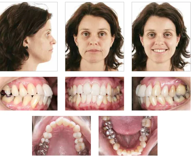
Figure 1 - Initial facial and intraoral photographs.
HISTORY AND ETIOLOGY
Female patient, Caucasian, searched for orthodon-tic treatment at age 44, in good general health with no signiicant medical history. The main complaint was related to crowding in the upper arch, and especially in the lower arch, as well as the gingival recessions, which were increasing over the years (Fig 1). There was a his-tory of caries and unsatisfachis-tory restorations in several teeth. No esthetic complaints were reported. In func-tional occlusion analysis, it was found that the right and let lateral guides were performed by the irst upper and lower premolars. The gingival recession of tooth #14 was, possibly, due to occlusal overload. Despite no functional guides were present, there were no signs or symptoms of temporomandibular disorders. No orth-odontic intervention had been performed before.
DIAGNOSIS
arch in the premolars and molars region, with a tendency to crossbite. The lower midline was shited 1 mm to the let side, and the upper and lower incisors and lower right canine were projected in relation to their apical bases. Tooth #43 was labially positioned with the long axis me-sially displaced, and presented marked gingival recession (Figs 1 and 2). The panoramic radiograph reported the presence of third molars, with the lower ones mesially tipped. Periapical radiographs revealed a regular alveolar bone loss in the maxilla and mandible, and suggested ex-ternal root resorption in the apical third of the teeth #31 and #41. Interproximal radiographs demonstrated excess of restorative material in several teeth. The cephalometric diagnosis conirmed the labial protrusion of the upper in-cisors (1-NA = 32° and 7.5 mm) as well as the lower ones (1-NB = 25° and 7 mm) (Figs 3, 4 and 5).
TREATMENT OBJECTIVES
Orthodontic treatment aimed to eliminate the ante-rior dental discrepancy, correcting the crowding of up-per and lower incisors, aligning and leveling the teeth without jeopardizing the facial proile; establishing es-thetically favorable and functionally efective overjet and
overbite, properly positioning the teeth on their apical bases and contributing to improve periodontal health. The extraction of premolars could result in lattening of the facial proile, aggravated by facial changes due to age; however, the treatment without extractions would increase the lack of lip seal, and contribute to the wors-ening of gingival recession and a greater tendency to re-lapse.8 Through the diagnosticsetupthe possibility of a lower incisor extraction was evaluated, because it is one of the most valuable orthodontic records to determine if a lower incisor should be extracted.1,3,11,22,24 Prior to ortho-dontics, the patient would be referred to the periodontist for free gingival grat in the teeth with accentuated gin-gival recession, preventing its intensiication and creating a thicker marginal gingiva.25 The shape of the upper arch should be improved by expanding the molar and pre-molar regions, which tended to cross, favoring a greater illing of the buccal corridor and broadening the smile. The occlusion key of the right and let molars and let ca-nine would be kept, while the Class I relationship in the right canine should be achieved. Inadequate restorations would be replaced at the end of orthodontic treatment, aiming periodontal health and occlusal stability.
TREATMENT PLAN
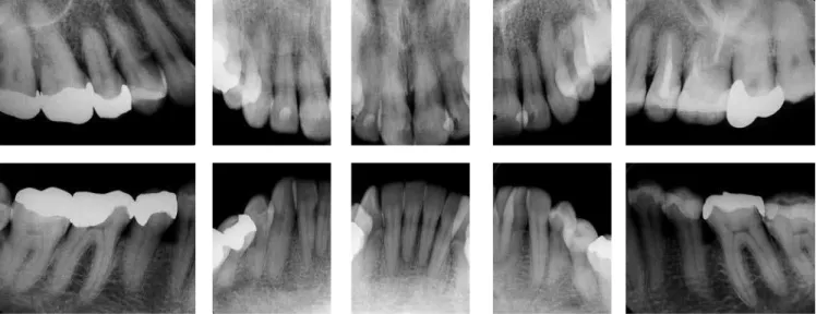
Performing the diagnostic setup was essential for the decision of the lower incisor extraction, besides help-ing to visualize treatment outcome and determine the amount of interproximal stripping that would be per-formed on the upper incisors for proper intercuspa-tion.17,24 To indicate the treatment with incisor extrac-tion, some requirements that applied to this case were also considered: Class I molar relationship, mandibular crowding greater than 4.5 mm (in this case, it was 7 mm), slight or nonexistent maxillary crowding (in this case, it was 3 mm), balanced sot tissue proile, minimal or moderate overbite and overjet1,7,22,24 (Figs 1 and 2). However, before the beginning of orthodontic treat-Figure 3- Initial periapical radiographs.
Figure 4- Initial lateral cephalometric radiograph (A) and cephalometric tracing (B).
Figure 5- Initial panoramic radiograph.
ment, the patient would be referred to the periodontist to control periodontal health conditions and to plan the free grat surgery in teeth with more advanced gingival recession (#23 and #43). Only ater 60 days these teeth could be moved. For the maxillofacial surgeon, extrac-tion of third molars would be required, because these teeth were in unfavorable positions. Ater the initial procedures with the multidisciplinary team, orthodon-tic treatment would start with bonding the brackets on upper and lower dental arches, Straight-Wire system, Roth prescription, slot0.022 x 0.028-in — except in the #43 tooth, which would not receive a bracket until the space for its alignment in the arch was obtained. In-terproximal stripping in the #45 and #44 teeth, which presented excess of restorative material, were scheduled in order to optimize the space for the tooth #43. The i-nalization would be accomplished through coordinated rectangular arches with ideal torques and shapes, and the use of intermaxillary elastics for inal intercuspation. If necessary, it would be requested an occlusal adjust-ment with the general dentist for occlusion reineadjust-ment and replacement of initially inadequate restorations.
TREATMENT PROGRESS
As planned, prior to orthodontics, the patient was re-ferred to the periodontist for the control of periodontal health and conditions and free grat in the region of teeth #23 and #43 — to increase the thickness of the marginal gingiva because orthodontic movement could favor the increase of gingival recession and bone fenestrations.9,21,25 Ater 60 days, orthodontic movement in these teeth was permitted. Third molar extraction, which were in unfa-vorable positions, was also performed at this stage.
Then, brackets were bonded on the upper teeth, Straight-Wire system, Roth prescription, slot 0.022 x 0.028-in, on teeth #17 to #27. Then, stripping was performed on the upper incisors, with manual abrasive strips, to facilitate alignment, avoid black spacesbetween these teeth and achieve excellent incisal relationship, by controlling the overbite and reducing overjet — consider-ing that the new occlusal situation would promote articu-lation of six upper teeth with ive lower ones.7,20,24 Align-ment and leveling nickel-titanium 0.012-in and 0.014-in archewires were used followed by stainless steel round and 0.014-in, 0.016-in, 0.018-in and 0.020-in archewires. Finishing occurred with 0.019 x 0.025-in stainless steel rectangular archwires with ideal shape and torque.
In the mandibular arch a Straight-Wire ixed orth-odontic appliance, Roth prescription, slot 0.022 x 0.028-in was placed, except 0.022 x 0.028-in the tooth #43, which received bracket bonding ater opening of space for its alignment and correction of the long axis, which was markedly me-sial. Stripping was performed for removal of restorative material excess in teeth #45 (mesial) and #44 (distal), and to facilitate the alignment of the tooth #43. The extrac-tion of the lower let lateral incisor (#32) was required for being the incisor in the most ectopic position and with the most unfavorable periodontal conditions.6,14 The closure of the extraction space was conducted us-ing a passive stainless steel 0.018-in round archwire and through distal movement of tooth #31 and mesial move-ment of the teeth #41 and #42 with elastomeric chain and nickel-titanium open spring installed between the teeth #44 and #42. Posterior anchorage in the right and let sides was obtained by tying together the mo-lars and premomo-lars with metal ligatures. Ater obtaining space for tooth #43, bracket bonding was proceeded, a lower 0.018 x 0.025-in stainless steel base archwire with a bypass was made for this tooth and, for its alignment and leveling, a superimposed 0.012-in nickel-titanium sectioned archwire was initially used, followed by a 0.014-in archwire, evolving into continuous arches, for completion of this phase. The inishing was done with 0.018 x 0.025-in stainless steel rectangular archwire with ideal form and torques, coordinated with the upper arch. Light triangular 1/4-in intermaxillary elastics were used in the canines and premolars region. Throughout the treatment, the patient was accompanied by the perio-dontist, with appointments every three months. Ater verifying the achievement of the goals predeined in the initial planning, the ixed orthodontic appliance was re-moved, initiating the retention phase. A removable up-per wraparound retainer was used as well as a bonded lingual retainer, made with 0.038-in braided stainless steel wire. The use of the upper retainer plate was rec-ommended for 24 hours a day in the irst six months; 18 hours a day, in the following six months; 12 hours a day, for more six months; and then daily use at night.
TREATMENT EVALUATION
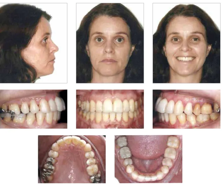
Figure 6 - Final intraoral and facial photographs.
of the axial inclination of the incisors resulted in sig-niicant improvement in dental esthetics and relected in the facial proile, with retraction of the lower lip, from a position 1 mm forward the S line (Steiner) to 0 mm, fa-voring passive lip sealing (Table 1). In the frontal photo-graph, the inal smile was more harmonious. The upper midline, which was angled, was corrected and became coincident with the middle of the lower central incisor, without esthetic commitment10,24 (Fig 6).
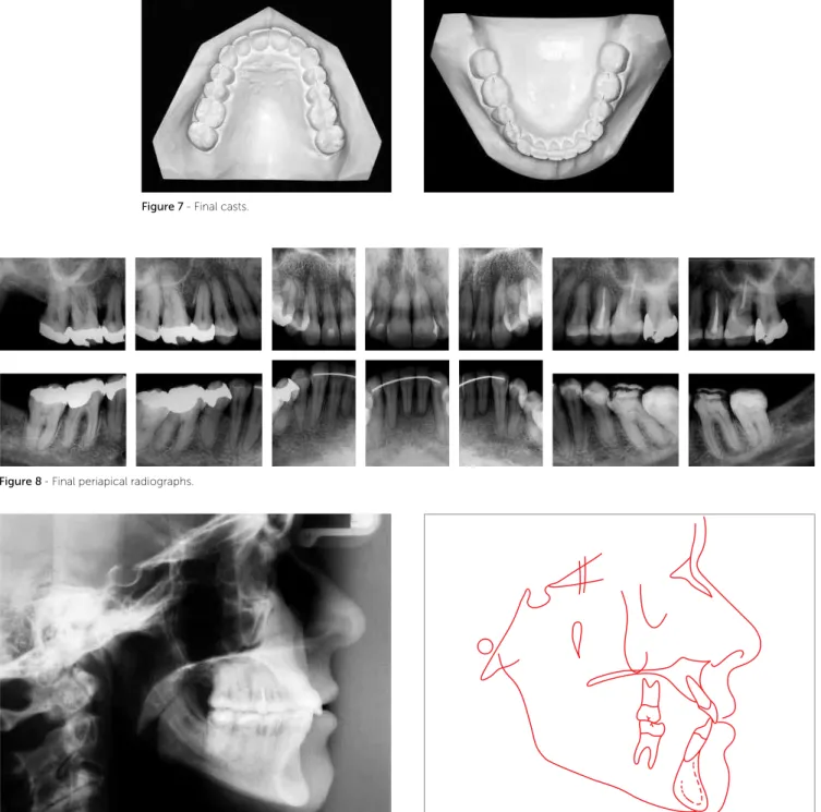
The periodontal health was markedly improved and the increase of overbite and overjet, which had its mea-sures reduced, allowed the establishment of a function-ally balanced occlusion (Fig 7).
The let and right molars and let canine keys of occlu-sion were maintained and the occluocclu-sion key on the right canine was achieved, resulting in right and let laterality
with disocclusion in the canines and without contacts in balance. The protrusive excursion resulted in adequate posterior disocclusion.
Total superimposition of cephalometric tracings il-lustrates the proile improvement with the change in the lower lip position, which made it more pleasant (Fig 10A). The partial superimpositions of the maxilla and mandible conirm the signiicant reduction in labial axial inclina-tion of the upper incisors and discrete uprighting of lower ones, with slight anchorage loss (Fig 10B).
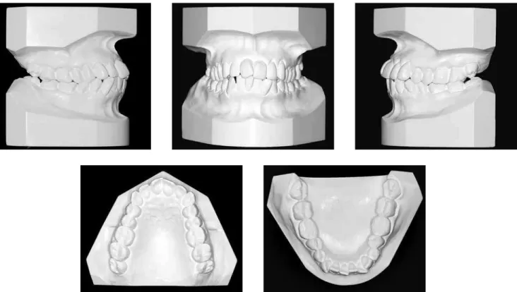
Figure 7 - Final casts.
Figure 8- Final periapical radiographs.
Figure 9- Final lateral cephalometric radiograph (A) and cephalometric tracing (B).
MEASURES Normal A B A/B diff.
Skeletal pattern
SNA (Steiner) 82° 80° 80° 0
SNB (Steiner) 80° 77° 77° 0
ANB (Steiner) 2° 3° 3° 0
Convexity angle (Downs) 0° 4° 3° 1
Y axis (Downs) 59° 59° 58° 1
Facial angle (Downs) 87° 86° 87° 1
SN-GoGn (Steiner) 32° 35° 33° 2
FMA (Tweed) 25° 28° 24° 4
Dental pattern
IMPA (Tweed) 90° 93° 98° 5
1.NA (degrees) (Steiner) 22° 32° 27° 5
1-NA (mm) (Steiner) 4 mm 7,5 mm 6 mm 1.5
1.NB (degrees) (Steiner) 25° 25° 27° 2
1-NB (mm) (Steiner) 4 mm 7 mm 6 mm 1
1
1- Interincisal angle (Downs) 130° 120° 120° 0
1-APo (mm) (Ricketts) 1 mm 5 mm 4 mm 1
Profile Upper lip – S line (Steiner) 0 mm -2 mm -2 mm 0
Lower lip – S line (Steiner) 0 mm 0 mm -1 mm 1
Table 1 - Summary of cephalometric measures.
Figure 10- Total (A) and partial (B) superimpositions of initial (black) and inal (red) tracings.
A B
Assessing the intercanine distance, it was found that there was a 1-mm reduction, and it can be said that the maintenance or reduction of this distance dur-ing mechanical extraction of incisors is advantageous10 compared to premolars, because there is a strong re-lationship between long-term stability of crowding correction and intercanine distance. It is believed that the treatment with extraction of an incisor and main-tenance of that distance or even decreasing it, in an-ticipation of a further natural decrease, provides
In the evaluation of inal periapical radiographs it was observed the absence of the upper and lower third molars, which were removed; and increasing of root apex rounding on the lower incisors (#41 and #31), which had already been observed in the initial radio-graphs (Fig 8). The improvement in axial inclination of #43 tooth, severely tipped mesially and out of position before treatment, draws attention to its repositioning in the arch and excellent periodontal recovery. The re-placement of inadequate restorations was requested at the end of treatment, but had not been completed yet.
FINAL CONCLUSIONS
The diagnosis and careful planning, with the help of the diagnostic setup,4 was essential for the decision of treatment with extraction of a lower incisor. Refer-ring the patient to the periodontist to perform gingival grat before orthodontic treatment enabled orthodontic
movement more safely and without injury to teeth al-ready compromised by periodontal recessions.21,25
Despite the diiculties or limitations that planning of cases with incisor extraction may result during orth-odontic treatment, provided properly conducted and evaluated — considering the particularities of each case —, it can be stated that the lower incisor extraction contributes efectively in the treatment of certain mal-occlusions, seeking excellence in orthodontic treatment outcomes (maximum function, esthetics and stability).13 The patient’s satisfaction by having her main complaint resolved relected also in increased self-esteem and gain of quality of life — beneits provided by orthodontics in the aspect of overall health.
Based on data from the literature and exempliied by the clinical report of this case, it can be concluded that the extraction of a lower incisor is a very efective thera-peutic approach in carefully selected situations.15
1. Bahreman AA. Lower incisor extraction in orthodontic treatment. Am J
Orthod. 1977;72(5):560-7.
2. Bernstein L. Edward H. Angle versus Calvin S. Case: extraction versus nonextraction. Historical revisionism. Part II. Am J Orthod Dentofacial Orthop. 1992;102(6):546-51.
3. Bolognese AM. Set-up: uma técnica de confecção. Rev SOB.
1995;2(8):245-9.
4. Bolton WA. Disharmony in tooth size and its relation to the analysis and treatment of malocclusion. Angle Orthod. 1958;28(3):113-30.
5. Brandt S, Sairstein GR. Diferent extractions for diferent malocclusions. Am J Orthod. 1975;68(1):15-41.
6. Canut JA. Mandibular incisor extraction: indications long-term evaluation. Eur J Orthod. 1996;18(5):485-9.
7. Faerovig E, Zachrisson BU. Efects of mandibular incisor extraction on anterior occlusion in adults with Class III malocclusion and reduced overbite. Am J Orthod Dentofacial Orthop. 1999;115(2):113-24.
8. Janson GRP, Canto GDL, Henriques JFC, Freitas MR, Mazziero ET, Toruño
JA. A importância da individualização no planejamento ortodôntico. Rev Dental Press Ortod Ortop Facial. 1998;8(2):31-45.
9. Holmes HD, Tennant M, Goonewardene MS. Augmentation of faciolingual
gingival dimensions with free connective tissue grafts before labial orthodontic tooth movement: an experimental study with a canine model. Am J Orthod Dentofacial Orthop. 2005;127(5):562-72.
10. Klein DJ. The mandibular central incisor, an extraction option. Am J Orthod Dentofacial Orthop. 1997;111(3):253-9.
11. Kokich VG, Shapiro PA. Lower incisor extraction in orthodontic treatment. Four clinical reports. Angle Orthod. 1984;54(2):139-53.
12. Kokich VO. Treatment of a Class I malocclusion with a carious mandibular incisor and no Bolton discrepancy. Am J Orthod Dentofacial Orthop. 2000;118(1):107-13.
REFERENCES
13. Lima CMF, Lacet E, Marques CR. Extração de incisivo inferior: uma opção terapêutica. Rev Dental Press Ortod Ortop Facial. 2005;10(4):47-59. 14. Little RM, Riedel RA, Artun J. An evaluation of changes in mandibular
anterior alignment from 10 to 20 years postretention. Am J Orthod Dentofacial Orthop. 1988;93(5):423-8.
15. Matsumoto MAN, Romano FL, Ferreira JTL, Tanaka S, Morizono EN. Extração de incisivo inferior: uma opção de tratamento ortodôntico. Rev Dental Press Ortod Ortop Facial. 2010;15(6):143-61.
16. Nef CW. The size relationship between the maxillary and mandibular anterior segments of the dental arch. Angle Orthod. 1957;27(3):138-47. 17. Owen AH. Single lower incisor extractions. J Clin Orthod.
1993;27(3):153-60.
18. Riedel RA, Little RM, Bui TD. Mandibular incisor extraction: postretention evaluation of stability and relapse. Angle Orthod. 1992;62(2):103-16. 19. Rosenstein SW. A lower incisor extraction. Aust Orthod J. 1976;4(3):107-9. 20. Sheridan JJ, Hastings J. Air-rotor stripping and lower incisor extraction
treatment. J Clin Orthod. 1992;22(4):187-204.
21. Tanaka O, Young Lon B, Tafarel IP, Siu Lon LF, OLiveira-Junior SR. A recessão e o enxerto gengival no tratamento ortodôntico. Orthod Sci Pract. 2008;1(1);37-47.
22. Telles CS, Urrea BEE, Barbosa CAT, Jorge EVF, Prietsch JR, Menezes LM, et al. Diferentes extrações em Ortodontia (sinopse). Rev SOB. 1995;2(2):194-9.
23. Tuverson DL. Anterior interocclusal relations. Part II. Am J Orthod. 1980;78(4):371-93.
24. Valinoti JR. Mandibular incisor extraction therapy. Am J Orthod Dentofacial Orthop. 1994;105(2):107-16.