355
Radiol Bras. 2011 Nov/Dez;44(6):355–359
Sonographic evaluation of temporomandibular joint internal
disorders
*
Avaliação ultrassonográfica dos distúrbios intracapsulares temporomandibulares
Carlos Fernando de Mello Junior1, Osmar de Cassio Saito2, Hélio Antonio Guimarães Filho3
Objective: To evaluate the sensitivity and specificity of high-resolution ultrasonography in the assessment of intra-capsular temporomandibular disorders. Materials and Methods: The authors have studied 38 patients (76 joints) with complaint of temporomandibular disorder. All the patients underwent ultrasonography and magnetic resonance imaging (gold standard for the evaluation) and the results were compared. Results: Among 24 joints demonstrating disc displacement at magnetic resonance imaging of patients at rest, 7 were confirmed at ultrasonography; in 13, the discs could not be visualized; and in 4, no sonographic abnormality was observed. In 48 joints, the articular discs could not be visualized at ultrasonography of patients at rest. Among them, 41 exhibited normal positioning at magnetic resonance imaging, and 7 exhibited anterior disc displacement. Morphological changes of the mandibular condyle were visualized in 13 joints at magnetic resonance imaging, and in 2 at ultrasonography. Conclusion: With the present study, the authors can conclude that ultrasonography offers high sensitivity and specificity in the diagnosis of the articular disc location with the patient at rest, either to analyze anatomical position or to analyze disc displacement. On the other hand, it does not offer significant results to analyze articular discs in patients with open mouth as well as to analyze disc/condyle morphological changes.
Keywords: Ultrasonography; Temporomandibular joint; Magnetic resonance imaging.
Objetivo: Avaliar a sensibilidade e a especificidade do exame ultrassonográfico de alta resolução para a avaliação dos distúrbios intracapsulares temporomandibulares. Materiais e Métodos: Estudamos 38 pacientes (76 articulações) com queixas de distúrbios temporomandibulares. Todos os pacientes realizaram exames de ultrassonografia e resso-nância magnética (padrão ouro para a avaliação) e os resultados obtidos foram comparados. Resultados: De 24 ar-ticulações evidenciando deslocamento discal com o paciente em repouso na ressonância magnética, 7 foram confir-mados pela ultrassonografia, em 13 não foram visualizados os discos e 4 estavam tópicos na ultrassonografia. Em 48 articulações, o disco articular não foi visualizado na ultrassonografia com o paciente em repouso. Destes, 41 apresen-tavam posicionamento normal na ressonância magnética e 7 apresenapresen-tavam deslocamento anterior. Alterações morfo-lógicas do côndilo mandibular foram visualizadas pela ressonância magnética em 13 articulações, identificadas pela ultrassonografia em 2 delas. Conclusão: Podemos concluir, no estudo, que o exame de ultrassonografia apresenta alta sensibilidade e especificidade para o diagnóstico da localização do disco articular com o paciente em repouso, tanto para a análise de seu posicionamento anatômico como nos casos de deslocamentos, não apresentando resul-tados significativos para a análise dos discos com o paciente com a boca aberta e para a análise de alterações mor-fológicas discais e condilares.
Unitermos: Ultrassonografia; Articulação temporomandibular; Imagem por ressonância magnética. Abstract
Resumo
* Study developed at Hospital das Clínicas da Faculdade de Medicina da Universidade de São Paulo (FMUSP), São Paulo, SP, Brazil.
1. PhD, Associate Professor, Division of Radiology, Course of Medicine, Universidade Federal da Paraíba (UFPB), João Pessoa, PB, Brazil.
2. PhD, MD, Physician Assistant at Hospital das Clínicas da Faculdade de Medicina da Universidade de São Paulo (FMUSP), São Paulo, SP, Brazil.
3. PhD, MD, Physician Assistant at Ecoclínica Diagnósticos, João Pessoa, PB, Brazil.
Mailing Address: Dr. Carlos Fernando de Mello Junior. Rua Valdemar Chianca, 365, ap. 1001, Bessa. João Pessoa, PB, Brazil, 58037-255. E-mail: [email protected]
Received March 26, 2011. Accepted after revision October 18, 2011.
Mello Jr CF, Saito OC, Guimarães Filho HA. Sonographic evaluation of temporomandibular joint internal disorders. Radiol Bras. 2011 Nov/Dez;44(6):355–359.
The arrival of magnetic resonance im-aging (MRI), with excellent resolution for the diagnosis of TMJ alterations, allowed the analysis of soft parts of the joint, as well as its lining cartilage and the articular disc(1). Magnetic resonance imaging is
cur-rently the gold standard for evaluation of intracapsular TMJ disorders.
Ultrasonography (US) may represent a useful option in cases of internal TMJ dis-orders and in the cases of patients with
INTRODUCTION
contraindications for MRI, such as those presenting with claustrophobia or with pacemakers.
This study was aimed at evaluating the sensitivity and specificity of high resolution US in the assessment of intracapsular TMJ disorders, attempting to establish param-eters and technical standards for analysis.
The temporomandibular joint
Temporomandibular joints are large bicondylar joints comprising the osseous components of the glenoid fossa and the mandibular condyle. It also comprises a flexible articular disc, attached by liga-ments and tendons which divide the articu-lar space in two compartments: the supe-rior and infesupe-rior compartments(2,3).
With the patient keeping the mouth closed, the mandibular condyle is located in the central region of the glenoid fossa and the position of the articular disc is con-sidered normal as its posterior portion is located between 12 and 1 o’clock on the articular surface of the mandibular condyle, as shown on Figure 1.
The articular disc presents a biconcave appearance in the middle, appearing like a “bow-tie” at MRI, although normal mor-phological variations may be observed. The middle of the disc, the intermediate zone, must be positioned at the anterosuperior aspect of the condyle with the patient keep-ing the mouth closed (Figure 1).
The collateral ligaments fix the disc me-dially and laterally. The superior and
infe-Studies indicate that the prevalence of disc displacement in asymptomatic
indi-viduals ranges between 12% and 34%(7).
Clinically, TMJ disorders related to the ar-ticular disc can range from clicking and/or opening limitation, observed in the early stages, to crepitus, lock jaw and progres-sion to osteoarthrosis, in advanced cases.
MATERIALS AND METHODS
Ultrasonography and MRI were per-formed in 38 patients presenting with intracapsular TMJ disorder.
A control group including 10 healthy voluntary individuals (20 joints), with no history of temporomandibular disorder was selected. All the volunteers have given their previous written consent. All of them un-derwent ultrasonography for characteriza-tion of the correct articular discs posicharacteriza-tion- position-ing that was later confirmed by MRI. In the control group, the physiological position-ing of the articular disc and its characteris-tics were demonstrated at US. The articu-lar disc presents intermediate and homoge-neous echogenicity, which allows the visu-alization of the posterior 2/3 of the disc with the patient keeping the mouth closed (Figure 3).
Overall, 76 joints were studied in 38 pa-tients, nine of them being men and 29, women, with ages ranging between 16 and 65 years and mean age of 33.13 years. All the patients submitted to the study were referred to the service with symptoms re-lated to TMJ, such as pain, clicking when opening the mouth and/or TMJ locking.
Figure 2. MRI. A: Sagittal section demonstrating habitual topography of the articular disc with the pa-tient at rest. B: Sagittal section showing physiological articular disc displacement after opening the mouth.
A B
Figure 1. MRI – sagittal T1-weighted image. The mandibular condyle is positioned in the middle of the glenoid cavity and the posterior portion of the articular disc is considered normal as it is located between 12 and 1 o’clock on the articular surface of the mandibular condyle (arrow).
rior retrodiscal ligaments posteriorly fix the disc, where they fuse with the posterior portion of the articular disc that contains a neurovascular bundle. The disc is anteri-orly attached to the tendinous portion of the lateral pterygoid muscle. With the opening of the mouth, the digastric muscle forces the inferior condylar displacement, anteri-orly and medially through the articular space, while the retrodiscal ligaments sta-bilize the disc, which can move up to 25 mm (Figure 2).
The etiologies for intracapsular TMJ disorders may be related to odontogenic disorders, infections or neoplasias, but in the greatest majority of cases they are re-lated to problems originated from the ar-ticular discs(4,5).
Figure 3. The articular disc presents intermediate and homogeneous echoge-nicity at US, allowing the visualization of the posterior 2/3 of the disc with the patient at rest.
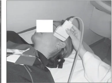
Figure 4. US transducer positioning for the study of TMJ articular disc.
Figure 5. Patient presenting complete anterior disc displacement. A: Ultrasonography demonstrating the non-visualization of the disc on the condylar surface (arrow). B: MRI demonstrating anterior articular disc displacement, which was not characterized at ultrasonography.
A B
The scans were performed with a HDI 5000 ATL apparatus (ATL, Philips Medi-cal Systems; Bothell, WA, USA), equipped with a 12.5 MHz transducer, after verbal detailed explanation on the procedure and after obtaining written consent from the patients and approval by the Human Re-search Ethics Committee of Universidade de São Paulo. All patients underwent MRI scans in a 1.0 or 1.5 T Philips apparatus, equipped with a TMJ coil, with sagittal and coronal T1- and T2-weighted sequences, without the utilization of paramagnetic contrast medium, since, according to the literature, such a procedure is considered
as the gold standard for the evaluation of TMJ disorders(1).
The US scans were performed with ob-lique axial sections, with the patients in dorsal decubitus (Figure 4), evaluating the following parameters: visualization or not of the articular disc with the mouth closed and after opening it, presence of joint effu-sion, morphological changes of the articu-lar disc, such as ruptures and degenerative processes, and in the mandibular condyle, such as the presence of osteophytes or ab-normalities on the articular surface.
The articular disc position was consid-ered normal when its posterior portion was
located between 12 and 1 o’clock on the ar-ticular surface of the mandibular condyle (Figure 4). In the present study, anterior displacement was considered whenever the posterior portion of the disc was located before the 12 o’clock position, the case was considered as being anterior displacement. Cases where the articular disc was not vi-sualized at US with the patient keeping the mouth closed were considered as being a disc displacement (Figure 5).
(test) were calculated and compared with the MRI results (gold standard) as follows: sensitivity, specificity, positive predictive value, negative predictive values and accu-racy. All the US scans were performed by an experienced radiologist and titular mem-ber of Colégio Brasileiro de Radiologia e Diagnóstico por Imagem.
RESULTS
Magnetic resonance imaging with the patients keeping their mouths closed dem-onstrated 24 TMJ with anterior disc dis-placement. In these same articulations, US demonstrated anterior displacement in 7, while in 13 the discs could not be visual-ized, and in 4, disc displacement was not observed.
In 48 joints, the articular disc could not be visualized at US with the patients keep-ing their mouths closed. Of those joints, 41 presented normal positioning at MRI and 7 presented anterior displacement.
Morphological alterations in the man-dibular condyle were visualized by MRI in 13 joints; on the other hand, US identified such alterations in 2 joints of this latter group. Articular effusion was visualized by MRI in 5 joints, and by US in only 1 joint. Magnetic resonance imaging demon-strated alterations in the articular discs, such as changes in signal intensity or loss of habitual morphology in 12 joints, which were not visualized at US.
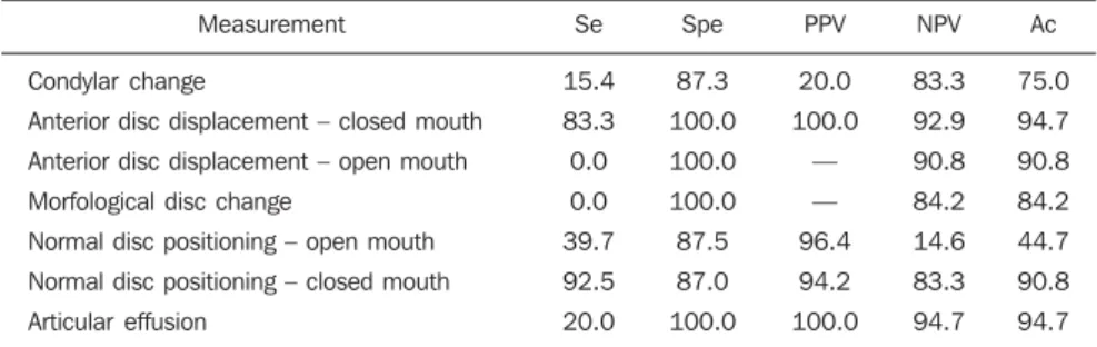
Table 1 shows the results from each one of the statistical measurements evaluated in the present study for each of the findings.
DISCUSSION
At MRI, 24 joints with anterior dis-placement of the articular disc were
iden-tified with patients keeping their mouths closed. In such joints, US demonstrated articular displacement in 7 cases, while in 13 cases the discs could not be visualized on the articular surface, and in 4 disc dis-placement was not observed at US. The authors considered that the non-visualiza-tion of the disc on the condylar surface with the patients at rest is related to the articu-lar disc displacement. Thus, US allowed the diagnosis of 20 (83.3%) among the 24 disc displacements observed in the present study. Such findings are similar to those in the studies developed by Emshoff et al.(8,9),
that demonstrated sensitivity and specific-ity around 90–95% for these parameters. Studies developed by Jank et al.(10) have
also demonstrated high sensitivity (90%) and specificity (84%) for the evaluation of the articular disc with the patient at rest.
Studies developed by Emshoff et al.(11)
have demonstrated accuracy > 90% in the evaluation of disc displacement by US. Similar results were obtained by Hayashi et al.(12), whose studies demonstrated
sen-sitivity and specificity of 83% and 96%, respectively.
The US findings related to condylar changes demonstrated that the method does not present yet significant sensitivity in the diagnosis of disorders related to the mor-phology and changes of the condylar cor-tical bone, a fact that was also observed by Emshoff et al.(13). Studies developed by
Brandlmaier et al.(14) have demonstrated
sensitivity of 87% and specificity of 20% in the diagnosis of TMJ osteoarthritis, in-dicating that US may be useful in the diag-nosis of the presence, but insufficient to diagnose absence of osteoarthritis.
The presence of articular effusion is an uncommon sign in asymptomatic pa-tients(15). More extensive articular effusion
may be observed in patients with articular dysfunction(16). The present study has not
included a significant number of patients to enable the establishment of parameters in relation to the sonographic diagnosis of presence of articular effusion, although recent studies report that US presents good sensitivity for such evaluation. Tognini et al.(17) have developed studies demonstrat-ing values sensitivity and specificity of about 75% for US in the detection of intra-articular fluid.
The method has not demonstrated sig-nificant sensitivity for the visualization of articular disc with the patient keeping the mouth open. In 48/76 joints (63%) the disc could not be visualized at US after the opening of the mouth. The disc could not be visualized either in 6 joints with open mouth in patients of the control group. Such finding is probably related to the medial displacement of the articular disc after opening the mouth, as the mandibu-lar condyle and the glenoid cavity do not allow appropriate ultrasound propagation, impairing the visualization of the articular disc.
Also, the method did not present signifi-cant sensitivity for the evaluation of mor-phological changes in the disc, as those iden-tified in 12 joints at MRI and in none at US. The present study demonstrates that, al-though still with limitations, the evaluation by US can become a useful option in the initial study of TMJ disorders in patients with contraindications for MRI, besides being more financially accessible, a fact with significant relevance in Brazil. In the future, further studies utilizing transducers with higher resolution may eventually al-low a more complete and accurate analy-sis of the joint.
CONCLUSIONS
Ultrasonography presented high sensi-tivity and specificity in the identification of TMJ articular disc displacement as com-pared with MRI that is the gold standard, and when the US scans were performed with the mouth closed. The results suggest that US can eventually be considered as an alternative method to detect the correct positioning or disc displacement in patients who cannot be submitted to MRI.
Table 1 Results comparison based on findings.
Measurement
Condylar change
Anterior disc displacement – closed mouth
Anterior disc displacement – open mouth
Morfological disc change
Normal disc positioning – open mouth
Normal disc positioning – closed mouth
Articular effusion Se 15.4 83.3 0.0 0.0 39.7 92.5 20.0 Spe 87.3 100.0 100.0 100.0 87.5 87.0 100.0 PPV 20.0 100.0 — — 96.4 94.2 100.0 NPV 83.3 92.9 90.8 84.2 14.6 83.3 94.7 Ac 75.0 94.7 90.8 84.2 44.7 90.8 94.7
The method has not presented satisfac-tory results in the characterization of articu-lar discs with patients keeping their mouths open as well as for detection of morpho-logical changes in articular discs and man-dibular condyles.
REFERENCES
1. Sano T. Recent developments in understanding temporomandibular joint disorders. Part 1: Bone marrow abnormalities of the mandibular condyle. Dentomaxillofac Radiol. 2000;29:7–10. 2. Ramos ACR, Sarmento VA, Campos PSF, et al.
Articulação temporomandibular – aspectos nor-mais e deslocamentos de disco: imagem por res-sonância magnética. Radiol Bras. 2004;37: 449– 54
3. Fritz J, Thomas C, Tzaribachev N, et al. MRI-guided injection procedures of the temporoman-dibular joints in children and adults: technique, accuracy, and safety. AJR Am J Roentgenol. 2009;193:1148–54.
4. Katzberg RW. Temporomandibular joint imaging. Radiology. 1989;170:297–307.
5. Sommer OJ, Aigner F, Rudisch A, et al.
Cross-sec-tional and funcCross-sec-tional imaging of the
temporoman-dibular joint: radiology, pathology, and basic bio-mechanics of the jaw. Radiographics. 2003; 23:e14.
6. Milano V, Desiate A, Bellino R, et al. Magnetic resonance imaging of temporomandibular disor-ders: classification, prevalence and interpretation of disc displacement and deformation. Dento-maxillofac Radiol. 2000;29:352–61.
7. Schmitter M, Kress B, Ludwig C, et al. Temporo-mandibular joint disk position assessed at coro-nal MR imaging in asymptomatic volunteers. Radiology. 2005;236:559–64.
8. Emshoff R, Jank S, Rudisch A, et al. Are high-resolution ultrasonographic signs of disc dis-placement valid? J Oral Maxillofac Surg. 2002; 60:623–8; discussion 628–9.
9. Emshoff R, Jank S, Rudisch A, et al. Error pat-terns and observer variations in the high-resolu-tion ultrasonography imaging evaluahigh-resolu-tion of the disk position of the temporomandibular joint. Oral Surg Oral Med Oral Pathol Oral Radiol Endod. 2002;93:369–75.
10. Jank S, Rudisch A, Bodner G, et al. High-resolu-tion ultrasonography of the TMJ: helpful diagnos-tic approach for patients with TMJ disorders? J Craniomaxillofac Surg. 2001;29:366–71. 11. Emshoff R, Jank S, Bertram S, et al. Disk
displace-ment of the temporomandibular joint: sonography
versus MR imaging. AJR Am J Roentgenol. 2002;178:1557–62.
12. Hayashi T, Ito J, Koyama J, et al. The accuracy of sonography for evaluation of internal derange-ment of the temporomandibular joint in asymp-tomatic elementary school children: comparison with MR and CT. AJNR Am J Neuroradiol. 2001; 22:728–34.
13. Emshoff R, Brandlmaier I, Bodner G, et al. Condylar erosion and disc displacement: detec-tion with high-resoludetec-tion ultrasonography. J Oral Maxillofac Surg. 2003;61:877–81.
14. Brandlmaier I, Rudisch A, Bodner G, et al. Tem-poromandibular joint internal derangement: de-tection with 12.5 MHz ultrasonography. J Oral Rehabil. 2003;30:796–801.