ABSTRACT
http://dx.doi.org/10.1590/1678-775720130546
Oral cavity infection: an adverse effect after the
treatment of oral cancer in aged individuals
Jie PAN1 2, Ning JIANG2
1- Department of Orthodontics, Shanghai Stomatological Disease Centre, Shanghai, China.
2- Department of Orthodontics, College of Stomatology, Ninth People’s Hospital, School of Medicine, Shanghai Jiao Tong University, Shanghai, China.
Corresponding address: Ning Jiang - Department of Orthodontics, College of Stomatology, Ninth People’s Hospital, School of Medicine, Shanghai Jiao Tong University - Shanghai 200011 - China - e-mail: [email protected]
!" #
O
bjective: The immune compromised patients after treatment of oral cancer may have a chance of infection by drug-resistant opportunistic microbes. We investigated the occurrence of opportunistic microorganisms in aged individuals receiving follow-up examinations after treatment of oral cancer in China. Material and Methods: These patients were used as test group and the respective age grouped healthy individuals as control group. In this study, the oral cavity microorganisms such as bacteria and yeast were taken for the analysis. After the screening of representative microorganisms, their aptitude of pervasiveness against drugs was studied. Here, we used antimicrobial agents which are common in clinical practice. We also performed studies to investigate the presence of toxin genes in methicillin-resistant S. aureus (MRSA). Results: The results indicate that the prevalence of drug-resistant microbes was more pronounced in oral cancer patients after initial treatment above 70 years old. The oxacillin resistance of S. aureus isolate compromise in elderly patients. Conclusions: This study reveals the occurrence of drug-resistant opportunistic microorganisms in oral cavity after treatment for oral cancer in aged individuals. Special attention should be directed to MRSA during the treatment of oral cancer, and to realize the fact of immune compromise in elderly patients.Keywords: Oral cancer. Dental infection control. Drug resistance. Opportunistic infections. Prevention and control.
INTRODUCTION
The term oral cavity refers to lips, buccal mucosa, alveolar ridges, retro molar trigone, hard of the tongue. Oral cancer or oral cavity cancer, a subtype of head and neck cancer, is any cancerous tissue growth located in the oral cavity24. The most
common oral cancer is squamous cell carcinoma (SCC) that affects the tissue lining of the oral cavity5. Oral cancer is the eleventh most common
cancer in the world with an estimated 267,000 cases and 128,000 deaths in around 2000, two-third of which occur in developing countries. The incidence of oral cancer is increasing in several parts of the world, particularly in Australia, Japan and parts of Europe. Oro-pharyngeal cancer is a !
cancer occurrence is particularly high in males. Incidence rates for oral cancer vary in men from 1 to 10 cases per 100,000 populations in many countries. Tobacco and alcohol are regarded as the major causes of oral cancer10.
The immune compromised patients after treatment of oral cancer may have a chance of infection by drug-resistant opportunistic microbes. Such microbes that originate in the oral cavity, representative microorganisms include
Staphylococcus aureus, Pseudomonas aeruginosa, and Candida albicans1,14.Pseudomonas aeruginosa
is increasingly recognized as an emerging opportunistic pathogen of clinical relevance. One of the most worrying characteristics of P. aeruginosa
is its low antibiotic susceptibility23. This low
S.No. Antibiotics Sensitivity dose
Resistance dose
$%&'*+ $%&'*+
1 Oxacillin <0.25 >2
2 Arbekacin <4 >8
3 Vancomycin <2 >8
4 Teicoplanin <8 >16
5 Linezolid <2 >4
6 Fluconazole <8 >64
7 <4 >32
8 Itraconazole <0.125 >1
9 Miconazole <0.5 >1
10 Amphotericin B <1 >2
11 Voriconazole <1 >4
12 Micafungin <1 >2
13 Imipenem/ cilastatin
<16 >16
14 Amikacin <32 >32
15 <4 >4
Figure 1- Antibiotics used to test the antimicrobial activity
encoded antibiotic resistance genes15.Most of these
organisms have become drug-resistant, which has diseases. The Candida species educe the infectious disease candidiasis, which causes infections such as oral thrush and vaginitis as well as life-threatening diseases, known as candidemia16. C. albicans are
yeast that normally inhabits the human mouth and skin, where it generally uneventfully coexists with a variety of other microorganisms. An infection occurs when the balance of bacteria in the body is disrupted, especially in immunocompromised
situations, allowing drug-resistant Candida
species to proliferate and overcome other healthy microorganisms11. Immunocompromised situations
are frequently seen in older individuals, infants, people infected with HIV, and individuals with cancer; oral cancer can reduce immunity in the maxillofacial region1,14. The principal treatments for
oral cancer are surgical excision, radiotherapy, and chemotherapy, employed alone or in combination13.
Currently, the systemic applications of antibacterial drugs have shown better results on curing diseases than local application, which could induce drug-resistant bacteria in the particular area2,3. In this study, we have investigated the
reason behind the frequent cause of oral infection after the treatment of oral cancer in elderly Chinese.
MATERIAL AND METHODS
Patient study
Out of several elderly patients (60–95 years old), 128 patients who had undergone treatment for oral-cavity related problems participated in the study at Ninth People’s Hospital, Shanghai Jiao Tong University. We have excluded patients with other systemic diseases like autoimmune disease or diabetes to avoid misleading of our parameter. The participants were divided into two groups: Group I - the patients undergone oral cancer treatment (n=93; 41 men, 52 women; average age 68.1±8.3 years) ranging from 1 month to 7 years after the initial treatment, 43 members were involved in species identification, remaining 50 members involved in drug-resistant test. These patients have been treated with surgery, chemo and/or radiotherapy; Group II - the control (n=35; 15 men, 20 women; average age 70.2±10.1 years), who had received treatment for oral cavities or with no history of any cancer treatment. The $ % & '* Hospital of Shanghai Jiao Tong University, according to Helsinki Declaration II, approved the study. Written informed consent was obtained from each participant.
Sample collection
Microbes were collected from the areas of surgery (Group I), tongue, gingiva, and palate by using sterilized dry cotton swabs. After wetting, the cotton swab was immediately put into an airtight sterilized test tube. To collect anaerobic microbes # deep area of the incision. Collected samples were immediately put into an anaerobic sample collection + < > was collected from each subject for the analysis.
Microbial cultivation
The microorganisms were cultured on nalidixic acid/cetrimide agar (Sigma-Aldrich Chemie GmbH,
?@ Pseudomonas
species, while Mannital Salt agar (Acumedia Manufacturers, USA) was used for the first screening of Staphylococcus species and Brilliance
Candida FK L $@
Candida species.
The nalidixic acid/cetrimide agar and Mannital Salt agar plates were incubated at 37°C under aerobic conditions for 2 days, while Candida-GS agar plates were incubated at 30°C for 3 days.
Microbial species detection and antimicrobial testing
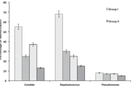
Figure 2- two species in same isolates were presented in two sets of group I & II in each species
Figure 3-
for species detection. Genomic DNA from each colony was obtained using a Wizard genomic extraction kit (Promega, Madison, WI, USA). For
Staphylococcus species, cells were treated with 1 mg/mL of lysostaphin in Tris-EDTA buffer (TE; 10 mM Tris-Cl, 1 mM EDTA, pH 8.0) at 37°C for 1 h + L # # F'Y@ with bacterial universal primers for 16S rDNA and 26S rDNA for fungus, followed by DNA sequencing. DNA sequencing was performed using an ABI Prism 3100 Genetic Analyzer (Applied Biosystems, Tokyo, Japan) with a Big Dye Cycle Sequencing reaction kit (AB Applied Biosystems). Identification of experimentally determined nucleotide sequences using sequence databases was performed by
Basic Local Alignment Search Tool (BLAST). The
antimicrobial activity was tested with respective antibiotics used commonly in clinical practice (Figure 1).
Statistical analysis
Statistical analyses were conducted using SPSS 15.0 (SPSS Inc., U.S.A.) and any differences at p<0.05 level were considered as statistically K were compared using independent student t-test.
RESULTS
,GHQWL¿FDWLRQRIPLFURRUJDQLVPV
The samples were collected from surgical scar, saliva, gingiva, and palate of 78 elderly Chinese (43 cases of group I and 35 cases of group II). K [#
Candida, Staphylococcus and Pseudomonas
Figure 4- Candida Species
=
Group I (n=43) Group II (n=35)
No. of positive samples
Percentage (%) No. of positive
samples
Percentage (%)
C. albicans 24 55.8 10 28.6*
C. glabrata 21 48.8 8 22.8*
C. krusei 12 27.9 6 17.1
C. guilliermondii 4 9.3 1 2.8
C. africana 5 11.6 2 5.7
!!!"#$%$'
Table 1-( Candida species and their percentage of occurrence
number of participants with these isolates.
Candida species
The universal primers for fungi, 26S rDNA
sequencing was used to identify the Candida
species (Figure 3). The Candida species isolated ]^ F @
C. albicans (43.6%), C. glabrata (37.2%), C. krusei
(23.1%), C. africana (9%) and C. guilliermondii
(6.4%). The number of Candida species isolated from group I were greater than that of group II. On comparison between the groups, the C. albicans
were found to be the dominant species in both group I (55.8%) and group II (28.6%). However, F`{{|@ between groups. Similarly, C. glabrata were found " difference between group I (48.8%) and group II (22.8%) participants (Table 1). However, no
C. africana
and C. guilliermondii species, rather very less groups (Table 1).
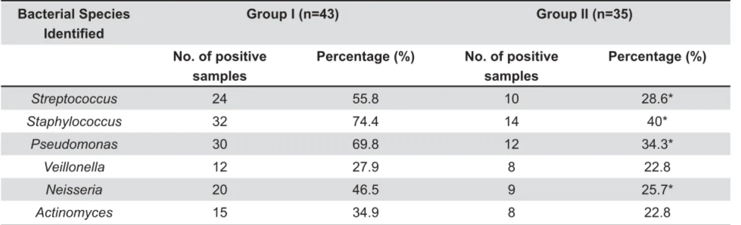
Bacterial species
The universal primers for bacteria, 16S rDNA } (Figure 4). The bacterial species isolated from ]^ F @
Staphylococcus (59%), Pseudomonas (53.8%),
Streptococcus (44%), Neisseria (37.2%),
Actinomyces (29.5%) and Veillonella (25.6), which are mostly found in the surgical scar and saliva. On comparison between two groups of isolates, the
Streptococcus, Staphylococcus, Pseudomonas and
Neisseria F`{{|@ in percentage of these species between groups I and II (Table 2). In Group I, the numbers of positive isolates of bacterial species were greater than that of the control group using traditional oral care methods. For Veillonella and Actinomyces, no groups (Table 2).
Occurrence of microorganism against respective antimicrobial agents
Antibiotics Percentage of drug-resistance in 25 cases
C. glabrata C. krusei
Sample # % Sample # %
Itraconazole 25 100 16 60
Miconazole 5 20 11 44
Fluconazole 5 20 8 32
2 8 7 28
Amphotericin B S - S
-Voriconazole S - S
-Micafungin S - S
-Table 3- Prevalence of Candida species against respective antibiotics
S=Susceptible
Bacterial Species =
Group I (n=43) Group II (n=35)
No. of positive samples
Percentage (%) No. of positive
samples
Percentage (%)
Streptococcus 24 55.8 10 28.6*
Staphylococcus 32 74.4 14 40*
Pseudomonas 30 69.8 12 34.3*
Veillonella 12 27.9 8 22.8
Neisseria 20 46.5 9 25.7*
Actinomyces 15 34.9 8 22.8
!!!"#$%$'
Table 2-(
the reason behind this dominant character, we studied the occurrence of microorganisms against the antimicrobial agents by categorizing the remaining group I participants (50 cases) with their history of treatment with commonly practiced antimicrobial agents (Table 1) after the treatment for oral cancer. These 50 participants were divided as 25 cases for the study against antifungal agents and remaining 25 against antibacterial agents.
The participants after the treatment of oral cancer were taking commonly practiced antifungal agents such as Itraconazole, Miconazole, L? |# % Voriconazole and Micafungin. The isolates were tested for the presence of Candida species. Except for the C. glabrata and C. krusei species, all other species were susceptible to the antifungal agents. Interestingly, Itraconazole had 100% resistance by
C. glabrata and 60% resistance by C. krusei species. The C. glabrata showed resistance to Miconazole F~{@ L? F~{@ | # F^@
However, C. krusei showed 44%, 32% and 28%
of resistance respectively. The other drugs were susceptible to all Candida species (Table 3).
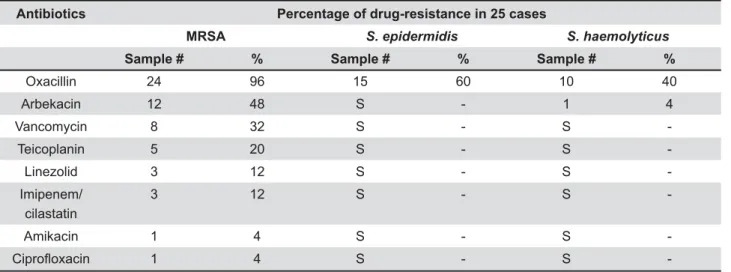
The antibacterial agents were tested for the drug-resistant activity of staphylococci subspecies, particularly on methicillin-resistant Staphylococcus aureus (MRSA), S. epidermidis
and S. haemolyticus. The tested agents are commonly practiced antibiotics such as Oxacillin, Arbekacin, Vancomycin, Teicoplanin, Linezolid, $ + Y" Surprisingly, MRSA showed drug-resistance to almost all antibiotics with variation in percentage of resistance. The MRSA showed the highest percentage of resistance to Oxacillin (96%) and Arbekacin. However, the lowest percentage (4%) of resistance was shown in relation to + Y" FK @ K species, S. epidermidis and S. haemolyticus,
Antibiotics Percentage of drug-resistance in 25 cases
MRSA S. epidermidis S. haemolyticus
Sample # % Sample # % Sample # %
Oxacillin 24 96 15 60 10 40
Arbekacin 12 48 S - 1 4
Vancomycin 8 32 S - S
-Teicoplanin 5 20 S - S
-Linezolid 3 12 S - S
-Imipenem/ cilastatin
3 12 S - S
-Amikacin 1 4 S - S
1 4 S - S
-Table 4- Prevalence of bacterial species against respective antibiotics
S=Susceptible
DISCUSSION
In this study, a prevalence of drug-resistant microorganisms in the oral cavity after treatment of oral cancer was performed in aged Chinese. The study was carried out in Ninth People’s Hospital, School of Medicine, Shanghai Jiao Tong University, Shanghai. The participants were the patients from this hospital who had undergone oral cancer treatment and others without any history of cancer but that could be patients who had undergone treatment for other oral diseases. This study was aimed to investigate the reason behind the frequent cause of oral infection after the treatment of oral cancer in elderly individuals.
Several lines of evidence support our views that there is a possibility of drug-resistant microbes present in immunocompromised patients, particularly after the treatment of oral cancer. In general, infections are commonly found in oral cancer patients after surgical excision of the tumor17–20. This might be due to wound exposure
during and after the operation, even if sutured, when microorganisms may infect oral regions, oropharynx, nasal cavity, and paranasal sinuses areas. Patients after oral cavity surgery often appear to have complications after bacterial infections, as well. Colonization of pathogenic bacteria in oral cavity is thought to increase the risk of infections such as pneumonia and bacteraemia4,7. It is therefore of high importance
the prevention from or cure for the infections. During the development of tumor, the tumor cells or soluble products produced by tumor cells inactivate lymphocytes and provoke immunosuppression in the body9,12.
The high percentages of microbial species
Candida, Staphylococcus, Streptococcus and
Pseudomonas species (Figure 2). The highest numbers of these species were found in patients after the treatment of oral cancer in comparison with the non-cancer patients (Tables 1 and 2). Similar results were obtained in a phase 1 clinical trial with the application of an antibacterial dressing spray in the prevention of post-operative infection in oral cancer patients25. Antimicrobial drugs
have been very helpful to prevent infections after surgery. People, however, sometimes misuse them and have antimicrobial drugs abuse. The abuse of antimicrobial drugs brings severe adverse effects to people, for example, allergy, toxic reaction, and opportunistic infections18. As a result, a new and
ideal preventive method is needed for people who have surgery to reduce the chances of bacterial infections.
In this study, after revealing the high percentage of microorganisms found particularly in post-treatment of oral cancer, we started to focus # microbes. The antimicrobial agents (Figure 1), which are common in clinical practice, were taken for this study. The isolates were tested for the presence of Candida species and subspecies of staphylococci, particularly on MRSA, S. epidermidis
and S. haemolyticus. Except for the C. glabrata
and C. krusei species, all other Candida species were susceptible to the antifungal agents (Table 3). The MRSA showed the highest percentage of resistance to Oxacillin (96%) and Arbekacin. The other species, S. epidermidis and S. haemolyticus,
periodontitis were mecA-positive (antimicrobial resistance)6. Another study, in the United States,
reported that the prevalence of MRSA organisms in the nasal and oral cavities of nursing home residents was 20–35%8. Furthermore, a Japanese
group investigated MRSA colonization in neonatal intensive care units and found that 207 (49.9%)
of 415 newborns had MRSA organisms22. When
compared with these results, the drug-resistance (96%) of MRSA against oxacillin was extremely high, possibly due in part to the tendency to prescribe long-term, high-dose antibiotic treatment in Japan21.
CONCLUSION
Our study revealed a group of opportunistic-microorganisms such as C. glabrata, C. krusei and subspecies of staphylococci, particularly Methicillin-resistant S. aureus (MRSA), S. epidermidis and
S. haemolyticus. Nevertheless, anti-tumor drugs used in tumor treatment will also lead to immune and bone marrow suppression. Therefore, post-treatment infections occurred13,14. Continuous
monitoring and a basic infection control strategy, including standard precautions, are important for older individuals, especially those receiving follow-up care for oral cancer. There is always a risk that they may become immunocompromised hosts easily susceptible to oral infections. Clinicians must also pay careful attention to opportunistic-microorganisms during the treatment of oral cancer, and to realize the fact of immune compromise in elderly patients.
REFERENCE
1- Belazi M, Velegraki A, Koussidou-Eremondi T, Andreadis D, Hini S, Arsenis G, et al. Oral Candida isolates in patients undergoing radiotherapy for head and neck cancer: prevalence, azole # ! Microbiol Immunol. 2004;19,347-51.
2- Belusic-Gobic M, Car M, Juretic M, Cerovic R, Gobic D, Golubovic V. Risk factors for wound infection after oral cancer surgery. Oral Oncol. 2007;43(1):77-81.
3- Cloke DJ, Green JE, Khan AL, Hodgkinson PD. McLean NR. L % ' 2004;57:556-60.
Y ' ' % common cause of persistent infections. Science. 1999;284:1318-22.
5- Crissman JD, Zarbo RJ. Dysplasia, in situ carcinoma, and progression to invasive squamous cell carcinoma of the upper aerodigestive tract. Am J Surg Pathol. 1989;13(Suppl. 1);5-16. 6- Cuesta AI, Jewtuchowicz VM, Brusca MI, Mujica MT, Rosa AC. Antibiotic susceptibility of Staphylococcus aureus isolates in oral mucosa and pockets of patients with gingivitis-periodontitis. Acta Odontol Latinoam. 2011;24,35-40.
7- Gosney MA, Preston AJ, Corkhill J, Millns B, Martin MV. Pseudomonas aeruginosa septicaemia from an oral source. Br Dent J. 1999;187:639-40.
8- Hall DL. Methicillin-resistant Staphylococcus aureus and infection control for restorative dental treatment in nursing homes. Spec Care Dentist. 2003;23,100-7.
9- Jewett A, Head C, Cacalano NA. Emerging mechanisms of immunosuppression in oral cancers. J Dent Res. 2006;85(12):1061-73.
10- Khan Z. An overview of oral cancer in Indian subcontinent and recommendations to decrease its incidence. 2012 [cited Nov. 10 2013] Available from: http://www.webmedcentral.com/ article_view/3626.
11- Li L, Redding S, Dongari-Bagtzoglou A. Candida glabrata: an emerging oral opportunistic pathogen. J Dent Res. 2007;86,204-15.
12- Mulder WM, Bloemena E, Stukart MJ, Kummer JA, Wagstaff J, Scheper RJ. T-cell receptor-zeta and granzyme B expression carcinoma. Gut. 1996;40:113-9.
13- Myers EN, Suen JY, Myers JN, Hanna EY. Cancer of the head and neck. 4th ed. Philadelphia: Saunders; 2003.
14- Napeñas JJ, Brennan MT, Bahrani-Mougeot FK, Fox PC, Lockhart PB. Relationship between mucositis and changes in oral # ! ! ! Pathol Oral Radiol Endod. 2007;103,48-59.
<| ' " bacteria. Clin Microbiol Infect. 2004;10:12-26.
16- Repetto EC, Giacomazzi CG, Castelli F. Hospital-related outbreaks due to rare fungal pathogens: a review of the literature from 1990 to June 2011. Eur J Clin Microbiol Infect Dis. 2012;31:2897-904.
17- Senpuku H, Sogame A, Inoshita E, Tsuha Y, Miyazaki H, Hanada N. Systemic disease in association with microbial species in oral # } # ~{{{< 18- Senpuku H, Tada A, Uehara S, Kariyama R, Kumon H. Postoperative infection by pathogenic micro-organisms in the oral cavity of patients with prostatic carcinoma. J Int Med Res. 2006;34(1):95-102.
19- Tada A, Hanada N, Tanzawa H. The relation between tube feeding and Pseudomonas aeruginosa detection in the oral cavity. J Gerontol A Biol Sci Med Sci. 2002;57:M71-2.
20- Tada A, Watanabe T, Yokoe H, Hanada N, Tanzawa H. Oral # # and type and quality of facilities for the bedridden. J Appl Microbiol. 2002;93:487-91.
21- Takeda S, Yasunaka K, Kono K, Arakawa K. Methicillin-resistant Staphylococcus aureus (MRSA) isolated at Fukuoka University Hospital and hospitals and clinics in the Fukuoka city area. Int J Antimicrob Agents. 2000;14:39-43.
22- Uehara Y, Kikuchi K, Nakamura T, Nakama H, Agematsu K, Kawakami Y, et al. Inhibition of methicillin-resistant Staphylococcus aureus colonization of oral cavities in newborns by viridans group streptococci. Clin Infect Dis. 2001;32:1399-1407.
23- Van Eldere J. Multicentre surveillance of Pseudomonas aeruginosa susceptibility patterns in nosocomial infections. J Antimicrob Chemother. 2003;51(2):347-52.
24- Werning, JW. Oral cancer: diagnosis, management, and rehabilitation. New York: Thieme; 2007.