INTRODUCTION
S
urgical resection of rectal cancer is still the only possibility of cure and is still regarded as the main form of treatment for many authors1.2. Theevolu-tion of the surgical technique, except for the access routes, reached its peak after finding that the total mesorectal excision and resection of the circumfer-ential margin significantly decrease local recurrence3.
In the 90s, it has become consensus that the treatment of adenocarcinoma of the rectum stages II and III would require, in addition to the operation, complementary chemotherapy and ra-diotherapy, after the discovery of their beneficial effects in reducing disease recurrence and increas-ing long-term survival rates4. Despite this evolution,
the treatment of rectal cancer remains challenging, since long-term survival has not evolved consistent-ly5. Eight large clinical series were published that
analyzed neoadjuvant therapy for rectal cancer. All these studies demonstrated a superiority of this therapeutic modality when compared with surgery performed in an exclusive manner, as well as in re-lation to the adjuvant therapy2.
Among the benefits of neoadjuvant radiotherapy and chemotherapy, there are: in-creased preoperative radiosensitivity of tissues due to the absence of surgical fibrosis, lower ex-posure of the small intestine to radiation, lower systemic toxicity, and decrease in lesions’ size, which increase resectability and the sphincter preservation rate6. As disadvantages we have:
the potential deficiency in the accurate determi-nation of pathologic stage, which may result in failure of the postoperative planning, the post-ponement of definitive surgical treatment, and possible increase in morbidity and operative mor-tality7. Currently, patients with resectable lower
Impact of neoadjuvant therapy in downstaging of lower rectal
adenocarcinoma and the role of pelvic magnetic resonance in staging
Impacto da terapia neoadjuvante na diminuição do estádio no adenocarcinoma de
reto baixo: papel da ressonância magnética da pelve na determinação do estádio
karina dagre Magri1; Fang Chia Bin, tCBC-sp1; Fernanda Bellotti ForMiga1; thiago da silveira Manzione, aCBC-sp2; Caroline
MerCi Caliari de neves goMes1; paulo de azeredo passos Candelári,tCBC-sp2; Jorge alBerto ortiz,tCBC-sp2; WilMar artur
klug1; José Mandia neto1; peretz CapelhuChnik, tCBC-sp1.
A B S T R A C T
Objective: to evaluate the effect of neoadjuvant therapy on the stage (TNM) of patients with rectal adenocarcinoma and validate the use of MRI as a method of determining locoregional stage. Methods: we conducted a retrospective study of 157 patients with lower rectum adenocarcinoma, whom we divided into two groups: Group 1, 81 patients (52%) who had undergone surgical treatment ini-tially, with the purpose to analyze the accuracy of locoregional staging by pelvic magnetic resonance imaging throug the comparison of radiological findings with pathological ones; Group 2, 76 patients (48%), who had been submitted to neoadjuvant therapy (chemothe-rapy and radiation) prior to definitive surgical treatment, so as to evaluate its effects on the stage by comparing clinical and radiological findings with pathology. Results: In group 1, the accuracy of determining tumor depth (T) and lymph node involvement (N) was 91.4% and 82.7%, respectively. In group 2, neoadjuvant therapy decreased the T stage, N stage and TNM stage in 51.3%, 21% and 48.4% of cases, respectively. Conclusion: neoadjuvant therapy in patients with rectal adenocarcinoma is effective in decreasing disease stage, and pelvic magnetic resonance imaging is effective for locoregional staging.
Keywords: Adenocarcinoma. Rectal Neoplasms. Neoadjuvant Therapy. Magnetic Resonance Imaging. Neoplasm Staging.
rectal cancer in stages II and III should be submit-ted to neoadjuvant therapy provided they do not have medical contraindications8.
Thus, the precise determination of the stage (TNM) is essential for the treatment to be well in-dicated2. As a general rule, Computerized
Tomogra-phy of the chest and abdomen is the choice for the detection of metastatic disease (M), and the pelvic magnetic resonance imaging (MRI) or the transrectal ultrasonography are better to determine the locore-gional stage (T and N)9. Conceptually, the ultrasound
exam is superior in the analysis of smaller and more superficial tumors when compared with MRI, which has better accuracy in larger tumors that extend be-yond the circumferential margin10,11.
The objectives of this study were to evalu-ate the effect of neoadjuvant therapy on the stage of patients with low rectal adenocarcinoma and to validate the use of MRI as a method of determining locoregional stage.
METHODS
We held a retrospective analysis of 157 medical records of patients diagnosed with lower rec-tal adenocarcinoma during the period from February 2005 to October 2012. This study was approved by the Ethics in Research Committee of the Irmandade da Santa Casa de Misericórdia de São Paulo, under number 109,338.
We divided patients into two groups ac-cording to the initial therapeutic approach: Group 1, patients initially referred to surgical treatment, on an elective basis, after preoperative staging; Group 2, patients who, after having their stage determined, were referred to neoadjuvant therapy prior to defin-itive surgical treatment. The operation in these cases was performed eight weeks after completion of the neoadjuvant therapy, without further staging by im-aging methods.
We performed preoperative staging by physical, proctologic and radiological examination, CT scan of the chest and upper abdomen to assess systemic disease (distant metastases) and pelvic MRI to evaluate locoregional involvement. The final stage
was determined by the pathological examination of surgical specimens, associated with pre- and in-traoperative findings. For the stage description, we adopted the system described by the American Joint Committee on Cancer12.
All imaging tests in this series were per-formed at the Radiology Service of the Irmandade da Santa Casa de Misericórdia de São Paulo, using MRI machines models Philips Intera 1.0T or Philips Achie-va 1.5T SE.
Depending on tumor location and intraop-erative conditions, the performed procedures were abdominal rectosigmoidectomy or rectal amputation with total mesorectal excision.
The chemotherapy regimen employed in pa-tients undergoing neoadjuvant therapy was 5-fluoro-uracil at a dose of 380 mg/m2 and Leucovorin 20 mg/
m2 for five consecutive days (D1 to D5) concurrent
with the first and fifth week of radiation therapy. The body surface area was obtained from the formula: Weight (kg)0.425 x Height (cm)0.725 x 71.84 /10.000.
Radiotherapy consisted of 28 sessions in five weeks and three days of 180cGy per session, to-tal 5040 cGy.
We excluded from the study patients with history of colorectal cancer surgery, those operated in the emergency department or undergoing pallia-tive surgery, and those who abandoned treatment.
We analyzed the variables gender, age at diagnosis, depth of tumor invasion in the rectal wall (T), lymph node involvement (N), presence of metas-tases (M), preoperative and final stages (TNM).
For the statistical analysis of the results we applied the Wilcoxon and McNemar tests to veri-fy possible differences between variables T, N, M and the stage of both groups. We did not compare Groups 1 and 2. We used a spreadsheet software for data organization and the IBM SPSS (Statistical Package for Social Sciences), version 21.0, to obtain the results.
RESULTS
and 76 (48%) to Group 2, in which the neoadjuvant therapy was performed before the operation.
The average age of Group 1 patients was 58.27 years ± 13.15, while in Group 2 it was 59.96 years ± 11.81.
In Group 1, 33 (41%) individuals were women, with a mean age of 58.63 years ± 13.44, and 48 men were (59%), mean age 58.02 years ± 13.09.
As for Group 2 patients, 37 (49%) were women, with a mean age of 59.56 years ± 12.62, and 39 men (51%), mean age of 60.33 years ± 11.15
Group 1 Results
The analysis of radiological and patholog-ical correlation of the T variable detected no statis-tically significant variation, with accuracy of 91% (Figure 1).
The analysis of radiological and patholog-ical correlation of the N variable detected no statis-tically significant variation, with accuracy of 83% (Figure 2).
The variable M remained constant, both pre and postoperatively. The correlation between clinical stage and the final stage showed agreement in 84% of cases. In 11% of cases, the stage was initially underestimated, and in 4%, overestimated. There was one case in which the lesion was not de-tected (Figure 3).
Group 2 Results
The analysis of the effect of neoadjuvant therapy on the variable T showed that there was gression in 51% of cases, and the pathological re-sponse (T0) occurred in 17% of cases (Table 1).
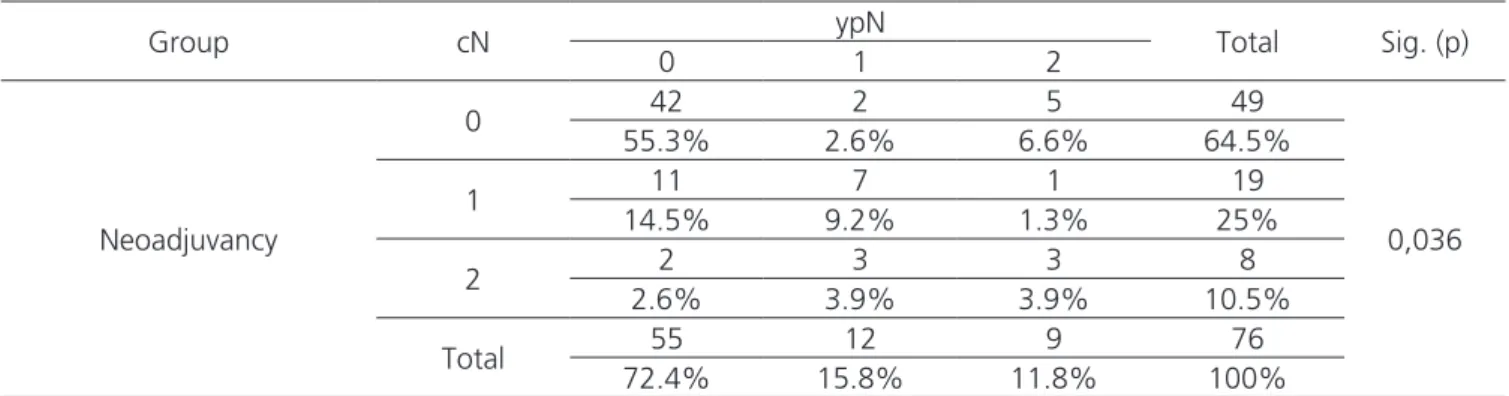
The analysis of the effect of neoadjuvant therapy on the variable N showed regression in 21% of cases (Table 2).
The analysis of the effect of neoadjuvant therapy on the variable M demonstrated that there was an increase in the occurrence of distant metas-tases of around 7%, with no statistical significance.
The analysis of the effect of neoadjuvant therapy on stage displayed a regression of 48.5%
Figure 1: Radiological and pathological correlation of changes in va-riable T in the 81 Group 1 patients. Source: ISCMSP, 2013.
Figure 2: Radiological and pathological correlation of changes in variable N in the 81 Group 1 patients. Source: ISCMSP, 2013.
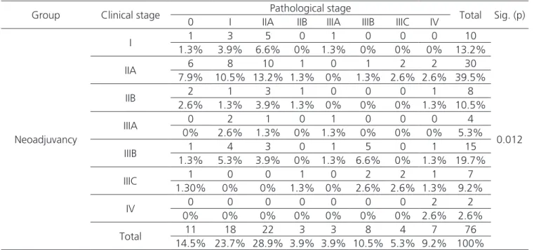
and a 20% increase. We observed a complete patho-logical response rate of 14.5%, which corresponds to 11 cases (Table 3). The exclusion of stages I and IV shows that stage regression occurs in 56% of cases, with complete pathological response in 16%.
DISCUSSION
The first large prospective randomized study that demonstrated the effectiveness of neoadjuvant chemotherapy and radiotherapy came from Germany in 2004. It randomized 823 patients to receive chemo-therapy and radiation preoperatively (421 cases) and postoperatively (402 cases). The authors found that the incidence of local recurrence at five years was 6% versus 13%, respectively. There was no significant in-crease in survival in five years between the groups13.
Although the optimal regimen of neoadju-vant treatment is not yet well defined, there is no doubt of its effectiveness, especially in the control of local
re-currence, and hence the increase in disease-free inter-val 2. There is a polarization between the European and
US institutions. In European publications, preference is mainly for short cycles of neoadjuvant radiotherapy, be-cause they have lower morbidity. In American studies, similar to what was done in this study, preference is given to cycles with longer duration, arguing that the reduction of tumor size is more efficient.
The intent of this study was to analyze, in a stratified manner, the effects of neoadjuvant therapy on the stage (TNM) and on their individual variables in Group 2 patients. These variables were determined in two specific moments: preoperatively, with the aid of pelvic MRI (for the determination of locoregional stage – T and N) and chest and abdomen CT scan (for detecting distant metastases – M); and in the postop-erative period, through histopathology data.
One might question maintaining the M variable in this study, since neoadjuvant therapy has essentially locoregional effects. However, its analysis Table 1: Variable T: comparison between radiological determination and histopathology in Group 2.
Group cT ypT Total Sig. (p)
0 1 2 3 4
Neoadjuvancy
1 1 0 0 1 0 2
< 0,001
1.3% 0% 0% 1.3% 0% 2.6%
2 2 1 4 6 0 13
2.6% 1.3% 5.3% 7.9% 0% 17.1%
3 8 1 13 21 1 44
10.5% 1.3% 17.1% 27.6% 1.3% 57.9%
4 2 0 1 10 4 17
2.6% 0% 1.3% 13.2% 5.3% 22.4%
Total 13 2 18 38 5 76
17.1% 2.6% 23.7% 5% 6.6% 100%
Source: ISCMSP, 2013. Teste dos Postos Sinalizados de Wilcoxon.
Table 2: Variable N: comparison between radiological determination and histopathology in Group 2.
Group cN ypN Total Sig. (p)
0 1 2
Neoadjuvancy
0 42 2 5 49
0,036
55.3% 2.6% 6.6% 64.5%
1 11 7 1 19
14.5% 9.2% 1.3% 25%
2 2 3 3 8
2.6% 3.9% 3.9% 10.5%
Total 55 12 9 76
72.4% 15.8% 11.8% 100%
is mainly for the correct determination of the stage, which depends critically on the three variables (T, N and M ). Furthermore, despite its systemic effects are still scarcely mentioned, some authors demonstrated that neoadjuvant chemotherapy may start the early systemic treatment of metastases, and be used as a marker of tumor response, which may enhance sub-sequent treatment14.
Group 1 corresponds to a period in which neoadjuvant therapy was not yet established in the service. From the end of 2007 on, patients who presented in clinical stage II or III (T3 N0 M0 or T1,2,3 N1,2 M0) have been submitted to neoadju-vant therapy.
The reason for analyzing Group 1 patients, of different treatment, was primarily to assess the quality in determining the clinical stage in our ser-vice, as it was the same throughout the sample of this series, both in Groups 1 and 2. Thus, the bias of overstaging or understaging, that MRI may po-tentially present, was eliminated. In Group 1, there was a significant correlation between the clinical and pathological stages, which occurred in 91% of cases for the variable T and 83% for the variable N.
For many authors, pelvic MRI is considered the most suitable technique for determining
locore-gional stage, due to its high sensitivity and specificity in the analysis of structures adjacent to the rectum, including the mesorretal fascia15. Likewise, it is the
only available technique for the proper assessment of the circumferential margin (CRM), currently con-sidered one of the most important prognostic factors of local recurrence. In a recent American publication, the authors concluded that CRM ≤ 1mm is an inde-pendent risk factor for local recurrence, equivalent to surgical safety margin; CRM≤2mm, on its turn, is associated with the occurrence of distant metasta-ses, regardless of tumor depth (T) and lymph node involvement (N)16.
According to Mortensen et al.17, the MRI
accuracy in determining tumor depth varies with the level of rectum wall involvement, as follows: T1 le-sions – 75%; T2 – 54%; T3 – 87%; and T4 – 86%. As for lymph node involvement, MRI’s accuracy is up to 85%15.
A meta-analysis published in 1997, involv-ing 26 publications with 1,976 patients, found that endorectal ultrasound has an accuracy of 88% in stage determination17. Among the disadvantages
re-lated to ultrasound, we can mention that is an op-erator-dependent examination; It has low sensitivity in distinguishing inflammatory thickening from trans-Table 3. Comparison between clinical and final stages in Group 2.
Group Clinical stage Pathological stage Total Sig. (p)
0 I IIA IIB IIIA IIIB IIIC IV
Neoadjuvancy
I 1 3 5 0 1 0 0 0 10
0.012
1.3% 3.9% 6.6% 0% 1.3% 0% 0% 0% 13.2%
IIA 6 8 10 1 0 1 2 2 30
7.9% 10.5% 13.2% 1.3% 0% 1.3% 2.6% 2.6% 39.5%
IIB 2 1 3 1 0 0 0 1 8
2.6% 1.3% 3.9% 1.3% 0% 0% 0% 1.3% 10.5%
IIIA 0 2 1 0 1 0 0 0 4
0% 2.6% 1.3% 0% 1.3% 0% 0% 0% 5.3%
IIIB 1 4 3 0 1 5 0 1 15
1.3% 5.3% 3.9% 0% 1.3% 6.6% 0% 1.3% 19.7%
IIIC 1 0 0 1 0 2 2 1 7
1.30% 0% 0% 1.3% 0% 2.6% 2.6% 1.3% 9.2%
IV 0 0 0 0 0 0 0 2 2
0% 0% 0% 0% 0% 0% 0% 2.6% 2.6%
Total 11 18 22 3 3 8 4 7 76
14.5% 23.7% 28.9% 3.9% 3.9% 10.5% 5.3% 9.2% 100%
mural tumor extension itself; bulky and stenotic le-sions are technically difficult to assess; its application in patients undergoing neoadjuvant therapy is still being determined, but the initial data are favorable to pelvic MRI9.
MRI is indeed an excellent method for as-sessing tumor invasion in the rectal wall, but the same can not be said with respect to lymph node involvement, since the literature data are not so encouraging. It has increasingly been given impor-tance to the morphological characteristics of peri-rectal lymph nodes, such as their heterogeneity and jagged edges, which are more predictive than their dimensions18. We should point out that 18% of
lymph node metastases occur in lymph nodes small-er than 5mm19.
This topic becomes even more import-ant regaring the new stage determination after the neoadjuvant therapy. The literature reveals that the current diagnostic methods, such as positron emis-sion tomography20 and high-resolution MRI21, are still
inconsistent in the evaluation of residual, clinically undetectable disease. In this series, we did not have a new stage determination because we believe that the ideal cancer treatment should be based on the initially set clinical stage, so that there would be no change in surgical planning. Nevertheless, we found 14.5% of complete pathological response, which makes us think about new treatment perspectives. Another very relevant aspect is the great complexity of performing MRI in the service where the study was conducted, due to high demand and high cost.
There is a lot of controversy in the literature regarding the ideal time interval to perform the oper-ation. Those defending shorter time intervals suggest that the operative difficulties are smaller due to lower incidence of adhesions and fibrosis arising from the pelvic radiation, allowing the realization of a more radical procedure; they also argue that the risk of dis-ease dissemination would be lower. Those defend-ing longer time intervals believe that the incidence of complete pathological response is higher. Tulchinsky
et al.22 found complete pathological response rates
of 35% in patients operated after seven weeks, com-pared with 17% in those operated before this
pe-riod. In our sample, the time interval between the completion of neoadjuvant therapy and surgery was eight weeks, and the complete pathological response rate was similar to the study of Rödel et al.23, which
reached 17%. However, stage regression in our study was 48.5%, an index similar to the one published by Kuriųet al.24 , of 40%.
This relatively below average index, with regards to the complete pathological response we obtained in our study, can be attributed to some ex-isting naming discrepancies in the literature to de-scribe the tumor behavior to neoadjuvant therapy. It is clear that often used terms, such as “downstag-ing”, “downsiz“downstag-ing”, “tumor regression”, may be wrongly employed. Isolated alterations in variables T, N or M may not necessarily be interpreted as stage reduction. Stage regression shall be determined by the combined analysis of variables (TNM)2. WE
should note that, by definition, the concept of com-plete pathological response should be translated as T0N0M0, ie no identification of tumor in the surgical specimen. The concept of complete clinical response, on its turn, is the absence of clinically detectable re-sidual disease after neoadjuvant treatment25.
We observed that neoadjuvant therapy re-gressed T stage in 51% of cases, and for variable N this index amounted to 21%. Despite this difference, both were significant from a statistical point of view. The relatively low response of N in relation to T is the one responsible for the great discussion generated around the therapeutic modality of expectant man-agement in the face of complete clinical response26,27.
There is no doubt that neoadjuvant ther-apy brings concrete benefits for patients with rec-tal adenocarcinoma, such as increased incidence of operations with sphincter preservation28, although
With the evolution of diagnostic imaging methods and advances in molecular biology, new neo-adjuvant therapy protocols will emerge in the near future to guide more individualized treatment modes, reduc-ing adverse effects and not delayreduc-ing surgical treatment, which is undoubtedly still the only curative therapy.
In conclusion, in patients with rectal adenocarcinoma neoadjuvant therapy and mag-netic resonance imaging of the pelvis are both effective, the former in stage reduction, and the latter as a method of determining locoregional stage.
REFERENCES
1. Lange MM, Martz JE, Ramdeen B, Brooks V, Boachie-Adjei K, van de Velde CJ, et al. Long-term results of rectal cancer surgery with a sys-tematical operative approach. Ann Surg Oncol. 2013;20(6):1806-15.
2. Kosinski L, Habr-Gama A, Ludwig K, Perez R. Shifting concepts in rectal cancer manage-ment: a review of contemporary primary rectal cancer treatment strategies. CA Cancer J Clin. 2012;62(3):173-202.
3. Heald RJ, Ryall RD. Recurrence and survival after total mesorectal excision for rectal cancer. Lan-cet. 1986;1(8496):1479-82.
4. NIH consensus conference. Adjuvant thera-py for patients with colon and rectal cancer. JAMA. 1990;264(11):1444-50.
5. O’Connell JB, Maggard MA, Ko CY. Colon can-cer survival rates with the new American Joint Committee on Cancer sixth edition staging. J Natl Cancer Inst. 2004;96(19):1420-5.
6. Willett CG, Warland G, Coen J, Shellito PC,
Compton CC. Rectal cancer: the influence of tumor proliferation on response to preoper-ative irradiation. Int J Radiat Oncol Biol Phys. 1995;32(1):57-61.
7. Holm T, Singnomklao T, Rutqvist LE, Ceder-mark B. Adjuvant preoperative radiotherapy in patients with rectal carcinoma. Adverse ef-fects during long term follow-up of two ran-domized trials. Cancer. 1996;78(5):968-76. 8. National Comprehensive Cancer Network. NCCN
Practice Guidelines for Colon and Rectal Cancer. NCCN Version 2010. Washington, DC; 2010. 9. Samee A, Selvasekar CR. Current trends in
staging rectal cancer. World J Gastroenterol. 2011;17(7):828-34.
10. Ceelen WP. Progress in rectal cancer treatment. ISRN Gastroenterol. 2012;2012:648183.
11. Marohn MRI. Endorectal ultrasound. Postgradute course syllabus. SAGES; 1997:126-53.
12. American Joint Committee on Cancer. AJCC Cancer staging manual. 6th Chicago, Ill; 2002.
13. Sauer R, Becker H, Hohenberger W, Rödel C, Wittekind C, Fietkau R, et al. Preoperative versus
R E S U M O
Objetivo: avaliar o efeito da terapia neoadjuvante, nos pacientes portadores de adenocarcinoma de reto, sobre o estádio (TNM) e val-idar o emprego da ressonância magnética como método de determinação do estádio locorregional. Métodos: estudo retrospectivo de
157 pacientes com diagnóstico de adenocarcinoma de reto baixo, que foram divididos em dois grupos: Grupo 1, 81 pacientes (52%), submetidos ao tratamento cirúrgico de princípio, cuja inalidade foi analisar a acurácia da determinação do estádio locorregional pela ressonância magnética da pelve, através da comparação entre os achados radiológicos e os achados anatomopatológicos; Grupo 2, 76 pacientes (48%), encaminhados à terapia neoadjuvante (quimioterapia e radioterapia), antes do tratamento cirúrgico deinitivo, com o intuito de avaliar seus efeitos sobre o estádio, através da comparação dos achados clínico-radiológicos com os anatomopa
-tológicos. Resultados: no grupo 1, a acurácia da determinação da profundidade da lesão (T) e do comprometimento linfonodal (N),
foram de 91,4% e 82,7%, respectivamente. No grupo 2, a terapia neoadjuvante diminuiu o estádio T, estádio N e o estádio TNM em 51,3%, 21% e 48,4% dos casos, respectivamente. Conclusão: a terapia neoadjuvante nos pacientes com adenocarcinoma de reto é
efetiva na diminuição do estádio e a ressonância magnética da pelve é eicaz na determinação do estádio locorregional.
postoperative chemoradiotherapy for rectal can-cer. N Engl J Med. 2004;351(17):1731-40. 14. Martin LK, Bekaii-Saab T. Optimizing
neoadju-vant therapy for rectal cancer with oxaliplatin. J Natl Compr Canc Netw. 2013;11(3):298-307. 15. Brown G, Radcliffe AG, Newcombe RG,
Dalli-more NS, Bourne MW, Williams GT. Preoperative assessment of prognostic factors in rectal cancer using high-resolution magnetic resonance imag-ing. Br J Surg. 2003;90(3):355-64.
16. Trakarnsanga A, Gonen M, Shia J, Goodman KA, Nash GM, Temple LK, et al. What is the significance of the circumferential margin in locally advanced rectal cancer after neoadjuvant chemoradiother-apy? Ann Surg Oncol. 2013;20(4):1179-84. 17. Mortensen LA, Leffers AM, Holck S, Bülow S,
Achiam M. Magnetic resonance imaging in the preoperative staging of rectum cancer. Ugeskr Laeger. 2009;171(35):2476-81.
18. Brown G, Richards CJ, Bourne MW, Newcombe RG, Radcliffe AG, Dallimore NS, et al. Morpho-logic predictors of lymph node status in rectal cancer with use of high-spatial-resolution MR im-aging with histopathologic comparison. Radiolo-gy. 2003;227(2):371-7.
19. Kim JH, Beets GL, Kim MJ, Kessels AG, Beets-Tan RG. High-resolution MR imaging for nod-al staging in rectnod-al cancer: are there any cri-teria in addition to the size? Eur J Radiol. 2004;52(1):78-83.
20. Hopkins S, Fakih M, Yang GY. Positron emission tomography as predictor of rectal cancer response during or following neoadjuvant chemoradiation. World J Gastrointest Oncol. 2010;2(5):213-7. 21. Chang GJ, You YN, Park IJ, Kaur H, Hu CY,
Ro-driguez-Bigas MA, et al. Pretreatment high-res-olution rectal MRI and treatment response to neoadjuvant chemoradiation. Dis Colon Rectum. 2012;55(4):371-7.
22. Tulchinsky H, Shmueli E, Figer A, Klausner JM, Rabau M. An interval >7 weeks between neoad-juvant therapy and surgery improves pathologic complete response and disease-free survival in patients with locally advanced rectal cancer. Ann Surg Oncol. 2008;15(10):2661-7.
23. Rödel C, Liersch T, Becker H, Fietkau R, Ho-henberger W, Hothorn T, et al. Preoperative chemoradiotherapy and postoperative chemo-therapy with fluorouracil and oxaliplatin ver-sus fluorouracil alone in locally advanced rectal cancer: initial results of the German CAO/ARO/ AIO-04 randomised phase 3 trial. Lancet Oncol. 2012;13(7):679-87.
24. Kuriu Y, Kokuba Y, Murayama Y, Komatsu S, Shiozaki A, Ikoma H, et al. Neoadjuvant chemo-radiotherapy for locally advanced rectal cancer. Gan To Kagaku Ryoho. 2012;39(12):1951-3. 25. Harb-Gama A, Perez RO, Julião GPS. Terapia
neo-adjuvante e neo-adjuvante no câncer de reto. Con-duta na resposta completa. In: Campos FGCM, Regadas FSP, Pinho M, editores. Tratado de Colo-proctologia. São Paulo: Atheneu; 2012. cap.30.2, p.455-62.
26. Hiotis SP, Weber SM, Cohen AM, Minsky BD, Paty PB, Guillem JG, et al. Assessing the pre-dictive value of clinical complete response to neoadjuvant therapy for rectal cancer: an analysis of 488 patients. J Am Coll Surg. 2002;194(2):131-5; discussion 135-6.
27. Habr-Gama A, Perez RO, Nadalin W, Sabba-ga J, Ribeiro U Jr, Silva e Sousa AH Jr, et al. Operative versus nonoperative treatment for stage 0 distal rectal cancer following chemo-radiation therapy: long-term results. Ann Surg. 2004;240(4):711-7; discussion 717-8.
28. Habr-Gama A, Perez RO, Kiss DR, Rawet V, Scanavini A, Santinho PM, et al. Preopera-tive chemoradiation therapy for low rectal cancer. Impact on downstaging and sphinc-ter-saving operations. Hepatogastroenterology. 2004;51(60):1703-7.
Received: 01/1022015
Accepted for publication: 08/03/2016 Conflict of interest: none.
Source of funding: none.
Mailing address: Karina Dagre Magri