ABSTRACT
http://dx.doi.org/10.1590/1678-775720130059
pH level and biocompatibility of white Portland
cement
Marina Angélica MARCIANO, Roberto Brandão GARCIA, Bruno Cavalini CAVENAGO, Paloma Gagliardi MINOTTI, !"#$!
Department of Operative Dentistry, Endodontics and Dental Materials, Bauru School of Dentistry, University of São Paulo, Bauru, SP, Brazil.
Corresponding address: Marina Angélica Marciano - Faculdade de Odontologia de Bauru - USP - Al. Octávio Pinheiro Brisolla, 9-75 - 17012-901 - Bauru - SP - Brazil - e-mail: [email protected]
%&'()*++4*;<"'"&%*=+4*;"">'?%)+@+4*D
O
bjectives: To investigate if there is a relation between the increase of bismuth oxide Material and Methods: White Portland cement (WPC) was mixed with 0, 15, 20, 30 and !"#$ ! "" the cements and immersed in Milli-Q water for 15, 30 and 60 days. After each period, the increase of the pH level was assessed. For the biocompatibility, two polyethylene "" %&'*+ " /!4'+ statistical analysis was performed using the Kruskal-Wallis, Dunn and Friedman tests for the pH level and the Kruskal-Wallis and Dunn tests for the biological analysis (p<0.05). Results: The results showed an increase of the pH level after 15 days, followed by a slight 4'+" # ## %7*$ ! # statistical differences were found among the groups in each period (p>0.05). The 15% 9; "# / 4' days (p<0.05). Conclusions: The addition of bismuth oxide into Portland cement did not affect the pH level and the biological response. The concentration of 15% of bismuth oxide # " concentrations evaluated.Keywords: Endodontics. Dental materials. Bismuth. Materials testing.
INTRODUCTION
Mineral Trioxide Aggregate (MTA) is widely used =#18,28,29
and has been suggested for vital pulp therapies, ! purposes22. Several studies have shown that
Portland cement contains chemical, physical and biological properties similar to MTA12,13, with the
exception of radiopacity10,16,25. Portland cement does
not have the minimum radiopacity recommended by GI'JL'NU/ 15 (3 mm equivalent of
Al). Thus, a radiopacifying agent is added in order to distinguish it from anatomical structures10.
Bismuth oxide (Bi2O3) is a heavy metal commonly used in pharmacological and chemical industries21.
Furthermore, this element is added to different endodontic materials as a radiopacifying agent, e.g., MTA, AH 26 and Sealer 2611,27,30. The effects
of bismuth oxide concentration in the biological and physicochemical properties of MTA have been questioned3,5,8. Previous studies have reported
interferences with cell growth and viability5,20, with
the hydration mechanisms of MTA2 and negative
effects on the compressive strength of the cement6.
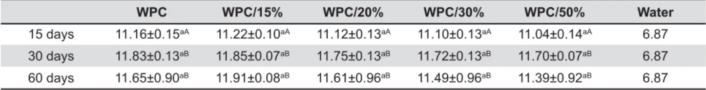
WPC WPC/15% WPC/20% WPC/30% WPC/50% Water
15 days 11.16±0.15aA 11.22±0.10aA 11.12±0.13aA 11.10±0.13aA 11.04±0.14aA 6.87
30 days 11.83±0.13aB 11.85±0.07aB 11.75±0.13aB 11.72±0.13aB 11.70±0.07aB 6.87
60 days 11.65±0.90aB 11.91±0.08aB 11.61±0.96aB 11.49±0.96aB 11.39±0.92aB 6.87
WPC=White Portland cement
Table 1- Means and standard deviations of the assessed pH in the evaluated periods. Different lowercase letters in rows
WPC WPC/15% WPC/20% WPC/30% WPC/50%
15 days 1.33 (1.03-1.43)aA 1.83 (1.36-2.10)aA 1.81 (1.16-2.20)aA 1.61 (0.83-2.13)aA 1.73 (1.36-2.36)aA
30 days 1.25 (1.20-1.30)aA 1.24 (0.96-1.56)aB 1.26 (0.96-1.70)aA 1.51 (1.33-1.73)aA 1.61 (1.23-2.23)aA
60 days 1.50 (1.00-1.56)aA 1.25 (0.83-1.53)aB 1.31 (0.90-1.53)aA 1.58 (1.26-1.80)aA 1.31 (1.06-1.86)aA
WPC=White Portland cement
Table 2- ! !
bismuth oxide in ProRoot MTA in comparison with Angelus MTA26. Duarte, et al.10 (2009) suggest that
this variance could be related to the method of proportion. Considering the high molecular weight of bismuth oxide (466.0 g/mol), the amount of this substance may vary, if the proportion included in "# 10.
To date, it is not completely clear if the presence of bismuth oxide as a radiopacifying agent affects the pH level or biological response of Portland-based cements. Considering that Portland cement is comparable to MTA, with regards to its physical16,
chemical4 and biological properties23, Portland
cement can be used to evaluate if the higher amounts of bismuth oxide are deleterious to the properties of the cement. The aim of the present study was to investigate if there is a relation between the increase of bismuth oxide and changes Portland cement.
MATERIAL AND METHODS
A portion of White Portland cement (WPC) (Irajazinho Votorantim, Cimento Rio Branco, Rio de Janeiro, RJ, Brazil) was mixed with 0, 15, 20, 30 and 50% bismuth oxide (BO) (Óxido de bismuto, Neon Comercial, São Paulo, SP, Brazil), proportioned in weight by using an electronic analytic balance (Mettler Toledo PG5002-S, Mettler Toledo, Barueri, SP, Brazil). The cements were divided into 5 groups, according to the bismuth oxide concentrations: Group 1) 100 g/WPC and 0g/BO; Group 2) 85 g/ WPC and 15 g/BO; Group 3) 80 g/WPC and 20 g/ BO; Group 4) 70 g/WPC and 30 g/BO; and Group
5) 50 g/WPC and 50 g/BO. The manipulation of the cements was performed using a total of 1 g of cement and 0.4 mL of distilled water.
pH assessment
A total of 0.038 g of manipulated cement, approximately, was placed into sterile polyethylene tubes (10 mm in length and 1 mm in internal diameter). Ten tubes were used for each group. { # "#! was immediately immersed into a glass flask containing 25 mL of Milli-Q water with a pH level corresponding to 6.87 (Purelab Option Elga DV25, Nova Analítica, São Paulo, SP, Brazil). To avoid any type of interference in the outcomes, all laboratory equipment was previously treated with 5% nitric acid. The measurement of the pH level was performed with a pH meter (B371, Micronal, São Paulo, SP, Brazil) and calibrated using solutions with pH levels corresponding to 4, 7, and 14. The assessments were performed after periods of 15, 30 and 60 days. The pH levels were initially (6.87) measured and compared with the proposed measurement periods.
Tissue reaction
Ninety adult male albino rats (Rattus norvegicus)
with approximately 300 g of body weight each were selected. The ethics committee of the institution in which the study was carried out, approved the use of the animals for research purposes (CEP 006-2009).
Figure 1- (A–C) Group 1 (0%WPC); (D–F) Group 2 (15%WPC); (G–I) Group 3 (20%WPC); (J–L) Group 4 (30%WPC); "# $ &'*+ / < > > ! (black arrow) is observed, presence of giant multinuclear cells (red arrow), areas with Portland cement particles involved ! ?K ? K / N > ? K ! < period were observed. Furthermore, at 30 days the presence of macrophage cells (black arrow) is predominant. Areas with Portland cement particles involved with giant cells are still present in this period (black circle). At 60 days, the chronic ! QK ?K ! ! R ? K
implanted subcutaneously in the dorsal region of the rats. The animals were divided into 5 groups (n=6) according to the cement implanted. Each animal received 2 implants of the same material. All the materials implanted were previously sterilized with ethylene oxide.
For the surgical procedures, the rats were anesthetized with a combination of ketamine and xylazin (Vet Brands International, Miramar, FL, USA) (0.05 mL/100 g). The tissue was disinfected with povidine (Tecnofarma Ind., Campinas, SP, Brazil) and two incisions were made through the skin using
were stained with hematoxylin and eosin. Four sections from each specimen were selected. Histological evaluations were carried out under a light microscope (Olympus, São Paulo, SP, Brazil) # ! # $ a quantitative evaluation of the inflammatory infiltrate, 30 microscopic fields were analyzed according to the classification adapted from Vosough Hosseini, et al.31 (2008): Grade 0 – Without
/G %U * U cells (25–125 cells); Grade 3 – Dense and severe %7/U cells). The measurements were repeated twice to ensure reproducibility. The median and range of the grades were calculated (Table 2).
Statistical analysis
As a result of absence of normal distribution, " {# ! the statistical analysis was performed using the nonparametric Kruskal-Wallis and Dunn tests to compare the pH level among the materials and the Friedman test to compare the periods for a same material (p<0.05). For the biological analysis, the Kruskal-Wallis and Dunn tests were performed (p<0.05).
RESULTS
+# represented in Tables 1 and 2, respectively. Considering the initial assessment of water, an increase of the pH level was observed after 15 days, followed by a slight increase after 30 ' { # increase of the pH level was observed from 15 to 30 days for all the tested cements (p<0.05). A comparison among the groups in each period revealed no statistically significant differences (p>0.05). Representative histological images of each group (horizontally) at 15, 30 and 60 days (vertically) (hematoxylin and eosin staining, 400x magnification) are presented in Figure 1. The " ! 15 days with the predominance of macrophage-like and multinucleated giant cells for all groups. At 60 ! " with isolated macrophage-like and multinucleated # # " # the groups in each experimental period (p>0.05). +/9; "# / 4' (p<0.05).
DISCUSSION
Bismuth oxide is added to MTA to provide radiopacity10. Variable amounts of bismuth oxide
were found in different commercial brands of MTA26. It was shown that high amounts of this
radiopacifying agent interfered with the physical and chemical properties of the Portland cement2,6.
However, the effects of bismuth oxide concentration on the pH levels and biological properties of Portland-based cements are not completely clear at the moment. The hypothesis tested in the present study was that the increases of bismuth oxide in Portland cement could decrease the pH levels and
In the present study, the concentrations of bismuth oxide added to Portland cement ranged from 0–50%. The proportions were calculated in weight and were selected according to previous investigations, that evaluated various compositions in relation to other properties7,14,32. High proportions
were tested to provide a critical condition of tissue exposition to bismuth oxide and interference in pH level.
High pH levels are expected for Portland-based cements due to the presence of calcium hydroxide in its composition4,16,30. The pH of calcium
silicate-based cements is around 8–12, which is considered adequate for tissue repair9,13. Elevated
concentrations of bismuth oxide added to Portland cement could decrease the amount of cement available to release hydroxyl ions, consequently decreasing the pH level9,24. A higher pH level
alteration was observed in the initial assessment (15 days) in relation to the water pH. There was an increase of the pH level from 6.87 to approximately 11, which is considered adequate30. It can be related
to the ion release that occurred during the setting time of the cements, as previously reported9.
After the setting time, the increase of the pH level assessed was slight (30 days). Following 60 !"# decrease of the pH level, but it remained at 11, approximately. A comparison among the groups " # differences (p>0.05). These results indicate that the addition of bismuth oxide did not interfere with the pH level of Portland cement.
=# materials due to their permanent contact with periapical tissues7. Ideally, the cements should not
promote irritation in the tissues or interfere with the repair process. Portland is a hydraulic cement due to its setting time and hardening by a chemical interaction with water1,17. It was previously shown
due to the fact that when bismuth is present in # !;=G= enough to react with all the molecules of the available bismuth. Camilleri, et al.5%U*
that bismuth oxide interfered with and inhibited cell growth in culture, suggesting that this substance presents toxic effect. Thus, the bismuth ions that remain un-reacted could possibly contact with the adjacent tissues and affect tissue repair or intensify
The subcutaneous implant method is widely = #7,14,25,31. This method was selected
to provide a dynamic system with defense cells and biotransformation19. Cell culture systems
have disadvantages related to the use of one cell lineage and the lack of cell turnover19. The
% #! ! giant cells, plasmocytes and polymorphonuclear * " 4+ " # evaluated, as previously reported7. There were
# # groups for inflammatory infiltrates for all the %7*{# " / WPC, from 15 to 30 and 60 days (p<0.05) (Table 2). These results are in agreement with previous studies, which suggest that bismuth oxide does not promote a chronic subcutaneous tissue response, genotoxic and cytotoxic effects7,14,32. The addition
of high amounts of bismuth is not appropriate due to the interference with the physical properties of the Portland cement6. Nonetheless, the quantity of
bismuth oxide did not present deleterious effects with the pH level or biocompatibility of the Portland cement, denying the two hypotheses tested.
CONCLUSION
The increment of bismuth oxide did not interfere with the pH level and intensity of chronic + concentration of 15% of bismuth oxide resulted # in comparison with the other concentrations evaluated.
ACKNOWLEDGMENTS
This work was supported by the Improvement of Higher Education Personnel - CAPES.
REFERENCES
1- American Society for Testing and Materials. ASTM C219: Standard terminology relating to hydraulic cement. West Conshohocken: ASTM; 2007.
2- Camilleri J. Hydration mechanisms of mineral trioxide aggregate. Int Endod J. 2007;40(6):462-70.
3- Camilleri J. Evaluation of the physical properties of an # =# U/4%4*U4/= 4- Camilleri J, Montesin FE, Brady K, Sweeney R, Curtis RV, Ford TR. The constitution of mineral trioxide aggregate. Dent Mater. 2005;21(4):297-303.
5- Camilleri J, Montesin FE, Papaioannou S, McDonald F, Pitt Ford TR. Biocompatibility of two commercial forms of mineral trioxide aggregate. Int Endod J. 2004;37(10):699-704.
6- Coomaraswamy KS, Lumley PJ, Hofmann MP. Effect of bismuth endodontic Portland cement-based (MTA-like) system. J Endod. 2007;33(3):295-8.
7- Coutinho-Filho T, De-Deus G, Klein L, Manera G, Peixoto C, Gurgel-Filho ED. Radiopacity and histological assessment of Portland cement plus bismuth oxide. Oral Surg Oral Med Oral Pathol Oral Radiol Endod. 2008;106(6):e69-77.
8- Cutajar A, Mallia B, Abela S, Camilleri J. Replacement of ### determination of physical properties. Dent Mater. 2011;27(9):879-91.
9- Duarte MAH, Minotti PG, Rodrigues CT, Zapata RO, Bramante CM, Tanomaru Filho M, et al. Effect of different radiopacifying agents on the physicochemical properties of white Portland cement and white mineral trioxide aggregate. J Endod. 2012;38(3):394-7. 10- Duarte MAH, Oliveira El Kadre GD, Vivan RR, Guerreiro Tanomaru JM, Tanomaru Filho M, Moraes IG. Radiopacity of portland cement associated with different radiopacifying agents. J Endod. 2009;35(5):737-40.
11- Ersev H, Schmalz G, Bayirli G, Schweikl H. Cytotoxic and # # eukaryotic and prokaryotic cells in vitro. J Endod.
1999;25(5):359-63.
12- Estrela C, Bammann LL, Estrela CR, Silva RS, Pécora JD. Antimicrobial and chemical study of MTA, Portland cement, calcium hydroxide paste, Sealapex and Dycal. Braz Dent J. 2000;11(1):3-9.
13- Holland R, Souza V, Murata SS, Nery MJ, Bernabe PF, Otoboni Filho JA, et al. Healing process of dog dental pulp after pulpotomy and pulp covering with mineral trioxide aggregate or Portland cement. Braz Dent J. 2001;12(2):109-13.
14- Hwang YC, Lee SH, Hwang IN, Kang IC, Kim MS, Kim SH, et al. Chemical composition, radiopacity, and biocompatibility of Portland cement with bismuth oxide. Oral Surg Oral Med Oral Pathol Oral Radiol Endod. 2009;107(3):e96-102.
15- International Organization for Standardization. ISO 6876:2012. Dental root canal sealing materials. Geneva: ISO; 2012.
16- Islam I, Chng HK, Yap AU. Comparison of the physical and mechanical properties of MTA and portland cement. J Endod. 2006;32(3):193-7.
/L= $! G# of concrete and concrete-making materials. Bridgeport: ASTM International; 2006.
18- Lee SJ, Monsef M, Torabinejad M. Sealing ability of a mineral trioxide aggregate for repair of lateral root perforations. J Endod. 1993;19(11):541-4.
20- Min KS, Chang HS, Bae JM, Park SH, Hong CU, Kim EC. The induction of heme oxygenase-1 modulates bismuth oxide-induced cytotoxicity in human dental pulp cells. J Endod. 2007;33(11):1342-6.
21- Neagoe AD. Bismuth. In: Merian E, Anke M, Ihnat M, Stoeppler M. Elements and their compounds in the environment: occurrence, analysis and biological relevance. 2nd ed. Weinheim: Wiley; 2004.
p. 671-83.
22- Parirokh M, Torabinejad M. Mineral trioxide aggregate: a comprehensive literature review - Part III: Clinical applications, drawbacks, and mechanism of action. J Endod. 2010;36(3):400-13.
23- Ribeiro DA, Duarte MA, Matsumoto MA, Marques ME, Salvadori DM. Biocompatibility in vitro tests of mineral trioxide aggregate and regular and white Portland cements. J Endod. 2005;31(8):605-7. 24- Santos AD, Moraes JC, Araújo EB, Yukimitu K, Valério Filho WV. Physico-chemical properties of MTA and a novel experimental cement. Int Endod J. 2005;38(7):443-7.
U=GG!G! !!{{ =# rat connective tissue. J Endod. 2006;32(8):776-80.
26- Song JS, Mante FK, Romanow WJ, Kim S. Chemical analysis of powder and set forms of Portland cement, gray ProRoot MTA, white ProRoot MTA, and gray MTA-Angelus. Oral Surg Oral Med Oral Pathol Oral Radiol Endod. 2006;102(6):809-15.
27- Tanomaru-Filho M, Chaves Faleiros FB, Saçaki JN, Hungaro Duarte MA, Guerreiro-Tanomaru JM. Evaluation of pH and calcium ion release of root-end filling materials containing calcium hydroxide or mineral trioxide aggregate. J Endod. 2009;35(10):1418-21.
28- Tanomaru-Filho M, Luis MR, Leonardo MR, Tanomaru JM, Silva LA. Evaluation of periapical repair following retrograde #" =# #" periapical lesions. Oral Surg Oral Med Oral Pathol Oral Radiol Endod. 2006;102(1):127-32.
29- Torabinejad M, Hong CU, Lee SJ, Monsef M, Pitt Ford TR. # ### =# dogs. J Endod. 1995;21(12):603-8.
30- Torabinejad M, Hong CU, McDonald F, Pitt Ford TR. Physical " =# 1995;21(7):349-53.
4/= # G! !GG! !#! G#{! "# ### UJ4%'*L/=L 32- Zeferino EG, Bueno CE, Oyama LM, Ribeiro DA. Ex vivo