ABSTRACT
http://dx.doi.org/10.1590/1678-775720140435
A titration model for evaluating calcium hydroxide
removal techniques
Mark PHILLIPS1, Scott McCLANAHAN2, Walter BOWLES2
1- Private practice, Duluth, MN, USA.
2- Division of Endodontics, University of Minnesota School of Dentistry, Minneapolis, MN, USA.
Corresponding address: Walter Bowles - Division of Endodontics - University of Minnesota School of Dentistry - 8-166 Moos Tower, 515 Delaware St SE - Minneapolis, MN 55455 - Phone: 612-624-9613 - Fax: 612-624-7960 - e-mail: [email protected]
Submitted: November 3, 2014 - Modiication: December 12, 2014 - Accepted: December 17, 2014
O
bjective: Calcium hydroxide (Ca(OH)2) has been used in endodontics as an intracanal medicament due to its antimicrobial effects and its ability to inactivate bacterial endotoxin. The inability to totally remove this intracanal medicament from the root canal system, however, may interfere with the setting of eugenol-based sealers or inhibit bonding of resin to dentin, thus presenting clinical challenges with endodontic treatment. This study used a chemical titration method to measure residual Ca(OH)2 left after different endodonticirrigation methods. Material and Methods: Eighty-six human canine roots were prepared for obturation. Thirty teeth were illed with known but different amounts of Ca(OH)2 for 7
days, which were dissolved out and titrated to quantitate the residual Ca(OH)2 recovered from each root to produce a standard curve. Forty-eight of the remaining teeth were illed with equal amounts of Ca(OH)2 followed by gross Ca(OH)2 removal using hand iles and randomized treatment of either: 1) Syringe irrigation; 2) Syringe irrigation with use of an apical ile; 3) Syringe irrigation with added 30 s of passive ultrasonic irrigation (PUI), or 4) Syringe irrigation with apical ile and PUI (n=12/group). Residual Ca(OH)2 was dissolved
with glycerin and titrated to measure residual Ca(OH)2 left in the root. Results: No method
completely removed all residual Ca(OH)2. The addition of 30 s PUI with or without apical ile
use removed Ca(OH)2 signiicantly better than irrigation alone. Conclusions: This technique
allowed quantiication of residual Ca(OH)2. The use of PUI (with or without apical ile)
resulted in signiicantly lower Ca(OH)2 residue compared to irrigation alone.
Keywords: Calcium hydroxide. Ultrasonic therapy. Glycerin. Therapeutic irrigation.
INTRODUCTION
Calcium hydroxide (Ca(OH)2) as an intracanal medicament (ICM) has been extensively studied and its clinical use well established3,17,23-26. In aqueous
solution Ca(OH)2 dissociates into calcium and hydroxyl ions. The large amount of hydroxyl ions liberated interferes with the bacterial cytoplasmic membrane integrity, largely by interruption of transfer of nutrients and destruction of phospholipids from unsaturated fatty acids10.
In vitro studies have demonstrated potential clinical concerns regarding the inability to fully remove calcium hydroxide. Residual Ca(OH)2 may interfere with sealer entrance into dentinal tubules and inhibit bonding of resin to dentin6. Additionally,
leakage may be increased with the use of calcium
hydroxide as an ICM13,14 or residual Ca(OH) 2 may
interfere with the setting of eugenol based sealers or MTA19,27.
A variety of Ca(OH)2 removal techniques have been studied. Irrigation-only techniques appear to result in poor Ca(OH)2 removal15, while use of a master apical ile or passive ultrasonic
irrigation (PUI) for Ca(OH)2 removal have been found efficacious12,22,28. For review of PUI, see
van der Sluise, et al.29 (2007). Quantiication of
residual Ca(OH)2 remaining in the root has been attempted by using digital images4,8,12,22, while
nonparametric grading systems have been used with digital photographic images in teeth with premade grooves11,30. Concerns exist, however,
debris vs. Ca(OH)2. More recently, spiral CT20 and
micro CT have been used to study calcium hydroxide removal31. These studies are all problematic in that
they cannot accurately detect residual Ca(OH)2 in actual teeth.
The technique proposed by Bramante allows reuse of specimens, and thus reduces inaccuracies due to different specimen anatomy5. With reuse of
the same specimens, however, cumulative effects of the chelating agent EDTA can occur and removal of residual Ca(OH)2 cannot be conirmed. Teeth used in this study had a standardized canal preparation and were only used once.
Visual identiication of residual Ca(OH)2,even
with SEM, is not accurate. Since Ca(OH)2 has a high pH (pH>12), this attribute may be used to identify residual Ca(OH)2 left in the root canal system by pH determination of known amounts retrieved from the canal compared to unknown amounts in canals after various irrigation methods have been used. The purpose of this study was to employ a chemical microtitration technique to test removal methods of Ca(OH)2 paste (Calasept®; JB Dental, Ridgeield,
Connecticut, USA) from the root canal system.
MATERIAL AND METHODS
This study was exempted by the Institutional Review Board of the University. Eighty-six extracted single canal maxillary and mandibular canines were stored in normal saline with 0.2% sodium azide. Samples were randomized into groups. Three teeth served as positive controls and three as negative controls. Thirty teeth served as standards.
Preparation of specimens
Teeth were decoronated at the cementoenamel junction and radiographed from the proximal and buccal view. The Pruett, Clement, and Carnes method was used to standardize curvature at
≤15°21. Two samples were excluded due to aberrant
anatomy. Root length was standardized at 17.5 mm. A glide path was established to a #25 Flex-O hand
ile (Dentsply-Maillefer, Johnson City, Tennessee, USA) and K3 nitinol iles (SybronEndo, Cuyahoga
Falls, Ohio, USA) were used to prepare each tooth according to manufacturers’ recommendations
to a 50/0.06 inal apical ile (FAF). Patency was established with size 20 Flex-O ile. Irrigation
with 1 mL of NaOCl 5.25% and recapitulation was
performed between iles. The length of the
Max-i-probe (Dentsply, Elgin, Illinois, USA) was set at 2 mm from the working length (WL). Thirty seconds of passive ultrasonic irrigation with 5.25% NaOCl was performed with a one minute soak using 17% EDTA
to remove smear layer. A inal 3 mL rinse of 5.2%
NaOCl was performed and all canals were dried with
paper points. Canals were then illed with Calasept®
[Ca(OH)2 based ICM] by inserting the syringe tip
until locked in, then loosening and backilling. Radiographs conirmed a dense Ca(OH)2 ill. Teeth were temporized with Fuji IX resin modiied glass
ionomer (GC Corporation, Tokyo, Japan) and placed
in an incubator (Precision Scientiic, Chennai, India)
at 37°C for one week in a humid environment.
Standards
In order to develop a standard curve using known amounts of Ca(OH)2, thirty teeth were selected. The identical radiographic and the preparation protocol were utilized for the standard teeth and for unknown samples, except that different known weights of Ca(OH)2 were added to the teeth (much smaller weights to replicate residual Ca(OH)2 after irrigation methods). After temporization and 7 days of incubation, the glycerin transfer and titration steps were performed in identical fashion to samples. A standard curve was generated by graphing the different known weights added to the teeth versus the pH recording after addition of known micromoles of HCL. Samples were titrated using 0.025, 0.05, 0.1, 0.5, 1, or 3 M HCL with 10 microliter aliquots using a micropipette. Usually titrations began with 10 microliters of 0.1 M. If the initial pH was higher, the operator may have used a higher molarity such as 0.2 M. If the initial pH of the mixture was lower (i.e. 11.0-11.4), the operator may have begun with a weaker concentration (i.e. 0.05 M HCL). pH measurements were recorded after each addition of HCL using a model HO4N-0001 micro pH electrode (Lazar Research Laboratories, Los Angeles, California, USA) and a Model 60 pH meter (Lazar Research Laboratories, Los Angeles, California, USA). After each aliquot addition of acid to the microcentrifuge tube, the tube was vortexed for 10 s. Adequate time was given for each pH measurement – this was approximately 10-60 s for the meter to equalize.
After creating a titration curve for each sample, linear regression was applied to each curve. The neutral point, pH=7, was selected for use in each regression curve. By solving for pH=7, cumulative micromoles of HCL added could be determined for each titration.
A second standard curve was calculated using only Ca(OH)2 in microcentrifuge tubes and not placed in teeth. In the second standard curve, small amounts of Calasept® was added to preweighed
Treatment groups
Samples were removed from incubator and
temporary illings removed. For each group (48 teeth; n=12/group), a #30 Flex-O ile and a #50 Flex-O ile were used for gross removal of Calasept®
before each irrigation technique: Group 1: Irrigation (NaOCl 5.2% 3 mL followed by EDTA 17% 3 mL.
A inal rinse of NaOCl 5.2% 5 mL was performed).
Group 2: Irrigation (as in group 1) with the addition of a K3 #50-0.06 taper instrumented to WL between
the irst two rinses. Group 3: Irrigation (as in group 1) with the use of PUI for 30 s between the irst
NaOCl and EDTA rinse. Group 4: consisted of NaOCl 5.2% 1.5 mL, use of a K3 #50-0.06 instrumented to
WL, 1.5 mL NaOCl, 30 s of PUI, and the inal EDTA
and NaOCl rinses as in previous groups.
Three teeth were selected for negative controls. These teeth were instrumented but canals were left empty. Negative control teeth were included in the experimental sample set and operator was also blinded to control teeth. Positive controls consisted of three microcentrifuge tubes with saturated solutions of Ca(OH)2.
Removal of residual calcium hydroxide
Any residual Ca(OH)2 remaining in the tooth after gross Ca(OH)2 removal was removed by the following manner: a preparation of 60% glycerin (Humco Corporation, Texarkana, TX): 40% distilled water at 40°C was placed into the canal with a Ultradent capillary tip (Ultradent Products, Inc., South Jordon, Utah, USA). PUI for 10 s was performed with a #15 Zipperer file (Roydent, Rochester Hills, Minnesota, USA) at 2 mm from
WL to help the remaining Calasept® dissolve. The
glycerin with dissolved Ca(OH)2 was removed using a narrow Ultradent tip and a 10 mL syringe, with the aliquots placed into a 1.5 mL microcentrifuge
tube (Fisher Scientiic, Pittsburgh, Pennsylvania,
USA). Aliquots were repeated until 100 microliters was obtained.
A single calibrated 20 microliter Pipetman micropipette (Gilson Inc., Middleton, Wisconsin, USA) was used for all titrations. Each microcentrifuge tube was labeled with a second random number to blind the operator. A titration curve was generated by adding 10 microliter aliquots of 0.025, 0.05, 0.1, 0.5, 1, or 3 M HCL using a micropipette. To ensure a good mix, each microcentrifuge tube
was vortexed with a Vortex Genie Mixer (Scientiic
Products, Evanston, Illinois, USA) for 10 s between additions and heated to 40°C±1° in a Hanau® low
temperature water bath (Teledyne Water Pick, Fort Collins, Colorado, USA). pH measurements were recorded after each addition of HCL using a model HO4N-0001 semi-micro pH electrode (Lazar Research Laboratories, Los Angeles, California, USA) and a Model 60 pH meter (Lazar Research Laboratories, Los Angeles, California, USA). Based on pilot studies, an algorithm describing which molarity to add based on the current pH was made. The micro pH electrode and meter were calibrated with standard pH solutions (Omega
Scientiic, Tarzana, California, USA). After each pH
measurement, the tip of the electrode probe was thoroughly rinsed with distilled water and wiped with a Kim Wipe® (Kimberley-Clark Professional,
Mississauga, Ontario, CA). All pH readings were
recorded. The chemical reaction between Ca(OH)2 and HCL is described by the equation: 2 HCl + Ca(OH)2→ CaCl2+2H2O7.
Standard deviations and means were calculated for each group, and pairwise comparisons were made using a Tukey-Kramer multiple comparisons adjustment.
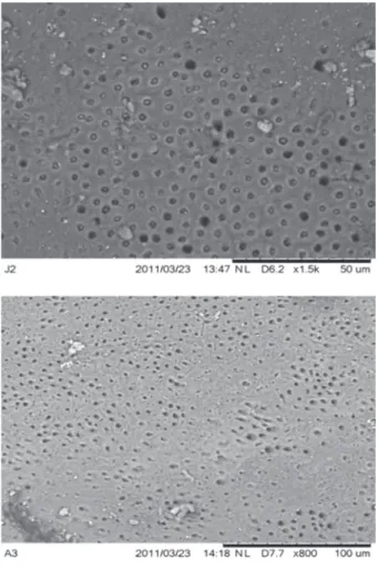
After the experiment, several representative teeth were split and SEM (Model TM3000 Table Top Microscope, Hitachi High-Technologies Corporation, Tokyo, Japan) was performed to visually assess the inside canal surface.
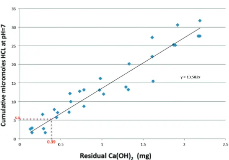
Figure 3- A titration curve was made from utilizing data points for an unknown sample titrated with HCL to pH 7.0. From the
µmoles used at pH 7.0, we can determine the amount Ca(OH)2 in our sample using the standard curve in Figure 2 Figure 2- Sample standard curve. Cumulative µmoles of HCL at pH=7 for each standard was plotted against initial known
weights (mg) of Ca(OH)2. For example, the vertical red dotted line indicates that 0.39 mg of Ca(OH)2 standard would take
5.3 µmoles of HCL to reach pH=7.0 (horizontal red dotted line). Using these standard known amounts, we can create a
RESULTS
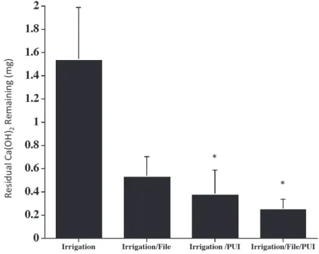
Figure 1 shows the means and standard deviations of residual calcium hydroxide after the various removal group techniques were applied.
The groups differed signiicantly [F(3,42)=4.47,
p=0.0082], indicating that there was a difference between the group means. The group 1 (irrigation
only) mean was signiicantly different than the means of groups 3 (PUI) and 4 (PUI + ile), p=0.0291
and p=0.0104, respectively. No other comparisons
were statistically signiicant. Negative controls [no
Ca(OH)2 added] were found to have near neutral pH measurements, while positive controls [fully Ca(OH)2 saturated 100 µL of glycerin] required larger amounts of HCL to achieve neutrality.
A standard curve is illustrated in Figure 2. An example of a titration of one of the unknown samples and the corresponding linear regression line and equation is shown in Figure 3. The dotted red line shows that at pH 7.0, it took 5.3 µmoles
of HCL to neutralize this sample. Based on the standard curve of known samples in Figure 2, we can determine from the µmoles of HCL exactly how much Ca(OH)2 was present in the sample [in this case using the y=13.582x linear regression formula, we now know y=5.3 and can solve for x as 0.39 mg of Ca(OH)2 in the unknown sample].
Examination of SEM’s taken after our study revealed some debris mixed in with the glycerin (see Figure 4).
DISCUSSION
This study found agreement with previous studies that no Ca(OH)2 removal technique successfully removed all the calcium hydroxide from the canal system12,16,20,28. This may pose a clinical
problem since residual calcium hydroxide interacts with eugenol in ZOE based sealers, leading to residual eugenol in the set product19. Thus Ca(OH)
2
may interfere with the obturation seal. The clinical implication of unset sealer is unclear and further clinical studies are needed to elucidate the effect of this interaction. Examination of SEMs taken after our study revealed some debris mixed in with the glycerin in the open canal. Perhaps the glycerin transfer failed to remove all the Calasept®, or the
glycerin transfer did remove all the Calasept®, and
what was seen mixed in with the remaining glycerin was actually dentinal debris. As part of the methods used in this experiment, collection of residual Ca(OH)2 included passive ultrasonic instrumentation between each transfer. This was done to ensure the Ca(OH)2 on the walls was incorporated into solution, and likely would have also incorporated more debris into the glycerin. One method to differentiate residual debris from Calasept® would
be to radiolabel Calasept® and apply removal
studies, which was not performed in this study. Then an autoradiographic analysis could be performed to determine “what we are actually looking at” after calcium hydroxide removal methods. Allison, et al.1
(1979) used such a technique with 45Ca to prove
that step back preparations had less leakage than serial preparation. It appears that some Ca(OH)2 is left in the canal during most irrigation methods, mainly in the dentinal tubules, but amounts of residual Ca(OH)2 in the open canal could be minimized using passive ultrasonic instrumentation. Removal of medicament trapped within the dentinal tubules was attempted with repeated dissolution using glycerin extracts and passive ultrasonic irrigation in the same root, but could not be removed. The results here are for comparative purposes, and demonstrate that even with ideal conditions and access (decoronated tooth, straight root), medicament still remains in the canal. This may interfere with the setting of eugenol based
sealers or MTA19,27.
One primary result of our study was that the 30 s use PUI (groups 3 and 4) produced better Ca(OH)2 removal than the irrigation-only group. This is in agreement with many studies supporting the effectiveness of PUI4,9,12,30,31. However, Lev, et al.18
(1987) found contrasting results in that 1 minute
of passive ultrasonic step back was not signiicantly
better in debris removal than irrigation and hand
iling alone. The 30 s time frame was adequate to achieve statistical signiicance. This is in contrast
to a study that examined debris removal after a 3 minute PUI time per canals (12 minutes per molar)2.
Another study utilizing sequential micro-CT scans studied Ca(OH)2 removal with PUI activation for 60 seconds per canal (4 minutes per molar)31. Our
results demonstrated two minutes (30 s per canal) of PUI for a typical four canal molar is adequate to remove most residual Ca(OH)2 from the root canal. This is in agreement with Sabins, et al.22 (2003) who found 30 s of PUI adequate to signiicantly reduce
debris levels in the mesial canals of mandibular molars22.
The agreement of our titration model with previous pixel or voxel-quantification studies confirms the accuracy of this technique for evaluating residual Ca(OH)2 in the canal. This is
very signiicant in that to our knowledge chemical
quantification of residual calcium hydroxide has never been performed. In contrast,
pixel-quantiication methods examine debris and Ca(OH)2
and, while they share many advantages, they also have limitations. Some potential issues with
pixel-quantiication methods include concerns with reuse
of teeth for repeated testing. Another concern is the need for very uniform canals – necessary so that teeth can be predictably split. One Ca(OH)2 removal study used mesial mandibular molar canals for uniformity in canal morphology12. While standard
preparation of canals was a part of this study, this measurement technique does not strictly require uniform canals. A titration technique, such as this, might be useful to evaluate Ca(OH)2 removal from the more variable-sized distal canal or C-shaped canals. In a clinical setting, non-uniform canals
may beneit the most from PUI.
A titration-quantiication method differs from
a voxel or pixel quantification method in that the former expresses results with a single value.
A pixel or voxel-quantiication method can give
results in terms of location of remaining debris or Ca(OH)2, while a chemical quantiication method gives a number for the entire canal. This may be a disadvantage for a titration technique in that it
may be more important to deine Ca(OH)2 removal speciically in one area, such as the apical one-third.
It is interesting to note a titration technique gives results in mg of remaining Ca(OH)2. Most studies
express results in percent, rather than actual weight of Ca(OH)2.
The titration technique is time consuming. Also, the Ca(OH)2 studied must be soluble in glycerin (or another solvent that can be titrated). Pilot studies indicated that Ultracal (Optident Ltd, International Develop Centre, West Yorkshire, UK) did not dissolve in glycerin. Further studies testing other solvents will allow more premixed Ca(OH)2 pastes to be studied with a chemical titration method.
CONCLUSION
This study tested a new approach to quantiication
of residual Ca(OH)2 by chemical microtitration. This model is novel and appears to be accurate and reliable, with several potential advantages. Adjuncts
to irrigation such as PUI for 30 s or the use of a inal apical ile were shown to improve Ca(OH)2 removal
from the open canal, within decoronated teeth with single straight canals, however the medicament was not fully removed from all teeth. Further studies utilizing the chemical titration method may help us understand residual Ca(OH)2 removal from diverse tooth types such as C-shaped molars and wide oval shaped canals.
ACKNOWLEDGEMENT
The authors deny any conlicts of interest and any inancial afiliations related to this study or its
sponsors.
REFERENCES
1- Allison DS, Weber CR, Walton RE. The inluence of the method of canal preparation on the quality of apical and coronal obturation. J Endod. 1979;5(10):298-304
2- Archer R, Reader A, Nist R, Beck M, Meyers W. An in vivo
evaluation of the eficacy of ultrasound after step-back preparation in mandibular molars. J Endod. 1992;18(11):549-52.
3- Baik EJ, Kum KY, Yun CH, Lee JK, Lee K, Kim KK, et al. Calcium hydroxide inactivates lipoteichoic acid from Enterococcus faecalis. J Endod. 2008;34(11):1355-9.
4- Balvedi RP, Versiani MA, Manna FF, Bifi JC. A comparison of two techniques for the removal of calcium hydroxide from root canals. Int Endod J. 2010;43:763-8.
5- Bramante CM, Berbert A, Borges RP. A methodology for evaluation of root canal instrumentation. J Endod. 1987;13(5):243-5. 6- Calt S, Serper A. Dentinal tubule penetration of root canal sealers after root canal dressing with calcium hydroxide. J Endod. 1999;25(6):431-3.
7- Cohen F, Lasfargues JJ. Quantitative chemical study of root canal preparations with calcium hydroxide. Endod Dent Traumatol. 1988;4:108-13.
8- Crumpton BJ, Goodell GG, McClanahan SB. Effects on smear layer and debris removal with varying volumes of 17% REDTA after rotary instrumentation. J Endod. 2005;31(7):536-8.
10- Estrela C, Sydney GB, Bammann LL, Felippe O Jr. Mechanism of action of calcium and hydroxyl ions of calcium hydroxide on tissue and bacteria. Braz Dent J. 1995;6:85-90.
11- Jiang LM, Verhaagen B, Versluis M, Langedijk J, Wesselink P, van der Sluis LW. The inluence of the ultrasonic intensity of the cleaning eficacy of passive ultrasonic irrigation. J Endod. 2011;37(5):688-92.
12- Kenee DM, Allemang JD, Johnson JD, Hellstein J, Nichol BK. A quantitative assessment of eficacy of various calcium hydroxide removal techniques. J Endod. 2006;32(6):563-5.
13- Kim SK, Kim YO. Inluence of calcium hydroxide intracanal medication on apical seal. Int Endod J. 2002;35(7):623-8. 14- Kontakiotis EG, Wu MK, Wesselink PR. Effect of calcium hydroxide dressing on seal of permanent root illing. Endod Dent Traumatol. 1997;13:281-4.
15- Lambrianidis T, Kosti E, Boutsioukis C, Mazinis M. Removal eficacy of various calcium hydroxide/chlorhexidine medicaments from the root canal. Int Endod J. 2006;39(1):55-61.
16- Lambrianidis T, Margelos J, Beltes P. Removal efficacy of calcium hydroxide dressing from the root canal. J Endod. 1999;25(2):85-8.
17- Law A, Messer H. An evidence-based analysis of the antibacterial effectiveness of intracanal medicaments. J Endod. 2004;30(10):689-94.
18- Lev R, Reader A, Beck M, Meyers W. An in vitro comparison of the step-back technique versus a step-back/ultrasonic technique for 1 and 3 minutes. J Endod. 1987;13(11):523-30.
19- Margelos J, Eliades G, Verdelis C, Palaghias G. Interaction of calcium hydroxide with zinc oxide-eugenol type sealers: a potential clinical problem. J Endod. 1997;23(1):43-8.
20- Nandini S, Velmurugan N, Kandaswamy D. Removal eficiency of calcium hydroxide intracanal medicament with two calcium chelators: volumetric analysis using spiral CT, an in vitro study. J Endod. 2006;32(11):1097-101.
21- Pruett JP, Clement DJ, Carnes DL Jr. Cyclic fatigue testing of nickel-titanium endodontic instrument. J Endod. 1997;23(2):77-85.
22- Sabins RA, Johnson JD, Hellstein JW. A comparison of the cleaning eficacy of short-term sonic and ultrasonic passive irrigation after hand instrumentation in molar root canals. J Endod. 2003;29(10):674-8.
23- Safavi KE, Nichols FC. Effect of calcium hydroxide on bacterial lipopolysaccharide. J Endod. 1993;19(2):76-8.
24- Silva LA, Nelson-Filho P, Leonardo MR, Rossi MA, Pansani CA. Effect of calcium hydroxide on bacterial endotoxin in vivo. J Endod. 2002;28(2):94-8.
25- Siquiera JF, Lopes HP. Mechanisms of antimicrobial activity of calcium hydroxide: a critical review. Int Endod J. 1999;32(5):361-9.
26- Souza CA, Teles RP, Souto R, Chaves MA, Colombo AP. Endodontic therapy associated with calcium hydroxide as an intracanal dressing: microbiologic evaluation by the checkerboard DNA-DNA hybridization technique. J Endod. 2005;31(2):79-83. 27- Stefopoulos S, Tsatsas DV, Kerezooudis NP, Eliades G. Comparative in vitro study of the sealing eficiency of white vs grey ProRoot mineral trioxide aggregate formulas as apical barriers. Dent Traumatol. 2008;24(2):207-13.
28- Taşdemir T, Celik D, Er K, Yildirim T, Ceyhanil KT, Yeşilyurt C. Eficacy of several techniques for the removal of calcium hydroxide medicament from root canals. Int Endod J. 2011;44(6):505-9. 29- Van der Sluis LW, Versluis M, Wu MK, Wesselink PR. Passive ultrasonic irrigation of the root canal: a review of the literature. Int Endod J. 2007;40(6):415-26.
30- Van der Sluis LW, Wu MK, Wesselink PR. The evaluation of removal of calcium hydroxide paste from an artiicial standardized groove in the apical root canal using different irrigation methodologies. Int Endod J. 2007;40(1):52-7.