INTRODUCTION
The bond strength of different denture teeth to their denture bases can be high enough to cause tooth fracture without debonding (1). If the bond between the parts resists until the materials fail, the bond will have fulilled its functional requirements (2). Nevertheless, bond failures between plastic teeth and heat-polymerized denture base resins can occur (3), and remain a major problem in prosthodontic practice (4). The bond between acrylic denture teeth and denture base materials remains unreliable, inconsistent and unpredictable (2,5). Previous surveys report that 26% to 33% of denture repairs are due to debonded teeth (6,7), frequently causing distress and cost for the patients (6).
The following causes of tooth debonding are well known: excessive stress, fatigue, insuficient tooth cleaning during denture base acrylic resin placement, wax and tinfoil
The Role of Surface Treatments on the Bond
between Acrylic Denture Base and Teeth
Lauro Egídio BRAGAGLIA1
Luiz Henrique Maykot PRATES2 Maria Cristina Marino CALVO3
1Private Practice, Florianópolis, SC, Brazil
2Department of Dentistry, Dental School, Federal University of Santa Catarina, Florianópolis, SC, Brazil 3Department of Public Health, Dental School, Federal University of Santa Catarina, Florianópolis, SC, Brazil
The aim of this study was to compare the bond strength between acrylic denture base and teeth subjected to 6 surface treatments.
Ninety-six specimens were made with poly(methylmethacrylate) teeth bonded to a microwave-polymerized acrylic denture base material. The
specimens were distributed into 6 groups (n=16) according to surface treatments: CT - no treatment (control); MN - methylmethacrylate monomer etching; AO - 50-μm-particle aluminum oxide air abrasion; BR - glaze removal with a round bur; ST - surface grinding with an aluminum oxide abrasive stone; group CV - cavity preparation (diatorics). The control and surface-treated groups were subjected to a
compressive load at 45o angle to the long axis of the teeth. Data were analyzed by one-way ANOVA, followed by Scheffé’s test (p<0.05). Bond strength means and (SD) in kgf for groups were: CT: 18.19 (7.14), MN: 18.34 (5.28), AO: 23.82 (5.40), BR: 23.30 (4.79), ST: 25.39 (7.80) and CV: 17.48 (7.17). There was statistically signiicant difference (p=0.037997) only between ST and CV. In conclusion, ridge lap surface grinding with an aluminum oxide abrasive stone provided the highest bond strength, though it differed signiicantly only when
compared to diatorics. The other surface treatments provided similar bond between the acrylic denture base and teeth.
Key Words: artiicial teeth, acrylic resins, tooth-resin bond, ridge lap treatment.
substitute contamination, defective properties of materials (8-10) and inappropriate heat-polymerizing technique (11).
Many factors have been investigated as to their inluence on the bond strength between artiicial teeth and denture base, such as ageing, ridge lap grinding, bonding agents, solvents or monomer-polymer solution application, surface grooving, tooth material, cross-linking agent concentration, denture base material, separating medium, impurities or wax contamination, thermocycling, microwave polymerization and polym-erization temperature rise (1-4,8,11-19). The American Dental Association (A.D.A.) speciication #15 deines standards concerning synthetic resin teeth, including the minimum bond strength between artiicial teeth and denture base materials and a bond test method (20).
Most experimental tests use lat and ground tooth surfacesbonded to denture base, or the original ridge lap area of the teeth without any denture base material coverage
in proximal, buccal and lingual surfaces (1,3,4,7,9,12-16). These conditions are not realistic and, moreover, the applied load direction is often different from clinical conditions. The aim of this study was to evaluate the inluence of surface treatments on the ridge lap area of poly(methylmethacrylate) (PMMA) acrylic denture teeth on their bond strength to a microwave-polymerized PMMA denture base material. The null hypothesis tested was that surface treatment on the ridge lap area of acrylic denture teeth inluences their bond strength to the denture base material.
MATERIAL AND METHODS
Acrylic Resin Pattern for Specimen Standardization
Two PMMA maxillary central incisors (one left and one right) with no cross-linking or iller particles (model A14 and 1C shade; Ivoclar Vivadent, Schaan, Liechtenstein) were used to form 2 acrylic dies for speci -men standardization. A line was drawn surrounding the cervical area, 1 mm above the bottom (ridge lap base) of the teeth. A drawing compass (Mod. 9000; Trident S/A, Itaipui, SP, Brazil) and a digital caliper accurate to the nearest 0.01 mm (727 Series; Starrett Ind. Com. Ltda., Itu, SP, Brazil) were used to draw the lines. The teeth were placed on a cone of wax (Pink Wax #7; Epoxiglass Chemical Products Ind. Com. Ltda., São Paulo, SP, Brazil) supported by a PVC tube 1 cm high and 1.87 cm diameter. The long axis of the teeth was perpendicular to the PVC tube bottom. Each tooth was gently embedded in wax until it reached the line previ-ously drawn. The angle between wax surface and teeth long axis was established in 45 degrees all around the cervical area with a stainless steel spatula. Each set (tooth, wax cone and PVC tube) was embedded in a microwave polymerization lask (Vipi-STG; Dental Vipi Ltda., Pirassununga, SP, Brazil). Routine procedure was used to wax removal. Denture base material (Vipi Wave, Dental Vipi Ltda.) was handled in a glass vessel, and left for 20 min to reach dough stage. The moulds were illed with denture base material and pressed under 1,200 kgf for 2 h in a hydraulic press (VH; Midas Dental Products Ltda., Araraquara, SP, Brazil), and heat-polymerized in a domestic microwave oven (CMS180; Consul, São Paulo, SP, Brazil) for 20 min at 80 watts followed by 5 min at 400 W. After cooling at room temperature, the models were delasked, inished, and inspected at ×5 magniication with a magnifying glass (TGB-390, Tasco
Sales Inc., Hong Kong) for any inaccuracy near the teeth.
Artiicial Tooth Cleaning
Ninety-six PMMA maxillary central incisors (48 left and 48 right teeth) were removed from the wax plates and residual wax was cleaned with dry cotton. The teeth were then cleaned with a wax remover (Remox; Dental Vipi Ltda.) and rinsed in boiling water for 10 s in order to ensure complete wax removal.
Inclusion in Polyvinyl Siloxane Moulds
Six moulds of polyvinyl siloxane putty material (Express; 3M/ESPE, St. Paul, MN, USA) were made over each PMMA model (left and right). These moulds were lasked in microwave lask (Vipi) with type II dental stone. Each lask provided 6 specimens, one per group, according to the same tooth side. After stone setting under 500 kgf for 1 h in a hydraulic press (VH), the lasks were opened and all stone surfaces were coated with sodium alginate tinfoil substitute (Vipi-Film; Dental Vipi Ltda.).
Surface Treatments of the Acrylic Denture Teeth
The 96 artiicial teeth were divided into 6 groups, according to the following surface treatments: CT - no treatment (control); MN - the tooth bases were etched twice with a methylmethacrylate monomer 10 min before acrylic resin packing and just before packing; AO - sur -face treatment with 50-μm-particle aluminum oxide air abrasion (Bio-Art; São Carlos, SP, Brazil) with 4.9 kgf/ cm2 air pressure at 1 cm distance, for 10 s; BR - ridge lap glaze removal with a 2.3-mm-diameter #8 round bur (KG Sorensen, Barueri, SP, Brazil) at low speed; ST - ridge lap glaze removal with an aluminum oxide abrasive stone (Schelble Burs; Petrópolis, RJ, Brazil); CV - a diatoric cavity (2 mm deep x 2.3 mm diameter) was prepared with #8 round bur (KG Sorensen) at low speed. Each tooth of the CV group was placed in a polyvinyl siloxane putty material custom support, attached to a milling machine (1000 N; Bio-Art, São Carlos, SP, Brazil), in order to provide cavities with standardized sizes and positions.
Specimen Preparation
cervical area were left exposed. The teeth received ridge lap treatments before insertion in the moulds, except for CT, which received no treatment, and MN, in which the monomer was applied after insertion of the teeth in the mould. The denture base material (Vipi Wave) was handled, pressed and heat-polymerized as previously described. After lask cooling at room temperature, the specimens were delasked and labeled. Finishing was made with sandpaper discs, and the specimens were examined with a ×5 magniication glass. Any inaccuracy in denture base material near the teeth was removed with sandpaper discs.
This process was repeated 8 times for the right tooth lask and 8 times for the left tooth lask. The specimens were stored in distilled water at room temperature for 7 days.
Bond Strength Testing and Statistical Analysis
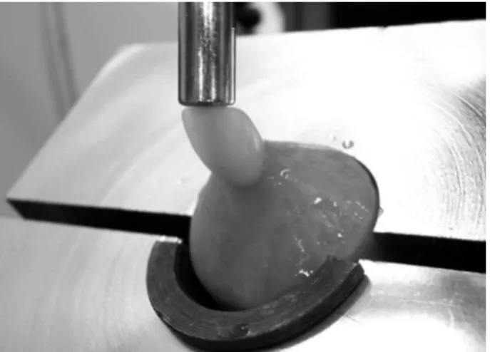
In order to measure the bond strength between the artiicial teeth and the denture base material, the specimens were ixed to a 45o angulated metal split sup-port (Fig. 1), attached to the test machine (Instron 4444; Instron Corp., Canton, MA, USA). A compressive load was applied on the incisal line by means of a cylindrical pin, at a 45o angle, with a crosshead speed of 0.5 mm/ min until fracture. The recorded ultimate failure load in Newtons (N) was converted into kgf. The bond surface was not calculated due to the complexity of curve and irregular shape of original teeth’s ridge lap and cervi -cal area. Resulting data were analyzed statisti-cally by
one-way ANOVA followed by Scheffé’s test (a=0.05). The failure modes were assessed with a magnify-ing glass (×5 magniication), accordmagnify-ing to the followmagnify-ing classiication: 1) Adhesive - when the separation oc -curred at the tooth/denture base interface; 2) Cohesive in PMMA denture base - when the denture base material remained bonded to the dislodged teeth, covering it completely; 3) Cohesive in the tooth - when total tooth base remained bonded to the denture base; 4) Cohesive in the tooth associated with cohesive in the denture base; and 5) Mixed - when signiicant areas of adhesive and cohesive failures occurred simultaneously.
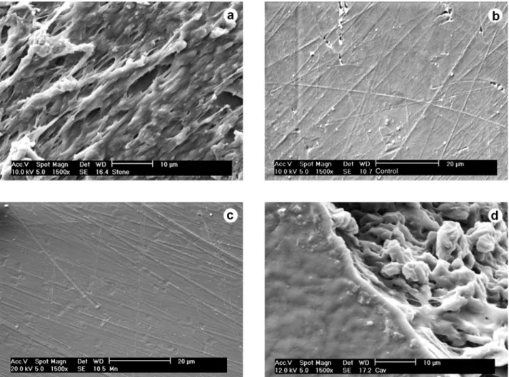
Six extra teeth received the surface treatments applied to the experimental groups and were prepared for scanning electron microscopy in order to verify the resulting ridge lap surface patterns with illustrative purposes. These specimens were coated with a 300 Å golden layer (Bal-Tec SCD 005, Bal-tec Co., USA) and were examined with a scanning electron microscope (Phillips SEM XL30, Phillips, Eindhoven, Netherlands) operating in a range of 10 kV and 20 kV.
RESULTS
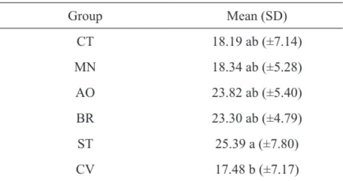
Table 1 and Figure 2 present the mean load (kgf) required for specimen bond to failure. There was statis -tically signiicant difference only between ST and CV (p=0.037997). The null hypothesis that surface treatment on the ridge lap area of acrylic denture teeth inluences their bond strength to the denture base material was partially rejected, since only ST and CV means (extreme means) differed signiicantly from each other, and were statistically similar to intermediary means, obtained in the other groups (CT, MN, AO and BR).
Regarding the failure modes, only cohesive fail-ures in the tooth associated with cohesive in the denture base (n=89), and mixed failures (n=7) occurred.
DISCUSSION
Factors affecting bond strength between plastic teeth and denture base have been investigated with dif-ferent testing methods and the resulting data have been used to suggest technical procedures that enhance this bonding. However, few studies apply methods with load direction (2,4,12,16) and specimen design (2) similar to clinical conditions, producing data that may not be clini-cally representative. Lab research on bonding between
Figure 1. Oblique view of specimen ixed to a 45o angulated
artiicial teeth and denture base usually employs testing methods with only one original or modiied tooth surface contacting the denture base material. The most com-mon surface is ridge lap base with no cervical coverage (1,3,4,7,12-16). The A.D.A speciication #15 test is an example of method with no cervical coverage (20). This may not be the most realistic clinical condition, which is why the present study used an alternative method.
Cervical coverage of artiicial teeth by denture base material in bond strength tests may be an in-convenience because the study variable (e.g.: surface treatment) may suffer the interference of mechanical retention. Nevertheless, it is important to ratify that partial covering of cervical surface of teeth (“neck”) is
a common practice, and occurs in most of the complete and partial dentures because of technical and esthetic reasons. Moreover, according to the indings of a pilot study conducted by our research group, cervical cover-age of plastic teeth enhances the bond strength between acrylic denture teeth and base material, possibly reducing the signiicance of other variables already known, like surface treatment and wax contamination.
The lack of signiicant difference among the mean values of groups CT, MN, AO, ST, CV and BR (except between ST and CV) suggests that the inluence of surface treatments may be minimized by mechani-cal retention resulting from tooth cervimechani-cal coverage. However, it is important to emphasize that failure mode analysis showed that mechanical retention due to cervical coverage was not the only factor responsible for bond strength. All specimens, even those with mixed failure, had some regions of cohesive failure in the tooth and/or Table 1. Bond strength mean values (kgf) and standard deviations
between the acrylic teeth and the denture base material.
Group Mean (SD)
CT 18.19 ab (±7.14) MN 18.34 ab (±5.28) AO 23.82 ab (±5.40)
BR 23.30 ab (±4.79)
ST 25.39 a (±7.80)
CV 17.48 b (±7.17)
CT: no treatment (control); MN - methylmethacrylate monomer etching; AO: 50-μm-particle aluminum oxide air abrasion; BR: glaze removal with a round bur; ST: surface grinding with an aluminum oxide abrasive stone; CV: cavity preparation (diatorics). Different
letters indicate statistically signiicant difference (Scheffé, p>0.05). Figure 2. Mean bond strengths recorded in the groups.
denture base material, with material remnants bonded to non-retentive ridge lap areas.
Tooth base roughening with cutting or abrasive rotary instruments or aluminum oxide air abrasion pro-vided slightly higher bond strength values than those achieved without surface modiication, or with cavity preparation or monomer etching, though without statisti-cal signiicance. As far as bond strength is concerned and considering the unavoidable clinical cervical coverage, in principle, choosing CT, MN, BR or AO is apparently indifferent. These results are similar to those of previous studies (8,12,15,16) and contrary to others (4,14). Dif -ferences may be due to the materials and methodology employed. However, it is likely that roughened surfaces provide a wider contact area with denture base resin and greater micromechanical retention, justifying the
slightly higher bond strength tendency in these groups. SEM analysis showed that surface treatment with aluminum oxide air abrasion (Fig. 3-A), round bur (Fig. 3-B) or abrasive stone (Fig. 4-A) resulted in higher sur -face roughness compared to no treatment (Fig. 4-B) and monomer etching (Fig. 4-C). The teeth treated with dia -torics showed rough surface inside the cavity (Fig. 4-D). The worst results of this study occurred in CV (cavity) group. It may be a consequence of non-treated surfaces tendency to lower bond strength associated with lower cohesive strength of denture base material, which had failured cohesively in all CV specimens, keeping diatorics illed with denture base resin. Also, it is possible that the sharpness of the cavity borders collaborates to stress concentration, thus, leading base material to failure in this area. High impact denture
resins might provide different results.
The tensile loads used in many artiicial tooth bond strength studies are not representative of real conditions either. The expulsive anatomic shape of anterior teeth and the direction of occlusal forces make the occurrence of signiicant tensile forces over these teeth unlikely. On the other hand, shear and compressive loads are much more plausible clinically, especially the angulated load applied by the authors. Similar load direction has previously been used by other researchers (2,4,12,16,17).
The proposed method can be further investigated using different denture base materials, tooth brands and materials, surface treatments, polymerizing parameters, thermocycling, and alimentary chemical solvents, among other variables not evaluated in this study.
Within the limits of this study, it may be concluded that regarding bond strength between acrylic teeth and denture base material, signiicant differences were found only between the surface treatments diatorics (worst results) and roughening with an aluminum oxide stone (best results). The groups treated with round bur, abrasive stone or air abrasion showed numerically higher bond strength than the other groups, but without statistical signiicance. After tooth debonding, most failures were cohesive in the teeth (buccal portion) associated with cohesive in the denture base material (palatal portion).
RESUMO
Este estudo comparou a resistência de união entre base de pró -tese e dentes de resina acrílica submetidos a 6 tratamentos de
superfície. Noventa e seis espécimes foram feitos com dentes de poli(metilmetacrilato) unidos a uma resina para base de prótese polimerizada por energia de microondas. Os espécimes foram
distribuídos em seis grupos (n=16) de acordo com o tratamento
de superfície: CT – controle, superfície não alterada; MN – apli
-cação de monômero de metilmetacrilato; OA – jateamento com partículas de óxido de alumínio de 50 μm; BR – remoção do brilho supericial com broca esférica; PE – asperização com pedra abrasiva de óxido de alumínio e; CV – confecção de cavidade. Os
grupos foram submetidos a uma carga compressiva em ângulo
de 45 graus com o longo eixo dos dentes. Os resultados foram analisados por meio da ANOVA de um fator, seguido do teste de Scheffé (p<0,05). As médias de resistência (kgf) dos grupos foram: CT: 18,19 (7,14), MN: 18,34 (5,28), OA: 23,82 (5,40), BR: 23,30 (4,79), PE: 25,39 (7,80) e CV: 17,48 (7,17). Houve diferença estatisticamente signiicante apenas entre os grupos PE e CV (p=0,037997). Pode-se concluir que a asperização da base
do dente com pedra abrasiva proporcionou a maior resistência de
união, embora com diferença estatisticamente signiicante apenas em relação à confecção de uma cavidade na base do dente. Os
demais tratamentos de superfície proporcionaram valores sem
diferenças estatisticamente signiicativas.
REFERENCES
1. Thean HP, Chew CL, Goh KI. Shear bond strength of denture teeth to base: a comparative study. Quintessence Int 1996;27:425-428. 2. Zuckerman GR. A reliable method for securing anterior denture
teeth in denture bases. J Prosthet Dent 2003;89:603-607. 3. Morrow RM, Matvias FM, Windeler AS, Fuchs RJ. Bonding of
plastic teeth to two heat-curing denture base resins. J Prosthet Dent 1978;39:565-568.
4. Barpal D, Curtis DA, Finzen F, Perry J, Gansky SA. Failure load
of acrylic resin denture teeth bonded to high impact acrylic resins.
J Prosthet Dent 1998;80:666-671.
5. Cunningham JL, Benington IC. Bond strength variation of syn
-thetic resin teeth in dentures. Int J Prosthodont 1995;8:69-72. 6. Vallittu PK, Lassila VP, Lappalainen R. Evaluation of damage to
removable dentures in two cities in Finland. Acta Odontol Scand 1993;51:363-369.
7. Darbar UR, Huggett R, Harrison A. Denture fracture-a survey. Br Dent J 1994;176:342-345.
8. Huggett R, John G, Jagger RG, Bates JF. Strength of the acrylic denture base tooth bond. Br Dent J 1982;153:187-190.
9. Clancy JM, Boyer DB. Comparative bond strengths of light-cured,
heat-cured, and autopolymerizing denture resins to denture teeth.
J Prosthet Dent 1989;61:457-462.
10. Clancy JM, Hawkins LF, Keller JC, Boyer DB. Bond strength and
failure analysis of light-cured denture resins bonded to denture
teeth. J Prosthet Dent 1991;65:315-324.
11. Schneider RL, Curtis ER, Clancy JM. Tensile bond strength of
acrylic resin denture teeth to a microwave - or heat - processed
denture base. J Prosthet Dent 2002;88:145-150.
12. Cardash HS, Liberman R, Helft M. The effect of retention grooves in acrylic resin teeth on tooth denture-base bond. J Prosthet Dent 1986;55:526-528.
13. Barbosa DB, Barão VA, Monteiro DR, Compagnoni MA, Marra J. Bond strength of denture teeth to acrylic resin: effect of
thermocy-cling and polymerisation methods. Gerodontology, 2008;25:237-44. 14. Vallittu PK. Bonding of resin teeth to the polymethyl methacrylate
denture base material. Acta Odontol Scand 1995;53:99-104. 15. Cunningham JL, Benington IC. An investigation of the variables
which may affect the bond between plastic teeth and denture base
resin. J Dent 1999;27:129-135.
16. Saavedra G, Neisser MP, Sinhoreti MAC, Machado C. Evaluation
of bond strength of denture teeth bonded to heat polymerized
acrylic resin denture bases. Braz J Oral Sci 2004;3:458-464. 17. Takahashi Y, Chai J, Takahashi T, Habu T. Bond strength of denture
teeth to denture base resins. Int J Prosthodont 2000;13:59-65. 18. Saavedra G, Valandro LF, Leite FP, Amaral R, Ozcan M, Bottino
MA, et al.. Bond strength of acrylic teeth to denture base resin after
various surface conditioning methods before and after
thermocy-cling. Int J Prosthodont 2007;20:199-201.
19. Nishigawa G, Maruo Y, Okamoto M, Oki K, Kinuta Y, Minagi S,
et al.. Effect of adhesive primer developed exclusively for
heat-curing resin on adhesive strength between plastic artiicial tooth and acrylic denture base resin. Dent Mater J 2006;25:75-80. 20. American Dental Association. Revised ANSI/ADA speciication 15
for synthetic resin teeth. Am Dent Assoc 1985;119-131.