r e v b r a s o r t o p .2 0 1 4;4 9(3):305–308
w w w . r b o . o r g . b r
Case Report
Ameloblastoma: a clinical and therapeutic analysis on
six cases
夽
,
夽夽
Frederico Barra de Moraes
a,∗, Rhanderson Miller Nascimento Cardoso
b,
Sinara Vieira Rodrigues
b, Marcus Vinícius Ferreira Dutra
b, Uiara Rios Pereira
b,
Thiago Raphael Sousa Alencar Borges
baDepartamento de Ortopedia e Traumatologia, Hospital das Clínicas, Universidade Federal de Goiás, Goiânia, GO, Brazil
bFaculdade de Medicina da Universidade Federal de Goiás, Goiânia, GO, Brazil
a r t i c l e
i n f o
Article history: Received 1 May 2013 Accepted 7 May 2013 Available online 25 April 2014
Keywords:
Ameloblastoma/etiology Ameloblastoma/diagnosis Ameloblastoma/surgery Mandibular neoplasms
a b s t r a c t
Ameloblastomas are odontogenic tumors that are locally invasive and slow-growing. Their etiology is still not well defined, but the forms of treatment have been widely discussed because of the possibility of tumor recurrence and postoperative complications. In this study, six patients who were diagnosed with ameloblastoma in the mandibular region and were treated in the Department of Orthopedics and Traumatology of Hospital das Clínicas, Federal University of Goiás, between 1958 and 1963, were evaluated. The radiological, clini-cal and therapeutic characteristics were evaluated. There was no predominance regarding gender in the sample studied. The symptoms most often presented by the patients were pain and tumor formation. The radiological characteristics with greatest incidence were multilocular lesions and the treatment used for all the patients was radical surgery. There was no recurrence over the minimum follow-up period of one year and six months.
© 2014 Sociedade Brasileira de Ortopedia e Traumatologia. Published by Elsevier Editora Ltda. All rights reserved.
Ameloblastoma: uma análise clínica e terapêutica de seis casos
Palavras-chave:
Ameloblastoma/etiologia Ameloblastoma/diagnóstico Ameloblastoma/cirurgia Neoplasias mandibulares
r e s u m o
Os ameloblastomas são tumores odontogênicos, localmente invasivos e de crescimento lento. Sua etiologia ainda não foi bem definida e as formas de tratamento são ampla-mente discutidas, por causa de possíveis recidivas do tumor e complicac¸ões pós-operatórias. Neste trabalho, foram avaliados seis pacientes diagnosticados com ameloblastoma na região mandibular e tratados no Departamento de Ortopedia e Traumatologia do HC-UFG, de 1958 a 1963. Foram avaliadas as características radiológicas, clínicas e terapêuticas. Não houve predomínio em relac¸ão ao gênero na amostra estudada. Os sintomas mais apre-sentados pelos pacientes foram dor e tumorac¸ão. As características radiológicas de maior
夽Please cite this article as: de Moraes FB, Cardoso RMN, Rodrigues SV, Dutra MVF, Pereira UR, Borges TRSA. Ameloblastoma: uma análise clínica e terapêutica de seis casos. Rev Bras Ortop. 2014;49:305–308.
夽夽
Work performed in the Department of Orthopedics and Traumatology, Hospital das Clínicas, Universidade Federal de Goiás, Goiânia, GO, Brazil.
∗ Corresponding author.
E-mail: frederico [email protected] (F.B. de Moraes).
306
r e v b r a s o r t o p .2 0 1 4;4 9(3):305–308incidência são de uma lesão multilocular e o tratamento usado em todos os pacientes foi o cirúrgico radical. A recidiva foi nula em um tempo mínimo de um ano e sete meses de seguimento.
© 2014 Sociedade Brasileira de Ortopedia e Traumatologia. Publicado por Elsevier Editora Ltda. Todos os direitos reservados.
Introduction
Odontogenic tumors are neoplasms derived from the cells responsible for odontogenesis.1 According to the tissue ori-gin, they are classified as epithelial, mesodermal or mixed. Ameloblastomas are the commonest tumors of epithelial ori-gin and account for around 23% of odontogenic tumors.2
Ameloblastomas were first described by Cusack in 1827 apud Chagas et al.3 They are locally aggressive and highly infiltrative, and have a high recurrence rate that has been esti-mated to be around 50%. Despite these characteristics, they are neoplasms that only rarely undergo metastasis.4
They are generally asymptomatic in their initial stages, which have the implication that they are only diagnosed later on, when the tumors have already reached a large size. The commonest symptoms are swelling, pain and local discomfort.3,5
The objective of this study was to report on and discuss the clinical-radiological characteristics of six patients with ameloblastomas who were attended at Hospital das Clínicas, Federal University of Goiás (HC-UFG).
Methodology
The medical files of six patients who were attended in the Department of Orthopedics and Traumatology (DOT) of HC-UFG over a five-year period (1958–1963) were reviewed. This was therefore a retrospective descriptive study. All the information relating to age, sex, clinical manifestations, tumor location, time when the symptoms started, radiologi-cal characteristics, form of treatment, length of follow-up and recurrences was studied.
The locations were divided between the mandible and the maxilla and, for locations in the mandible, it was investi-gated whether the tumor affected the body, angle or ramus of the mandible, or some combination of these. According to the number of radiolucent compartments of the lesion, the tumors were classified as unilocular or multilocular; in the latter case, they would take on a “honeycomb” or “soap bub-ble” appearance.6–9 The surgical treatment was classified as radical or non-radical. According to this division, treatment described as non-radical consisted of enucleation or curet-tage, while radical treatment consisted of complete or partial surgical resection of the tumor.10
Results
The group was composed of three men and three women. Their mean age was 37.5 years (range: 25–50). The tumor was located in the mandible in all the patients. Two cases involved
only the body of the mandible; one, the body and angle; and two, the body, angle and ramus.
The commonest symptoms presented were complaints of increased mandibular volume, i.e. tumor growth and pain (spontaneous or during mastication), which were present in all the patients. Additional symptoms that were present in only two patients are: limitation of movement and formation of a fistula into the oral cavity with discharge of purulent and bloody content.
The time that had elapsed from the beginning of the symp-toms to the consultation at HC-UFG ranged from one to eight years, for five patients who had not had any previous treat-ment. There was one specific case in which the symptoms started 24 years before the consultation in this hospital, but this was a differentiated case because the patient had already gone through two non-radical treatments (curettage), with tumor recurrence.
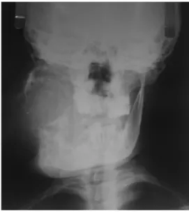
There were no descriptions of radiography in the medi-cal files of the two patients. The radiographs available on the other patients had a multilocular appearance (Fig. 1). It could be seen in all the cases that these tumors had reached enor-mous sizes and were described in accordance with parameters used at that time, as presenting the size of a “lime” or of a “large orange”. These measurements would be equivalent to approximately 4 cm×5 cm×5 cm for the smaller tumors and 8 cm×8 cm×9 cm for the larger tumors. This explains why the treatment used in all the cases was surgical resection of the tumor, i.e. radical treatment.
Five patients were followed up after the surgical treat-ment. The length of follow-up ranged from one year and seven months to seven years and two months. None of these patients presented recurrence (Table 1 and Fig. 2).
r e v b r a s o r t o p .2 0 1 4;4 9(3):305–308
307
Table 1 – Clinical characteristics of the patients with ameloblastoma.
Sex F M F F M M
Age 48 32 38 32 25 50
Location Mandible Mandible Mandible Mandible Mandible Mandible
Part of mandible Body Body and angle Body, angle and ramus Body, angle and ramus
Body and angle Body
Clinical manifestations
Tumor growth and pain
Tumor growth and pain
Tumor growth, pain, limitation of movement and formation of fistula containing pus and blood
Tumor growth, pain, limitation of movement and formation of fistula containing pus and blood
Tumor growth and pain
Tumor growth and pain
Time elapsed since start of symptoms
24 years 1 year and 6
months
1 year 8 years 5 years 5 years
Radiological characteristics
Multilocular – Multilocular – Multilocular Multilocular
Treatment Radical Radical Radical Radical Radical Radical
Length of follow-up
4 years and 11 months
– 2 years and 6 months 1 year and 7
months
6 years and 11 months
7 years and 2 months
Recurrence N – N N N N
Approximate size
“Lime”
4 cm×5 cm×5 cm
– “Large Bahia orange”
7 cm×7 cm×8 cm
“Large avocado” 8 cm×8 cm×9 cm
– “Small orange”
6 cm×6 cm×7 cm
Fig. 2 – After the operation.
Discussion
The ratio between the sexes was 1:1 in this study. This is in accordance with the literature, in which it is usually shown that there is no notable relation to gender regarding occur-rences of ameloblastoma.1,3,8,11,12In a study conducted on 116 patients, the ratio between the male and female genders was 1.2:1,5 and a single study in which this ratio was analyzed found that it was 2:1, with predominance among males.13
Regarding the patients’ ages, the mean was 37.5 years. This is also confirmed in the literature, where these tumors have been shown to be predominantly in adults, generally in their fourth or fifth decade of life.3,12
Ameloblastomas have been found located in the mandible in around 80% of the cases and in the maxilla in the remain-ing 20%.3,8,11,12,14 In our study, all of them were located in the mandible, and this is close to the data found by Kim and Jang,5who in a study on 71 cases observed occurrences of ameloblastomas in the mandible in 93.9% of them. In the same study, the location was only in the body of the mandible in 60.6% of the cases and in the body and angle in 2.8% of the cases, while there were no cases located simultaneously in the body, angle and ramus of the mandible.5Tumors that
are more voluminous may affect the adjacent soft tissues in an infiltrative manner, to the point of promoting erosion and reabsorption of the tooth roots.1
Our sample included two cases in which only the body of the mandible was affected; one, the body and angle; and two, the body, angle and ramus. This was probably due to the long period of evolution. The tumors had already reached large pro-portions, which thus explains why the body, angle and ramus were simultaneously affected, which had not been reported in other studies.
The clinical manifestations most commonly presented by the patients were tumor growth (100%) and pain (100%), fol-lowed by limitations on movement (33.3%) and formation of fistulas with drainage of pus and/or blood (33.3%). The authors of the studies analyzed are unanimous in stating that the vast majority of ameloblastomas are slow growing and therefore only rarely manifest with signs other than local tumor growth, which is the commonest finding.3,5,13 Medeiros et al.14 also cited Neville et al.16to affirm that these tumors are only rarely painful, unless they become secondarily infected, and that signs or symptoms of nerve impairment are uncommon, even in large tumors. Thus, the high prevalence of pain in the cases reported may be indicative of associated infectious processes, particularly when it is taken into consideration that in some cases there was a great hiatus of time between the beginning of the symptoms and seeking medical assistance.
The characteristic most commonly found through radio-graphic analysis is the multilocular pattern (65.4%), as shown in the study on 52 cases by Saddy et al.15In our study, the multilocular pattern also predominated. However, even with a clearly determined radiographic appearance, the definitive diagnosis of ameloblastoma should be sought by correlating this with histopathological examination of the lesion.3
308
r e v b r a s o r t o p .2 0 1 4;4 9(3):305–308numbers of recurrences. The unicystic form accounts for around 14% of the cases; it is less invasive and does not present large numbers of recurrences. The peripheral form is rare, accounting for less than 1% of the cases, and it only affects soft tissues surrounding the region of the teeth.16
Ameloblastomas present only a few symptoms at a late stage, which makes it difficult to identify these tumors at their early stages.9It has been shown that when individuals realize that they have this tumor, or a healthcare professional notices it, the tumor already presents a considerable volume. Another obstacle is that in many cases, the patients perceive the tumor growth as having the consistency of bone, but end up seeking medical care only after the condition has evolved for some time, which may even be years later. In this case series, all the patients were individuals of low social class and had difficul-ties in accessing healthcare services. For this reason, these tumors had evolved over a longer time and therefore had large dimensions. It has been reported that infiltration of adja-cent soft tissues may occur in cases of tumors that are more voluminous.1One of these cases had even evolved from the beginning of the symptoms eight years earlier and was the one with the greatest tumor growth in terms of dimensions, comparable with the size of a large avocado.
The treatment for ameloblastomas is surgical and may be radical or non-radical. Radiotherapy is not indicated, since the lesions are radioresistant. Non-radical treatment is generally used for unicystic tumors. However, according to Nakamura et al.,17this treatment method, which includes marsupializa-tion and enucleamarsupializa-tion followed by appropriate bone curettage, has been shown to be very efficient and has reduced the need for surgical resection and thus reinforced the indications for non-radical treatment for ameloblastomas.
In turn, radical treatment implies total removal of the lesion, generally with a safety margin of one to two centimeters,1,13and is more indicated for lesions that are more aggressive, such as in cases of multicystic ameloblastoma or even in unicystic cases with infiltrating characteristics. In our cases, we chose radical surgical treatment for all the patients mainly because of their late diagnosis, with tumors that already had large dimensions. In this case series, rad-ical therapeutic management did not present recurrence in the five cases that were followed up. One of these cases had even presented recurrences subsequent to curettage per-formed previously at another service.
Regarding the length of follow-up, our cases did not show any recurrence over a minimum period of one year and seven months. However, it should be taken into consideration that two of the cases were followed up for less than three years. Most studies have evaluated recurrence over a mean period of four to five years.13Nevertheless, since all of our cases were treated with full surgical resection and with a mean length of follow-up of four years and seven months, i.e. concordant with other studies,18,19we can consider that the lack of recurrence in our series was a valid result.
Conflicts of interest
The authors declare no conflicts of interest.
r e f e r e n c e s
1. Sá AC, Zardo M, Paes Júnior AJ, Souza RP, Neme MP, Sabedotti I, et al. Ameloblastoma da mandíbula: relato de dois casos. Radiol Bras. 2009;37(6):465–8.
2. Avelar RL, Antunes AA, Santos TS, Andrade ESS, Dourado E. Tumores odontogênicos: estudo clínico-patológico de 238 casos. Rev Bras Otorrinolaringol. 2008;74(5):668–73. 3. Chagas JF, Toledo Júnior JI, Pascoal MBN, Pascoal MI, Aquino
JL, Campos JLG, et al. Ameloblastomas: aspectos clínicos e terapêuticos. Rev Bras Cir Cabec¸a Pescoc¸o. 2007;36(3):159–62. 4. Ciment LM, Ciment AJ. Malignant ameloblastoma metastatic
to the lungs 29 years after primary resection. Chest. 2002;121(4):1359–61.
5. Kim SG, Jang HS. Ameloblastoma: a clinical, radiographic, and histopathologic analysis of 71 cases. Oral Surg Oral Med Oral Pathol. 2001;91(6):649–53.
6. Swan JS. The teeth, jaws, and salivary glands. In: Juhl JH, Crummy AB, Kuhlman JE, editors. Paul & Juhl’s essentials of radiologic imaging [versão digital]. 7th ed. Philadelphia: Lippincott Williams & Wilkins; 1998. p. 13–31.
7. Ogunsalu C, Daisley H, Henry K, Bedayse S, White K, Jagdeo B, et al. A new radiological classification for ameloblastoma based on analysis of 19 cases. West Indian Med J. 2006;55(6):36–41.
8. Martinez CR, Barros RM, Orué NR, Oliveira JGP, Monteiro JCC. Ameloblastoma: estudo clínico-histopatológico. Rev Cir Traumatol Buco-Maxilo-Fac. 2008;8(2):55–60.
9. Martins MD, Rosa Junior OA, Martins MAT, Bussadori SK, Fernandes KPS. Ameloblastoma: revisão de literatura. ConScientia e Saúde. 2007;6(2):269–78. Disponível em: http://www4.uninove.br/ojs/index.php/saude/article/viewFile/ 1108/886
10. Gardner DG, Pecak AM. The treatment of ameloblastoma based on pathologic and anatomic principles. Câncer. 1980;46(11):2514–9.
11. Martins RH, Andrade J, Sobrinho, Rapoport A, Rosa MP. Histopathologic features and management of ameloblastoma: study of 20 cases. São Paulo Med J. 1999;117(4):171–4.
12. Costa DO, Ecard MB, Oliveira SP, Silva LE, Dias EP, Lourenc¸o SQC. Estudo retrospectivo dos casos diagnosticados como ameloblastoma no Servic¸o de Anatomia Patológica do Hospital Universitário Antônio Pedro entre 1997 e 2007. J Bras Patol Med Lab. 2008;44(6):441–7.
13. Lagares DT, Cossío PI, Guisado JMH, Pérez JL. Mandibular ameloblastoma. A review of the literature and presentation of six cases. Med Oral Patol Oral Cir Bucal. 2004;10(3):231–8. 14. Medeiros M, Porto GG, Laureano Filho JR, Portela L,
Vasconcellos RH. Ameloblastoma em mandíbula. Rev Bras Otorrinolaringol. 2008;74(3):478.
15. Saddy MS, Chilvarquer I, Dib LL, Sandoval RL. Aspectos clínicos, radiográficos e terapêuticos do ameloblastoma. Rev Pós Grad. 2005;12(4):460–5. Disponível em:
http://www.fo.usp.br/revistas/rpg/EDICOES/RPG-4 09 684.pdf 16. Neville BW, Damm DD, Allen CM, Bouquot JE. Patologia oral &
maxilofacial. 2nd ed. Rio de Janeiro: Guanabara; 2004. 17. Nakamura N, Higuchi Y, Mitsuyasu T, Sandra F, Ohishi M.
Comparison of long-term results between different
approaches to ameloblastoma. Oral Surg Oral Med Oral Pathol Oral Radiol Endod. 2002;93(1):13–20.
18. Olaitan AA, Arole G, Adekeye EO. Recurrent ameloblastoma of the jaws: a follow-up study. Int J Oral Maxillofac Surg. 1998;27(6):456–60.