REVISTA
BRASILEIRA
DE
REUMATOLOGIA
www . r e u m a t o l o g i a . c o m . b r
Original
article
Colonoscopic
evaluation
in
patients
with
ankylosing
spondylitis
Haim
Cesar
Maleh
a,∗,
Blanca
Elena
Rios
Gomes
Bica
b,
José
Ângelo
de
Souza
Papi
c,
Mário
Newton
Leitão
de
Azevedo
c,
Antônio
José
de
Vasconcellos
Carneiro
caClinicalMedicine,UniversidadeFederaldoRiodeJaneiro,RiodeJaneiro,RJ,Brazil
bServiceofRheumatology,HospitalUniversitárioClementinoFragaFilho,UniversidadeFederaldoRiodeJaneiro,RiodeJaneiro,RJ,Brazil
cDepartmentofClinicalMedicine,MedicineSchool,UniversidadeFederaldoRiodeJaneiro,RiodeJaneiro,RJ,Brazil
a
r
t
i
c
l
e
i
n
f
o
Articlehistory:
Received7June2013 Accepted12March2014 Availableonline21August2014
Keywords:
Ankylosingspondylitis Colonoscopy
Histologicalanalisys Intestinalalteration
Intestinalinflammatorydisease
a
b
s
t
r
a
c
t
Introduction:Patientswithankylosingspondylitiscanhaveintestinalinflammatorylesions,
thustheuseofcolonoscopyforsuchpatientsshouldbedefined.
Objectives:Toassessthegrossintestinalcolonoscopicchangesandmicroscopic
histopatho-logicfindingsofpatientswithankylosingspondylitis;tocorrelatethecolonoscopicand histopathologicfindings;andtostudytherelationshipofthehistopathologicfindingswith extra-articularmanifestationsofthedisease,HLA-B27,BASFIandBASDAI.
Methods:Thisis across-sectionalstudyof22 patientswithankylosingspondylitis.The
patientsunderwentclinicalassessment,BASDAIandBASFIapplication,bloodcollectionfor HLA-B27measurement,andcolonoscopywithbiopsyoffourintestinalsegments(terminal ileum,rightandsigmoidcolons,andrectum).
Results:Abnormalcolonoscopic resultswereobtained in 13 (59.1%)patients, themajor
abnormalitybeing intestinal polyps.The groups ofnormalandabnormal colonoscopic results(n=9andn=13,respectively)werehomogeneousregardingage,BASFI,BASDAI,and categoricalvariables,andtheP-valueshowednosignificantdifferencebetweengroups.The histopathologicalfindingsrevealedabnormalbiopsiesin81%,90.9%,90.9%and86.4%for terminalileum,rightcolon,sigmoidcolon,andrectum,respectively.Thehistopathologic resultsshowednostatisticallysignificantassociationwiththeextra-articular manifesta-tions,BASFI,BASDAIandHLA-B27positivity.
Conclusions:Thehistologicalanalysisofthefourintestinalsegmentsevidenced
inflamma-torylesionsinpatientswithnormalandabnormalcolonoscopicresults,independentlyof bowelsymptomatologyandtherapyusedinthetreatmentofthebasaldisease.
©2014ElsevierEditoraLtda.Allrightsreserved.
∗ Correspondingauthor.
E-mail:[email protected](H.C.Maleh). http://dx.doi.org/10.1016/j.rbre.2014.03.020
Avaliac¸ão
colonoscópica
em
pacientes
com
espondilite
anquilosante
Palavras-chave:
Espondiliteanquilosante Colonoscopia
Análisehistológica Alterac¸ãointestinal
Doenc¸ainflamatóriaintestinal
r
e
s
u
m
o
Introduc¸ão: Pacientescomespondiliteanquilosantepodemapresentar-secomlesões
infla-matóriasintestinais,e,porisso,deveserdefinidoousodacolonoscopiaparataispacientes.
Objetivos:Avaliarasalterac¸õescolonoscópicasintestinaismacroscópicaseachados
histopa-tológicos microscópicos de pacientes com espondilite anquilosante; correlacionar os achadoscolonoscópicosehistopatológicos;eestudararelac¸ãodosachadoshistopatológicos comasmanifestac¸õesextra-articularesdadoenc¸a,HLA-B27,BASFIandBASDAI.
Métodos: Esteéumestudotransversalde22pacientescomespondiliteanquilosante.Os
pacientespassaram por umaavaliac¸ão clínica,BASDAIe BASFI,coletade sanguepara determinac¸ãodeHLA-B27, ecolonoscopiacombiópsiadequartosegmentosintestinais (íleoterminal,cólondireito,cólonsigmoideereto).
Resultados: Resultadoscolonoscópicosanormaisforamobtidosem13(59,1%)pacientes,
eaprincipalanormalidadefoiapresenc¸adepóliposintestinais.Osgruposderesultados colonoscópicosnormaiseanormais(n=9e n=13,respectivamente)foramhomogêneos noquedizrespeitoàidade,BASFI,BASDAI,evariáveiscategóricas,eovalorPnão rev-eloudiferenc¸asignificativaentregrupos.Dosresultadoshistopatológicos,81%tiveramuma biópsiaanormaldoíleoterminal,90.9%tiveramumabiópsiaanormaldocólonsigmoide, eabiópsiaretalestavaanormalem86.4%.Osachadoshistopatológicosrevelarambiópsias anormaisem81%,90.9%,90.9%e86.4%paraoíleoterminal,cólondireito,cólonsigmoide ereto,respectivamente.Osresultadoshistopatológicosnãorevelaramassociac¸ão estatisti-camentesignificativacomasmanifestac¸õesextra-articulares,BASFI,BASDAIepositividade paraHLAB27.
Conclusões: A análise histological dos quatro segmentos intestinais evidenciou lesões
inflamatóriasempacientescomresultadoscolonoscópicosnormaiseanormais, indepen-dentementedasintomatologiaintestinaledotratamentousadoparaadoenc¸abasal.
©2014ElsevierEditoraLtda.Todososdireitosreservados.
Introduction
Ankylosingspondylitis (AS) is a chronic inflammatory dis-ease characterized by involvement of the axial skeleton, entheses and, less commonly, peripheral joints. AS is cause of inflammatory back pain due to involvement of the sacroiliac joints and the spine, which can result in ankylosis.1 In addition to damage to the axial skeleton
and peripheral joints,AS may also present extra-articular manifestations,suchasuveitis andcardiacand pulmonary manifestations.2
SubclinicalintestinalinflammationcanoccurinAS,which canbeobservedthroughileocolonoscopyin25%-49%ofcases. Microscopiclesions,detectedbyintestinalbiopsy,are more frequentthan gross lesions,being observedin 50%-60% of patients and histologically classified as acute or chronic.3
Chronic injuries are more common, occurring in 80% of patientsandwithsimilarcharacteristicstotheileocolitisof Crohn’sdisease(CD).Itissuggestedthattheintestinalmucosa plays an important role in the pathogenesis of spondy-loarthritides,throughthepossiblepermeabilitytoexogenous antigens.3,4
Theaimofthis studywas toevaluatetheprevalenceof bowelinflammationinpatientswithASthroughcolonoscopy, besides possible associations with clinical and functional aspectsinherenttotheunderlyingdisease.
Patients
and
methods
Studydesign
Across-sectionalstudywasconductedwith22patients diag-nosed withASfollowedatthe spondyloarthritisoutpatient clinic,HospitalUniversitarioClementinoFragaFilho(HUCFF), UniversidadeFederaldoRiodeJaneiro(UFRJ).Allparticipants readandsignedtheInformedConsentForm(ICF)provided, inaccordancewiththestandardsoftheinstitution’sResearch EthicsCommittee(REC).
Inclusioncriteria
Patients diagnosed with AS defined by New York Criteria (1984).5
Exclusioncriteria
Proceduresperformed
Data,suchasage,gender,race,extra-articularmanifestations ofthedisease,amongothers,werecollectedfromtheclinical evaluationperformedwithpatients.
Tocharacterizetheclinicalactivityofthediseaseandof physicalworkcapacityofthesample,patientsweresubmitted totheuseoftwoinstruments:theBathAnkylosingSpondylitis FunctionalIndex(BASFI)andtheBathAnkylosingSpondylitis DiseaseActivityIndex(BASDAI),6whichaddressissues
refer-ringtoclinicalsymptomsofthedisease,aswellaslimitations forspecificactivities,allowingtheirgraduation.
Examinations
BloodcollectionswereperformedforHLA-B27determination intheClinicalAnalysisLaboratory,UFRJ,withafastingperiod of8hours,usingthesurfacephenotypingtechniqueandthe cellulartechniquewithanti-HLAB27/B7byflowcytometry.
Patientsunderwentcolonoscopywithbiopsy,inorderto assessthepresenceofintestinallesionsinthecolon; concomi-tantly,histopathological analysisofbowel was carried out, throughthecollectionofbiopsymaterialin4pre-definedsites: terminalileum,ascendingcolon,colonsigmoidandrectum. Inflammatorylesionswereclassifiedbyinflammationdegree (mild,moderateandsevere).
Statisticalanalysis
To test the significance ofthe tests mentioned above, the following statistical methods were applied:1. To compare numericaldatabetweenindependentsamples,Student’sttest or the Mann-Whitneytest (non-parametric)was used. The variancehomogeneitywas testedbytheLevene’stest;2. To compareproportions(categoricaldata)thechi-squared(2)or
Fisher’sexacttestwasused;3.Alogisticregression analysis wasperformedtoidentifytheindependentbaselinevariables thatinfluence(orpredict)thechangeofthecolonoscopy out-come.
Non-parametricmethodswereused, becausesome vari-ables did not present a normal distribution (Gaussian distribution)duetothewidedispersionandoftherejection ofthehypothesisofnormality,accordingtothe Kolmogorov-Smirnovtest. A level of5% as a criterion for determining significancewas adopted. Thestatistical analysis was per-formedwithstatisticalsoftwareIBMSPSSStatistics,version 19.
Results
Ofthe 22 patients included, 19 (86.4%) were male, and 11 patients (50%)were classified asCaucasians (Table 1). The mean age of the study population was 45.9±10.9 years (Table2).Withregard topathological featuresofASinthe samplestudied, 6patients(27.3%)hadsomeextra-articular manifestation,anditsmostprevalentexpressionwasanterior uveitis,presentin5patients(22.7%)(Table1).
Asforthecharacterizationofclinicaldiseaseactivity,as wellasthefunctionalcapacityofthepatients,thesamplewas
Table1–Generaldescriptionofbasalcategorical variables.
Variable n %
Gender
Female 3 13.6
Male 19 86.4
Race(caucasians) 11 50.0
HLA-B27
Positive 9 40.9
Undetermined 4 18.2
Negative 9 40.9
Presentedextra-articularchange 6 27.3
Presentedocularchange 5 22.7
Presentednokidneychange 22 100.0
Presentedcardiacchange 1 4.5
Presentednoneurologicalchange 22 100.0
Presentednopulmonarychange 22 100.0
Colonoscopy
Normal 9 40.9
Abnormal 13 59.1
Sigmoidcolonbiopsy
Normal 2 9.1
Abnormal 20 90.9
Mildinflammation 17 77.3
Moderateinflammation 2 9.1
Severeinflammation 1 4.5
Terminalileumbiopsy
Normal 4 18.2
Abnormal 17 81.0
Mildinflammation 4 18.2
Moderateinflammation 5 22.7
Severeinflammation 8 36.4
Rightcolonbiopsy
Normal 2 9.1
Abnormal 20 90.9
Mildinflammation 17 77.3
Moderateinflammation 2 9.1
Severeinflammation 1 4.5
Rectalbiopsy
Normal 3 13.6
Abnormal 19 86.4
Mildinflammation 14 63.6
Moderateinflammation 4 18.2
Severeinflammation 1 4.5
subjectedtoinstrumentsforevaluationofthedisease,among them,respectively,BASDAIandBASFI.ThemeanBASDAIwas 3.6±1.5;andBASFIshowedameanof4.6±1.8(Table2).
All patients underwent blood collection with HLA-B27 dosage,and9patients(40.9%)werepositiveforthismarker. The remainingsamples showed negative or indeterminate HLAB27(Table1).
Table2–Analysisofcategoricalandnumericvariablesaccordingtochangeincolonoscopyexamination.
Categoricalvariable Colonoscopy,normal(n=9) Colonoscopy,abnormal(n=13) P-value
n % n %
Male 8 89% 11 85% 0.774
Caucasians 5 56% 6 46% 0.665
HLA-B271negative 5 56% 4 31% 0.489
Absenceof:
Extra-articularchange 8 89% 8 62% 0.157
Ocularchange 8 89% 9 69% 0.279
Cardiacabnormality 9 100% 12 92% 0.394
Abnormalresults
Sigmoidcolonbiopsy 8 89% 12 92% 0.784
Terminalileumbiopsy 7 88% 10 77% 0.549
Rightcolonbiopsy 8 89% 12 92% 0.784
Rectalbiopsy 8 89% 11 85% 0.774
methotrexatebeingthe mostused incombination therapy, followedbysulfasalazine.
Whenevaluatingpatientsundergoingcolonoscopy,9had normalresults, and 13 had abnormalresults, formingtwo groups:normalcolonoscopy(n=9)andabnormalcolonoscopy (n=13).
Inpatientswithabnormalcolonoscopy,themain colono-scopicfinding wasthepresenceofintestinalpolyps,which were present in 6 patients. Of the 6patients with intesti-nalpolyps,5showedpolypsclassifiedasofthesessiletype, and1showedapedunculatedpolyp.Themostcommon loca-tionofpolypswasinthesigmoidcolon.Thehistopathological analysisofpolypsexcludedthepresenceofmalignancy,and only 1 patient had a tubular adenoma. Comparing the 2 groups formed(normal and abnormalcolonoscopy), it can beseen that,withrespecttothebaselinenumerical analy-ses,bothgroupsshowedhomogeneityintheaspectsrelated toage,BASFIandBASDAI(Table2).AccordingtotheP-value presented,thenumericalvariablesshowednosignificant dif-ferencebetweenthecolonoscopyresults.Asfortheanalysisof categoricalvariables,whencomparingthe2groups,onecan alsonoticethatbothwerehomogeneous.
Amongthe22patients,17(81%)hadalterationinthe ter-minal ileum biopsy, and in 8 (36.4%) there was a marked inflammation. Twenty patients (90.9%) had alteration in the right colon biopsy, and 17 (77.3%) had histopathology consistent with mild inflammation. The histopathological evaluationofthe sigmoid colonshowed abnormality in20 patients(90.9%),and17(77.3%)wereclassifiedaspresenting mild inflammation. In the rectum, the analysis by biopsy showedchangesin19patients(86.4%),withmild inflamma-tionin14patients(63.6%)(Table1).
By analyzing the sigmoid colon biopsy, 2 patients had results considered normal and 20 patients had an abnor-malreport,withprevalenceofamildinflammationpattern (Table3).Therewasnostatisticalsignificancebetweenthe2 groupsanalyzed,asthegroupwithabnormalbiopsyhada pre-dominantnumberofpatients.Allpatientswhohadsometype ofinflammationinthe sigmoidcolonbiopsy showed alter-ationsalsointherightcolon(p=0.000)andrectal(p=0.000) biopsiesatthelevelofsignificanceof5%.
Withrespecttothehistologicalevaluationofthe termi-nalileum,1patientwasexcludedfromtheanalysisbecause no biopsy of that site was performed; 4patients had nor-malresults,and 17 wereconsidered abnormal(Table4).In the group withabnormal biopsies, it wasmainly observed anaccentuatedmicroscopicinflammation.Accordingtothe P-valuepresented,thevariablesshowednosignificant differ-encebetweentheresultsoftheterminalileumbiopsies.
Intheanalysisoftherightcolonbiopsy,20patientshad their biopsy classified asabnormal,with preponderanceof mildinflammation,andthisshouldbeconsideredasthemost frequenttype ofinflammationduringthe group’s analysis. Patients with any typeof inflammationduring right colon biopsy showedabnormalities inthe rectalbiopsy(P=0.000) atthelevelofsignificanceof5%.AccordingtotheP-value pre-sented,thebaselinenumericalandcategoricalvariableswere notstatisticallysignificantintheanalysisforthe groupsof rightcolonbiopsy(Table5).
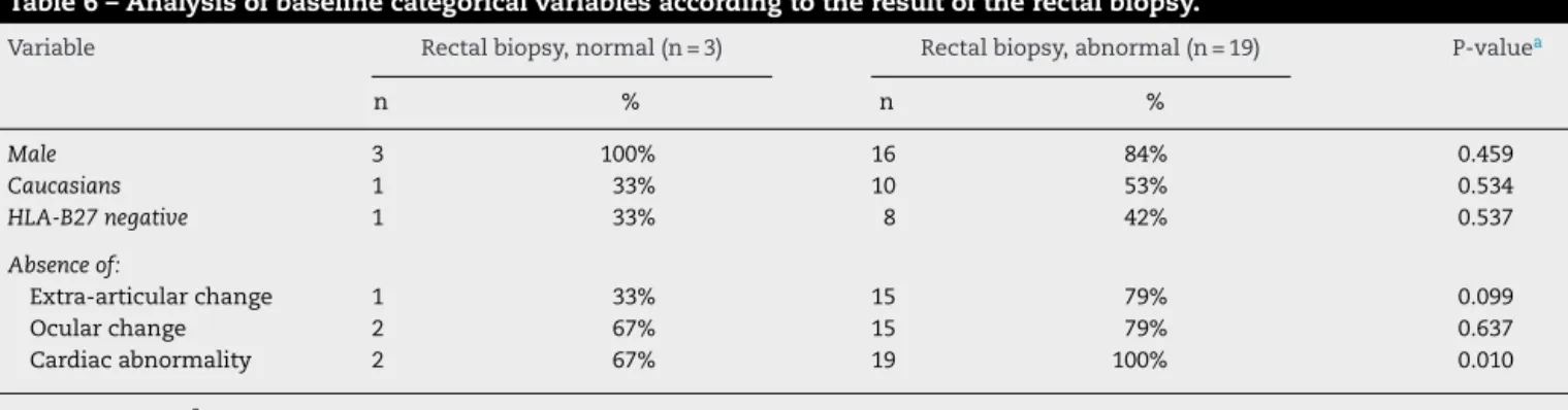
Inthe evaluationoftherectal biopsies,19 patients pre-sented biopsy with an altered result, and 3 patients were classifiedasnormalresults(Table6).Therewaspredominance ofmildinflammation.AccordingtotheP-valueshown,there isatendencyfortheabnormalgroupofgreaterBASDAIs com-paredtothe normalgroup.Inaddition,thereisatrendfor fewerextra-articularchanges(P=0.099)ata10%levelforthose patients who had sometypeofinflammationinthe rectal biopsy,contrarytothegrosscolonoscopicfindings.
Discussion
ASaffectsmenandwomenataratioof2:1aroundthe3rd decadeoflife,7and90%ofpatientsarepositiveforHLA-B27;8
Inthisstudy,theprevalenceofASwasalsohigherinmen ver-suswomen,occurringataratioof6:1,butHLA-B27positivity wasfoundin40.9%ofpatients.VanPraetetal.13evaluated65
patientswithASundergoingcolonoscopy;theseauthorsalso showednoassociationofmicroscopicintestinalinflammation withpositivityforthisantigen.
Table3–Analysisofbaselinecategoricalvariablesaccordingtotheresultofthesigmoidcolonbiopsy.
Variable Sigmoidcolonbiopsy,normal(n=2) Sigmoidcolonbiopsy,abnormal(n=20) P-valuea
n % n %
Male 2 100% 17 85% 0.556
Caucasians 1 50% 10 50% 1.000
HLA-B27negative 1 50% 8 40% 0.783
Absenceof:
Extra-articularchange 1 50% 15 75% 0.449
Ocularchange 2 100% 15 75% 0.421
Cardiacabnormality 1 50% 20 100% 0.001
Abnormalresults
Terminalileumbiopsy 1 50% 16 84% 0.241
Rightcolonbiopsy 0 0% 20 100% 0.000
Rectalbiopsy 0 0% 19 95% 0.000
a Chi-Squared(2)test.
Table4–Analysisofbaselinecategoricalvariablesaccordingtotheresultoftheterminalileumbiopsy.
Variable Terminalileumbiopsy,normal(n=4) Terminalileumbiopsy,abnormal(n=17) P-valuea
n % n %
Male 4 100% 14 82% 0.364
Caucasians 1 25% 9 53% 0.314
HLA-B27negative 1 25% 7 41% 0.215
Absenceof:
Extra-articularchange 3 75% 12 71% 0.861
Ocularchange 3 75% 13 76% 0.950
Cardiacabnormality 4 100% 16 94% 0.619
Abnormalresults
Terminalileumbiopsy 3 75% 16 94% 0.241
Rightcolonbiopsy 3 75% 15 88% 0.496
Rectalbiopsy
a Chi-Squared(2)test.
patients,with90%ofthesetestingpositiveforHLA-B27.9–11
Inthepresentstudy,ourdatawerenearthoseinthe litera-ture,and27.3%ofpatientshadsometypeofextra-articular manifestationofthedisease,anterioruveitisbeingthemore prevalentandpresentin5patients(22.7%).Ofthese5patients, 4weremale,and4werepositiveforHLAB27.In2006, Rud-waleitstudiedcolonoscopiesofpatientswithASandshowed
thatsubclinicalgrossintestinalinflammationwaspresentin 25%-49%ofpatients;3however,inthisstudythesubclinical
grossinflammatorylesionscharacterizedbythepresenceof ilealinflammationwerefoundinonly2patients(9.09%).
It is known that inpatients with AS and submitted to colonoscopy,themicroscopicinflammatorylesions,detected in50%-60%ofcasesbyintestinalbiopsy,aremoreprevalent
Table5–Analysisofbaselinecategoricalvariablesaccordingtotheresultoftherightcolonbiopsy.
Variable Rightcolonbiopsy,normal(n=2) Rightcolonbiopsy,abnormal(n=20) P-valuea
n % n %
Male 2 100% 17 85% 0.556
Caucasians 1 50% 10 50% 1.000
HLA-B27negative 1 50% 8 40% 0.783
Absenceof:
Extra-articularchange 1 50% 15 75% 0.449
Ocularchange 2 100% 15 75% 0.421
Cardiacabnormality 1 50% 20 100% 0.001
Abnormalresults
Rectalbiopsy 0 0% 19 95% 0.000
Table6–Analysisofbaselinecategoricalvariablesaccordingtotheresultoftherectalbiopsy.
Variable Rectalbiopsy,normal(n=3) Rectalbiopsy,abnormal(n=19) P-valuea
n % n %
Male 3 100% 16 84% 0.459
Caucasians 1 33% 10 53% 0.534
HLA-B27negative 1 33% 8 42% 0.537
Absenceof:
Extra-articularchange 1 33% 15 79% 0.099
Ocularchange 2 67% 15 79% 0.637
Cardiacabnormality 2 67% 19 100% 0.010
a Chi-Squared(2)test.
ifcomparedtogrosslesions,beingusuallyasymptomatic.3
In 2009, Hascelik et al.12 studied 25 patients with a
diag-nosisofAS, which underwent colonoscopywithbiopsy.In theiranalysis,theseauthorsalsofoundahigherprevalence ofhistologicalbowelinflammatorylesions,andmicroscopic intestinalinflammationwasfoundin20of25patients(80%), withmoreprevalenceintheileum.In2013,VanPraetetal.13
evaluated65patientswithASandshowedmicroscopic intesti-nallesionsin46.9%oftheirpatients,particularlyofthechronic typeandwithapreferentiallocationintheileum.
Corroborating datafrom the literature,in ourstudy the microscopicinflammatorylesionswerealsomoreprevalent thangrosslesions,beingpresentinall4intestinalsegments analyzed,eveninpatientswithnormalcolonoscopicfindings. In our analysis, we observed a predominance of chronic-typelesionswithsomedegreeofinflammationinover80% ofthebiopsiesofeachintestinalsegment(Table1), exceed-ing the data on the prevalence of intestinal inflammation inpreviouslyreportedstudies.Mild inflammation predomi-natedinthesigmoidcolon,rightcolonandrectum,evenin patientstakingdisease-modifyingdrugsandanti-TNF␣
med-ications.Severeinflammationpredominatedintheterminal ileum, irrespective ofthe therapyused forAS. Thisfact is anadditional element that supportsone ofthe pathophy-siologicaltheoriesofAS,whichsuggeststhattheintestinal mucosaisconsideredasainitialpathologicalsiteofthe dis-ease,occurringlocalpresentationofpathogenicantigenstoT lymphocytesCD8,withtheirsubsequentcirculationtojoints throughadhesivemolecules.3,14
In AS, in patients with chronic inflammatoryintestinal lesionsevidencedbyhistologicalanalysisofthecolon,there isastrongerassociationbetweenthepresenceofintestinal inflammationwithclinicalactivityofAS.3,4,12VanPraetetal.13
evaluated65patientswithASshowingassociationbetween microscopicintestinalinflammationwithdiseaseactivity,as measuredbyBASDAI.Thisfactwasnotproveninouranalysis, whereasignificantstatisticalrelationshipbetweenthe sug-gestiveclinicalparametersofdiseaseactivityandfunctional capacityofpatients(i.e.BASFIandBASDAI)wasnotconfirmed bytheresultspresented.
Contrary towhat weobserved inour analysis, Hascelik studied25patientswithadiagnosisofAS,12whounderwent
colonoscopywith biopsy and a preliminary assessment of demographicandclinicalparameters,includingBASDAIand BASFI.Inhisanalysis,Hascelikalsofoundahigherprevalence
ofhistologicalinflammatorybowellesionswithpresenceof microscopic intestinalinflammationin 80% ofcases,more prevalent in the ileum. Furthermore, patients with gross intestinallesionshadhigherdiseaseactivity,characterizedby BASDAI.
Thesmall sizeofoursample wasalimiting factor that may justify,inthis study,the absenceofastatistically sig-nificantcorrelationamongtheclinicalcharacteristics ofAS (assessedbyBASFIandBASDAI),andlaboratorydata(HLA-B27 positivity)withthe histologicaldatapresented,which sug-gestedthe presenceofmicroscopicintestinalinflammation inintestinalsegmentsexaminedbycolonoscopy.
Asregardstheinfluenceofmedicationsintheinductionof colonicinflammatorylesions,theliteratureisscarceinterms ofdatashowingtheinvolvementofDMARDSand anti-TNF drugs.Onepointthatshouldbequestionedwouldbethefact thattheuseofanti-TNFagentsismaskingthecolonoscopy and histology reports, bypreventing the emergence of the characteristiclesions,sincethistypeofmedicationalsohas itsindicationforthetreatmentofpatientswithCDand ulcer-ative rectocolitis (URC).There are no datain theliterature addressingtheinfluenceofanti-TNFinbothgrossand micro-scopicintestinallesionsinpatientswithAS.
Conclusion
Inthepresentstudy,thecolonoscopicchangesinpatientswith ankylosingspondylitisarethesameasthosefoundinthe gen-eralpopulation,confirmingthepolypoidlesionsasthemost prevalent,andnotjustifyingtheroutineuseofcolonoscopy. Thebowelhistologicalanalysisshowedinflammatorylesions inallfoursegmentsbiopsied,regardlessofthecolonoscopic report,intestinalsymptomatologyandtherapyusedforthe underlyingdisease.
Conflicts
of
interest
Theauthorsdeclarenoconflictsofinterest.
r
e
f
e
r
e
n
c
e
s
Espondiloartropatias:EspondiliteAnquilosanteeArtrite PsoriásicaDiagnósticoeTratamento-PrimeiraRevisão.Rev BrasReumatol.2007;47:243–50.
2. SieperJ,BraunJ,RudwaleitM,BoonenA,ZinkA.Ankylosing spondylitis:anoverview.AnnRheumDis.2002;61Suppl 3:iii8–18.
3. RudwaleitM,BaetenD.Ankylosingspondylitisandbowel disease.BestPractResClinRheumatol.2006;20:451–71. 4. DeKeyserF,ElewautD,DeVosM,DeVlamK,CuvelierC,
MielantsH,etal.Bowelinflammationandthe spondyloarthropathies.RheumDisClinNorthAm. 1998;24(4):785–813,ix-x.
5. vanderLindenS,ValkenburgHA,CatsA.Evaluationof diagnosticcriteriaforankylosingspondylitis.Aproposalfor modificationoftheNewYorkcriteria.ArthritisRheum. 1984;27:361–8.
6. TorresTM,CiconelliRM.InstrumentosdeAvaliac¸ãoem EspondiliteAnquilosante.RevBrasReumatol.2006;46:52–9. 7. BraunJ,SieperJ.Ankylosingspondylitis.Lancet.
2007;369:1379–90.
8. SieperJ,RudwaleitM,KhanMA,BraunJ.Conceptsand epidemiologyofspondyloarthritis.BestPractResClin Rheumatol.2006;20:401–17.
9.ZeboulonN,DougadosM,GossecL.Prevalenceand characteristicsofuveitisinthespondyloarthropathies:a systematicliteraturereview.AnnRheumDis.2008;67: 955–9.
10.LinderR,HoffmannA,BrunnerR.Prevalenceofthe spondyloarthritidesinpatientswithuveitis.JRheumatol. 2004;31:2226–9.
11.MartinTM,SmithJR,RosenbaumJT.Anterioruveitis:current conceptsofpathogenesisandinteractionswiththe
spondyloarthropathies.CurrOpinRheumatol.2002;14: 337–41.
12.HascelikG,OzB,OlmezN,MemisA,YorukG,UnsalB,etal. Associationofmacroscopicgutinflammationwithdisease activity,functionalstatusandqualityoflifeinankylosing spondylitis.RheumatolInt.2009;29:755–8.
13.VanPraetL,VandenBoschFE,JacquesP,CarronP,JansL, ColmanR,etal.Microscopicgutinflammationinaxial spondyloarthritis:amultiparametricpredictivemodel.Ann RheumDis.2013;72:414–7.