228
Rev Dor. São Paulo, 2016 jul-sep;17(3):228-31
ABSTRACT
BACKGROUND AND OBJECTIVES:Gunshot wounds may
have fatal results. Even when not causing major injuries to soft and hard tissues, there may be other severe problems. his stu-dy aimed at reporting the case of a projectile located in the left infratemporal fossa and at discussing treatment, risks and com-plications.
CASE REPORT:Male patient, 18 years old, sufered a gunshot wound which has penetrated the face by the left zygomatic re-gion, and was lodged in the homolateral infratemporal fossa. his has caused jaw function impairment and pain. Foreign body was surgically removed by preauricular access and patient was then submitted to physiotherapy. After treatment, temporoman-dibular joint function was reestablished and esthetic results were considered excellent without sequelae.
CONCLUSION:Management of patients hit by projectiles is more complex when these are located in an area of diicult access and close to noble structures. Radiographic techniques obtained by means of diferent planes allow the accurate location of the object. here may be major deformity and functional incapacity, especially if the facial nerve is afected during bullet trauma or during surgery. Although there were no nervous injuries, func-tional impairment of orofacial structures was decisive to indicate the surgical procedure. Surgical removal of the projectile from the infratemporal fossa, combined with postoperative physiothe-rapy, has shown to be an efective treatment.
Keywords: Infratemporal fossa, Projectile, Rehabilitation, Tem-poromandibular joint.
Surgical treatment of projectile in the infratemporal fossa. Case report
Tratamento cirúrgico de um projétil na fossa infratemporal. Relato de caso
Eduardo Grossmann1, Luciano Ambrosio Ferreira2
1. Universidade Federal do Rio Grande do Sul, Porto Alegre, RS, Brasil.
2.Faculdadede Ciências Médicas e da Saúde, - Suprema, Hospital Maternidade herezinha de Jesus, Juiz de Fora, MG, Brasil.
Submitted in April 12, 2016.
Accepted for publication in June 20, 2016. Conlict of interests: none – Sponsoring sources: none.
Correspondence to:
Coronel Corte Real, 513 – Petrópolis 90630-080 Porto Alegre, RS, Brasil. E-mail: [email protected]
© Sociedade Brasileira para o Estudo da Dor
RESUMO
JUSTIFICATIVA E OBJETIVOS: Ferimentos causados por arma de fogo podem ter resultados fatais. Mesmo que a bala não cause grandes lesões para os tecidos moles e duros, outros pro-blemas graves podem ocorrer. O objetivo deste estudo foi relatar o caso de um projétil localizado na fossa infratemporal esquerda, discutir o tratamento cirúrgico, seus riscos e complicações.
RELATO DO CASO: Paciente do gênero masculino, 18 anos, sofreu um ferimento por arma de fogo que penetrou na face pela região zigomática esquerda, alojando-se na fossa infratemporal homolateral. Esse ocasionou comprometimento da função man-dibular e dor. O corpo estranho foi removido cirurgicamente por meio do acesso pré-auricular e o paciente foi posteriormente submetido a sessões de isioterapia. Após o tratamento, foi re-estabelecida a função da articulação temporomandibular, a dor desapareceu e os resultados estéticos foram considerados excelen-tes, sem sequelas.
CONCLUSÃO: O manuseio dos pacientes acometidos por projétil torna-se mais complexo quando esse está localizado em uma área de difícil acesso e ao lado de estruturas nobres. Técnicas radiográicas, obtidas por meio de diferentes planos, permitem uma localização precisa do objeto. Grande deformidade e incapa-cidade funcional podem ocorrer, especialmente, se o nervo facial é afetado durante o trauma balístico ou durante o ato cirúrgico. Apesar de não haver lesões nervosas, o comprometimento fun-cional das estruturas orofaciais foi decisivo para indicar o pro-cedimento cirúrgico. A remoção cirúrgica do projétil da fossa infratemporal combinado com a isioterapia, pós-operatória, mostraram ser um tratamento eicaz.
Descritores: Articulação temporomandibular, Fossa infratempo-ral, Projetil, Reabilitação.
INTRODUCTION
It is diicult to calculate the actual incidence of facial gunshot injuries. In a retrospective study about body wounds caused by war projectiles, 6.9% of injuries afected the face1. he majority
of such maxillofacial gunshot wounds were caused by suicide at-tempts, mostly committed by young men1.
In general, facial gunshot injuries have fatal results. However, depending on the penetration trajectory, location and destruc-tion power, the projectile or its fragments may not cause direct damage to soft and hard tissues. However, it is recommended that maxillofacial gunshot wound victims should be transported to duly equipped surgery and trauma centers to revert facial, cra-nial and neurologic damage. Approximately 40% of cases require
CASE REPORT
229
Tratamento cirúrgico de um projétil na fossa infratemporal. Relato de caso
Rev Dor. São Paulo, 2016 jul-sep;17(3):228-31
emergency interventions2.
Sometimes, the option is not to remove fragments, which proba-bly will become encapsulated just requiring a scheduled clinical follow-up3. his is because in patients with retained projectiles,
lead (Pb) serum levels are of concern due to possible toxicity. he clinical relevance of this inding is not clear, but one should consider long-term poisoning, especially in children, elderly and systemically impaired patients. Furthermore, retained fragments may cause severe infections, such as meningitis if there is migra-tion of micro-organisms to central nervous system4.
Because of possible systemic, neurological and vascular com-plications, it is important to accurately locate the fragment. If surgical intervention is necessary, the most adequate access for projectile removal should be chosen.
his study aimed at reporting a case of therapeutic intervention related to the surgical removal of a projectile located in the left infratemporal fossa, in addition to discussing management, its risks and complications.
CASE REPORT
Male patient, 18 years old, sought for treatment ten days after being shot in the face by a projectile caliber 22. During anamne-sis, patient reported pain, limited mouth opening and diiculty to perform right laterality movement. Physical exam revealed in-jury in the left zygomatic area (Figure 1), the interincisal distance of which was 20.01mm. Patient presented no sensory, autono-mic or motor impairment during evaluation.
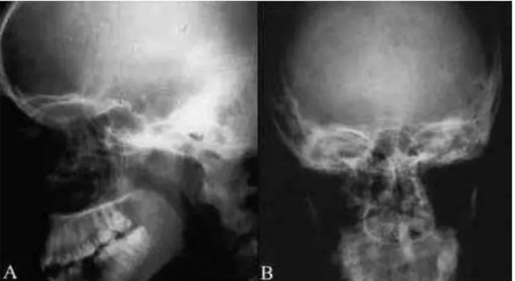
Lateral and forward radiographies showed the projectile located medially to the left mandibular condyle (Figures 2, A and B) within the infratemporal fossa.
Based on signs, symptoms and images, a surgery aimed at re-moving the projectile, followed by physiotherapy was proposed and the patient accepted the treatment by signing the free and informed consent term.
Patient underwent general anesthesia with right-sided
nasotra-cheal intubation. Furthermore, antisepsis with 2% chlorhexidine and apposition of the surgical area were performed, being the left ear and the left lateral corner of the eye visible and acoustic me-atus was tamponed with gauze. he preauricular area was then stained with methylene blue followed by local iniltration with 2% lidocaine chlorhydrate with vasoconstrictor (1:100,000). A 15-minute interval was maintained for anesthetic solution to act and then skin and connective tissue were incised toward the su-pericial layer of the temporal fascia. Susu-pericial temporal vessels and auriculo-temporal nerve were retracted anteriorly with the lap and use of 2 Senn-Miller retractors to avoid damaging them. Temporal fascia was obliquely incised in anteroposterior direc-tion over the zygomatic arch. hen, deep dissecdirec-tion was started to visualize the most supericial surface of the temporomandi-bular and capsular ligaments, respectively, with joint eminence palpation. he next step involved detachment with periosteum elevator downward, reaching superior head of lateral pterygoid muscle .With the same instrument, we proceeded in the ante-rior and infeante-rior direction to locate the infeante-rior head of lateral pterygoid muscle . he region between both heads was carefully explored with Matzenbauer scissors aiming at minimizing the chances of damaging the maxillary artery and also at locating the projectile. Projectile was removed with the help of a Halsted anatomic clamp (Figure 3). Layers were closed from inside to outside with Vycril 4-0. he skin was closed with 6-0 Polypropi-lene (ProPolypropi-lene) and protected with a gauze overlay. he suture was removed at intervals from the 5th to the 7th postoperative day.
Figure 1. Blunt injury (arrows) in left zygomatic area
Figure 2. A-Lateral radiography, presence of a projectile located in the infratemporal fossa; B- Forward radiography, the projectile is located in anatomical space corresponding the infratemporal fossa.
230
Grossmann E and Ferreira LA Rev Dor. São Paulo, 2016 jul-sep;17(3):228-31
Postoperative course went without complications and patient started physiotherapy 7 days after leaving hospital. Physiothera-py sessions were performed twice a week for three months. hese sessions involved 1.5 w/cm2 ultrasound on the left area for 5
minutes, associated to overlapping wooden spatulas, in addition to moist hot towels and passive stretching exercises for mouth opening, closing and lateral jaw movement. New postoperative radiographic exams, similar to previous exams, showed that the projectile was no longer there (Figure 4, A and B). Patient pre-sented no sensitive, autonomic or motor impairment. Follow up revealed that patient had mouth opening of 40.02mm, without pain and lateral jaw limitation.
Figure 4. Lateral (A) and forward (B) radiographs post-surgical
Note the absence of the projectile.
DISCUSSION
he literature reports that head and face are places commonly reached by gunshot injuries5,6. In general, there are major
de-formities and functional incapacity, especially when temporo-mandibular joint, facial nerve and deep skull and face structu-res are involved. A region possibly reached by this injury is the infratemporal fossa, which being close to the temporal fossa is delimited between surfaces of zygomatic, temporal, major wing of the sphenoid bone and ramus of mandible5. In this area, noble
structures of major functional importance are present such as la-teral and medial pterygoid muscles, the pterygoid venous plexus, the optic ganglion, the cord of tympani nerve, the mandibular nerve and one of its branches, and the lingual nerve, in addition to the maxillary artery, the pathway of which is supericial or deep below lateral pterygoid muscle5.
In case of injury of this fossa, there might be several compli-cations, including jaw displacement, limited mouth opening, impairment of excursive jaw movements, development of ante-rior open byte and temporomandibular ankylosis6. Other more
severe complications may also occur if immediate clinical and surgical measures are not taken, such as arterio-venous damage involving ptetygoid venous plexus and/or maxillary artery, invol-ved masticatory and/or facial muscles, joint ibrotic ankylosis, infection, parotid gland changes, decreased or absence of taste sensitivity, lateral and anterior tongue border hypoesthesia, in addition to pain while speaking.
Immediate risks and complications of gunshot wound were not conirmed during evaluation of patient’s clinical presentation,
however, surgery for projectile removal was indicated due to pain and functional impairment of jaw movements. he physical presence of the foreign body associated to local edema involving both left lateral pterygoid muscle heads, was clinically manifes-ted by pain at mouth opening and laterality movements to the right. If the projectile was stable in a soft or hard tissue structure not causing pain or masticatory impairment, a more conservative clinical approach associated to physiotherapy could be preferred7
being also recommended clinical and imaging follow up. he preauricular approach was chosen for allowing adequate lo-cal exposure, with excellent view of the region, for being esthe-tically favorable and acceptable, in addition to minimizing risks of facial nerve injury5. Despite the choice of the most adequate
surgical incision, the deep and unique location of the projectile between both heads made diicult its access. Insertions of this muscle in capsule, joint disk and neck of mandible structures could be impaired, resulting in other functional damages. Dis-section for access could also be inluenced by the anatomic varia-tion of this muscle, expressed by diferent ways of inservaria-tion or its disposition in three heads5.
Regardless of surgical access - endaural, preauricular or post--auricular – the risk of damaging the facial nerve should always be considered, especially its temporal branches or, less frequently the zygomatic branch and the trigeminal auriculotemporal ner-ve5,6. However, in this reported case, there has been no nervous
damage, that is, motor, sensitive or autonomic damage observed during postoperative follow-up.
As exploratory surgical alternative, trans-surgical endoscopy could have been used, which has as advantages the access to di-ferent body areas with less need of dissection, decreasing tissue morbidity as compared to open surgery8. A paper is mentioned
in the literature where authors have successfully removed a bullet from the infratemporal fossa by endoscopy with intraoral access8.
However, such approach requires experience and training of the medical team for its indication, in addition to availability of spe-ciic and expensive equipment, which has made unfeasible its use in this case.
Available imaging exam at the moment was not the most accura-te since the radiograph was unable to show the condition of ad-jacent soft tissues, in addition to ofering bidimensional images. Exams with higher diagnostic precision could evaluate injury--related tissue damages before surgery. Literature mentions the use of nuclear magnetic resonance, ultrasound imaging, compu-terized tomography and compucompu-terized tomography with tapered beam as accurate methods to locate projectiles or foreign bodies as a consequence of face wounds9. However, these devices are
expensive and usually are not available for public health assis-tance. So, as diagnostic alternative, we have used radiographic exposures in diferent planes (forward and lateral) to estimate the tridimensional location of the projectile, which has also provided the evaluation of surrounding bone structures.
231
Tratamento cirúrgico de um projétil na fossa infratemporal. Relato de caso
Rev Dor. São Paulo, 2016 jul-sep;17(3):228-31
herapeutic exercises to improve mouth amplitude with wooden spatulas have also promoted stabilization of results and restora-tion of proprioceprestora-tion10.
CONCLUSION
Projectile removal with preauricular approach associated to com-bined physiotherapeutic treatment has provided joint function reestablishment, pain elimination, an appropriate mouth ope-ning without facial sensitive, autonomic and motor sequelae.
REFERENCES
1. Norris O, Mehra P, Salama A. Maxillofacial Gunshot Injuries at an Urban Level I Trauma Center-10-Year Analysis. J Oral Maxillofac Surg. 2015;73(8):1532-9. 2. Maurin O, de Régloix S, Dubourdieu S, Lefort H, Boizat S, Houze B, et al.
Maxillo-facial gunshot wounds. Prehosp Disaster Med. 2015;30(3):316-9.
3. Farrell SE, Vandevander P, Schofstall JM, Lee DC. Blood lead levels in emergency department patients with retained lead bullets and shrapnel. Acad Emerg Med. 1999;6(3):208-12.
4. Brinson GM, Senior BA, Yarbrough WG. Endoscopic management of retained airgun projectiles in the paranasal sinuses. Otolaryngol Head Neck Surg. 2004;130(1):25-30. 5. Isolan GR, Rowe R, Al-Mefty O. Microanatomy and surgical approaches to the in-fratemporal fossa: an anaglyphic three-dimensional stereoscopic printing study. Skull Base. 2007;17(5):285-302.
6. Pires MS, Giongo CC, Antonello G de M, Couto RT, Filho R de O, Junior OL. An interesting case of gunshot injury to the temporromandibular joint. Craniomaxillofac Trauma Reconstr. 2015;8(1):79-82.
7. Oh DW, Kim KS, Lee GW. he efect of physiotherapy on post-temporomandibular joint surgery patients. J Oral Rehabil. 2002;29(5):441-6.
8. Nef LL, Liess BD, Chang CW. Transoral endoscopic removal of a bullet from the infratemporal fossa. Otolaryngol Head Neck Surg. 2008;138(1):113-4.
9. Daghfous A, Bouzaïdi K, Abdelkei M, Rebai S, Zoghlemi A, Mbarek M, et al. Con-tribution of imaging in the initial management of ballistic trauma. Diagn Interv Ima-ging. 2015;96(1):45-55.