M a i l i n g A d d r e s s : C l a u d i o M a g a l h ã e s R a n g e l • R u a I t a p e v a , 2 4 0 – 0 1 3 3 2 - 0 0 0 – S ã o P a u l o , S P - B r a z i l
E-mail: [email protected] Received on 10/13//04 • Accepted on 03/17/06
Aortic Stenosis and Coronary Disease. Analysis of Risk
Factors
Claudio Magalhães Rangel, Max Grinberg, Raul Cavalcante Maranhão, Laura Inês Ventura Instituto do Coração do Hospital das Clínicas – FMUSP - São Paulo, SP - Brazil
O
BJECTIVETo analyze clinical laboratorial aspects of the presence of coronary disease in patients with aortic stenosis and evaluate the infl uence of risk factors in the development of obstructive coronary disease.
M
ETHODSWe studied 65 patients who had severe aortic stenosis with an indication for surgery, ages 51 to 85 years, 40 of them women. The coronary angiography assessment resulted in two groups: 26 (40%) with obstructive coronary disease and 39 (60%) with no coronary artery lesion. Personal antecedents for coronary disease (smoking, dyslipidemia, diabetes mellitus, arterial hypertension, family antecedents, sedentarism, and alcoholism) were analyzed. Additionally, the following assessments were made: electrocardiogram, echocardiogram with Doppler, and laboratory tests (blood glucose, total cholesterol and fractions, triglycerides, Apo-A1 and B, fi brinogen, lipoprotein (a) and fraction of triglycerides and cholesterol removal in both groups.
R
ESULTSIn the age analysis, the group with obstructive coronary disease belonged to an older age range with statistical signifi cance (p<0.0001). Signs of ischemia of the anterior wall identifi ed on the electrocardiogram showed a signifi cant relationship with the obstruction of an anterior interventricular artery (p<0.002). The univariate analysis showed a significant difference between the groups regarding averages of the aortic (p= 0.041), HDL (p=0.042), and fi brinogen (p=0.047) gradients. The group with coronary disease presented an average gradient and HDL level lower than the group without obstructive coronary disease. For the fi brinogen variable, the average in the group with no coronary disease was lower compared to that of the coronariopathy group. The multivariate logistic regression analysis showed fibrinogen levels as an independent variable for coronary disease (p<0.039).
C
ONCLUSIONFibrinogen was an independent risk factor for the association between obstructive coronary disease and aortic stenosis.
K
EY WORDSAortic stenosis in adults is characterized by degenerative alterations of the valve leafl ets that encumber the proper emptying of the left ventricle, leading to the development of muscular hypertrophy because of chronic and progressive pressure overload of the left ventricle. The main causes of aortic stenosis are congenital, rheumatic, and degenerative or senile.
The expression “risk factor”1 describes characteristics
that may be found in healthy individuals, which are independently associated with the manifestation of a given disease. In this sense, a risk factor can be defi ned as any measurable trait or characteristic in an individual that may lead to a greater probability of his manifesting a certain disease2.
A meta-analysis of 33 studies showed a 37% prevalence of coronar y disease in patients with calcifi ed aortic stenosis3.There are still unanswered
questions concerning risk factors for coronary artery disease in aortic stenosis patients. In the analysis of risk factors for coronary disease, it is not possible to assert what degree of participation these factors have in the development of coronary disease associated with aortic stenosis. In analyzing risk factors in an aortic stenosis patient, it would be extremely important from a clinical point of view to know the probability of this patient presenting an associated coronary disease. The answers to these questions could help in identifying the probability of the aortic stenosis patient also bearing an associated coronariopathy. We could determine the participation of each risk factor, alone or in combination, in the development of coronary disease in aortic stenosis patients and diminish the morbidity/mortality of this association.
M
ETHODSSixty-fi ve patients participated in this research, 40 (61.5%) females, and 25 (38.5%) males. Patients were enrolled in the protocol according to the following inclusion criteria: presence of severe aortic stenosis, absence of any other valve disorder, age greater than or equal to 50 years, no previous cardiac surgery, and absence of clinically signifi cant renal, hepatic, hemic, or neoplastic disease.
For each patient selected for the protocol, a case report form was completed that included age, gender, body mass index, risk factors for coronary disease, anamnesis data, electrocardiogram, and echocardiogram with Doppler. At the end of the interview, dates were scheduled for laboratory tests and cardiac catheterism.
In the body mass index evaluation, anthropometric measurements of weight and height were used for the Body Mass Index (BMI) calculation using the formula BMI = weight (Kg) / height2(m2)4.
Selected risk factors were smoking (habit of smoking
more than fi ve cigarettes a day for at least six months)5,
HDL-cholesterol under 35 mg/dL, LDL-cholesterol over 130 mg/dL6, diabetes mellitus (blood glucose levels
over 140 mg/dL after 12-hour overnight fast)7, systemic
arterial hypertension (patients with systolic arterial pressure over 150 mmHg and diastolic pressure over 90 mmHg)8, family antecedents (parents or siblings
with history of coronary disease, and ages less than 55 years of age for men and less than 65 years of age for women)2, sedentarism (patients who reported activities
such as walking less than 30 minutes a week or sporadic participation in sports)2, and alcoholism (men with intake
of more than 4 doses a day or more than 20 doses a week and women with intake of more than 3 doses a day or more than 12 doses a week)2.
Anamnesis data were defi ned by functional class according to the New York Heart Association classifi cation9,
angina by the Canadian Cardiovascular Society10,syncope
and symptoms and signs compatible with congestive heart failure.
A 12-lead electrocardiogram was performed on all patients using Hewlett Packard equipment, model 1700, according to conventional criteria. Since there are classic electrocardiographic changes in aortic stenosis, we will analyze the association of anterior wall ischemia with signifi cant damage of the anterior descending artery. We will not examine causal relationship with other affected coronary arteries, since they may be confused with the classic eletrocardiographic alterations that are present in severe aortic stenosis.
The echocardiogram with Doppler was performed on all patients with ATL equipment, HDI 3000 model; the maximal aortic valvar gradient was analyzed by applying BERNOULLI’s modifi ed equation11, and the
ejection fraction (EF) of the left ventricle by means of the Cube Method12,13.
Blood test samples were drawn in the morning after a 12-hour fast. Measurements of blood glucose, triglycerides, total cholesterol and fractions, fi brinogen, apoA1, apoB and Lp (a) were carried out by the Clinical Laboratory of the Heart Institute. Also included in this study were the plasma removal kinetics of artifi cial chylomicrons, and this exam was performed by the Lipids Laboratory of the Heart Institute.
Cardiac catheterism was performed on all patients enrolled in the study as a pre-operative test in order to analyze coronary anatomy and evaluate the need for myocardial revascularization associated to aortic valve replacement. The test was carried out using the SONES and SHIREY technique14.Subjects were considered
coronary patients when they had at least one subepicardial artery with an atherosclerotic process causing more than 50% reduction of the vessel lumen in comparison to the closest normal segment.
on tables containing absolute (n) and relative (%) frequencies. The associations between these variables and the presence of coronary disease will be compared using the chi-square test, verisimilitude ratio test, or Fisher’s exact test15. For the analysis of the incidence of
coronary disease by age bracket, the ratio of verisimilitude test was used15.
Continuous variables are presented descriptively in tables containing means and standard deviation. The means of these variables as to the presence of coronary disease were compared with Student’s t-test16.Gradient,
blood glucose, triglycerides, Lp (a), and FTR (Ch) variables were submitted to logarithmic transformation for parameter analysis.
Variables that showed statistical signifi cance in the univariate analysis were used for the adjustment of a multiple logistic regression model with a stepwise variable selection procedure16.
P values of p < 0.05 were considered signifi cant.
R
ESULTSAges of the 65 patients who participated in the study varied between 51 and 85 years (mean 68), and 40 (61.5%) of the subjects were women. Coronary angiography resulted in 26 (40%) patients with obstructive coronary disease and 39 (60%) without obstructive coronary disease.
We observed a greater proportion of coronary disease incidences in the older age ranges. There was a difference (p<0.0001) between the age brackets, i.e., the 71-80 year range had a larger proportion of patients with disease than the other age ranges.
A correlation between anterior ischemia seen on the electrocardiogram and damage of the anterior interventricular artery was observed. Of the 17 patients with lesions in this artery, 11 showed signs of anterior ischemia on the electrocardiogram (64.71%) with p<0.002 (Fisher ’s test). There was no significant association between the groups with and without coronary disease as to gender, number of risk factors, family antecedents, systemic arterial hypertension, diabetes mellitus, dyslipidemia, smoking, sedentarism, and alcoholism. No signifi cant association was noted between the groups with and without coronary disease as to functional class, angina, syncope, and heart failure.
We did observe a signifi cant difference between the groups in term of means of the maximal gradient (p = 0.041), HDL cholesterol (p = 0.042), and fi brinogen (p = 0.047) variables. Patients with coronary disease had lower values for the mean gradient and HDL than those without coronary disease. With the fi brinogen variable, patients without coronary disease had lower mean levels when compared to those with coronary disease as is shown on Table 1.
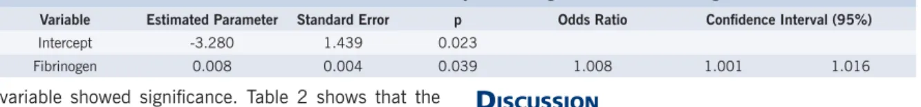
The influence of the parameters analyzed in the presence of coronary disease is described on Table 2. Only fi brinogen exerted a signifi cant infl uence (p = 0.039) on the presence of coronary disease.
For the presence of coronary disease in the logistic regression model, the explanatory variables gradient, HDL, and fi brinogen were considered. These variables showed statistical signifi cance in the univariate analysis. After the stepwise selection of variables, the fi brinogen
Table 1 - Mean and standard deviation of continuous variables per group (with and without coronary disease) and probability of signifi cance (p) of student’s t-test
Variable
Coronary disease p
+
-n Mean DP n Mean SD
Age in years 26 70.46 9.02 39 65.9 8.38 0.250
BMI 26 21.54 4.39 39 19.62 3.070 0.078
GRAD* mmHg 26 77.35 26.77 39 87.54 24.40 0.041
EF (%) 26 0.665 0.099 39 0.696 0.085 0.174
Glucose* mg/dL 26 125.65 64.68 39 104.67 21.27 0.084
T. C. mg/dL 26 213.27 39.48 39 206.23 33.87 0.445
H.D.L. mg/dL 26 41.85 13.37 38 47.79 13.23 0.042
L.D.L. mg/dL 25 143.8 37.07 38 134.13 28.84 0.064
V.L.D.L. mg/dL 25 26.28 10.08 38 24.21 10.45 0.439
TG* mg/dL 26 146.15 90.67 39 128.38 69.27 0.371
APO-A1 g/l 26 1.45 0.25 39 1.55 0.270 0.134
APO-B g/l 26 1.38 0.32 39 1.27 0.270 0.141
FIBRIN. mg/dL 26 377.96 72.16 39 341.82 69.45 0.047
Lp(a)* mg/dL 26 30.95 21.74 39 38.65 25.83 0.244
FTR (TG) min 26 0.036 0.021 39 0.040 0.023 0.553
FTR (Ch)* min 26 0.012 0.014 39 0.015 0.014 0.492
0 0.1 0.2 0.3 0.4 0.5 0.6 0.7 0.8 0.9
100 200 300 400 500 600
Fibrinogen (mg/dl)
Probability (coronary disease)
0 10 20 30 40 50 60 70 80 90 100
0 0.1 0.2 0.3 0.4 0.5 0.6 0.7 0.8 0.9
Percentage
Sensitivity Specificity
Table 2 - Variables used in model adjustment: gradient, hdl, fi brinogen
Variable Estimated Parameter Standard Error p Odds Ratio Confi dence Interval (95%)
Intercept -3.280 1.439 0.023
Fibrinogen 0.008 0.004 0.039 1.008 1.001 1.016
Chart 1 - Dispersion of sensitivity and specifi city of the logistic regression model
Chart 2 - Dispersion of probability of coronary disease by fi brinogen level. The pink line represents the 0.42 probability that corresponds to 57.7% sensitivity and 63.2% specifi city.
variable showed signifi cance. Table 2 shows that the estimated parameter for fi brinogen is positive, indicating a positive correlation as to the probability of coronary disease (the greater the value of fi brinogen, the greater the probability of coronary disease).
Chart 1 shows the distribution of sensitivity and specificity values for different coronar y disease probabilities. The maximum value point for both indices is the probability value of 0.42.
A 0.42 probability corresponds to a fi brinogen value of 370, that is, if the patients with values over 370 are classifi ed as coronary patients and those with values under 370 are considered not ill, we have a 57.7% sensitivity and a 63.2% specifi city (Charts 1 and 2)
D
ISCUSSIONThe incidence of coronary disease in patients submitted to aortic valve replacement is estimated between 7% and
66%17,18. A meta-analysis with 33 studies showed a 37%
prevalence of coronary disease in patients with calcifi ed aortic stenosis3. In our series, the 40% prevalence of
coronary disease proved to be within the average of the above mentioned range, as had also been verifi ed in well-conducted studies performed by LUND et al19 Mautner et
al20 and Paquay et al21.
The average age of 70 years was similar to those noted by other authors such as VEKSHTEIN22 and BESSONE23.
range in the coronariopathy patients. A certain correlation was observed between age and coronariopathy rate, where the older age bracket has a greater probability of coronary disease associated with aortic stenosis22,23. Our
highest age mean is in agreement with that found in most medical literature.
The presence of coronary disease in patients with aortic stenosis has been the focus of many studies, and some have suggested a correlation between risk factors and the presence of coronary disease in aortic valve disease patients18,24-26. The participation of risk
factors for coronary disease has not been adequately appreciated as a predictive factor of coronary disease in patients with aortic stenosis18,24,26,27. In some studies,
the absence of risk factors and angina were suffi cient to exclude coronary disease associated with valve disorders26. On the other hand, Acar et al28 and Pluta et
al18 found a high risk factor incidence in patients with
coronary disease associated with aortic stenosis, while Carstens et al29 and Exadactylos et al30 did not observe
a correlation between the presence of risk factors and the incidence of coronary disease associated with aortic stenosis. In our group of patients, the greater number of risk factors did not increase the incidence of coronary disease, a fact that concurs with the studies of Carstens29
and Exadactylos30.
Some authors24,31,32 have observed that the aortic
gradient tends to be smaller in patients with than in those without angina, especially when the angina is severe. This observation may be caused by the great prevalence of coronary disease associated with moderate aortic stenosis. Berndt et al33 demonstrated that the aortic gradient was
smaller in patients that had coronary disease, which was also a result noted in our study. Several authors18,24,28,31,32
had this same result, which raises the hypothesis that myocardial ischemia or infarct could potentially diminish the gradient through the aortic valve.
Several studies in literature34-38 observed that reduced
levels of HDL-c increase the risk of coronary disease, especially if triglyceride levels are also elevated. Our study showed higher HDL-c levels in patients with aortic stenosis without coronary disease, reinforcing the concept of a protection factor for coronary disease. This aspect identifi ed in our study is an important point because, when faced by a given patient’s situation, it allows us, along with other factors, to determine the probability of his presenting coronary disease associated with aortic stenosis.
The level of plasmatic fi brinogen has been shown to be a predictor of coronary disease in several prospective studies39,40. Seven prospective studies41,42 have noted
an increase in the incidence of coronary disease when fi brinogen levels are high. The participation of fi brinogen as a risk factor in the coronary disease of a patient with a valve disorder has not yet been reported in literature.
In our research, this correlation was statistically signifi cant on the univariate analysis (Table 1) and on the logistic regression model (Table 2). We studied it as an important factor in analyzing the probability of a patient with aortic stenosis having an associated coronary disease, since with the progressive increase of fi brinogen levels, the probability of this association also rises (Chart 2).
In conclusion, the observation of fi brinogen levels in clinical practice may be indicative of the increased prevalence of obstructive coronary disease in patients with aortic stenosis. This result was seen in our research, where the level of fi brinogen was an independent risk factor for the association of obstructive coronary disease and aortic stenosis, as the highest levels increased the probability of this association. This aspect, along with an analysis of the aortic gradient, age range, electrocardiogram, and HDL-c level, may be extremely important in the diagnosis and treatment of this group of patients, improving the clinical follow-up of this population.
R
EFERENCES1. Doyle J. Risk factors in coronary heart disease. N Y State J Med 1963;63:1317-20.
2. Wood D, De Backer G, Faergeman O, Grahan I, Mancia G, Pyörälä K. Prevention of coronary heart disease in clinical practice. Recomendation of the Second Joint Task Force of European and others Societies on Coronary Prevention. Eur Heart J 1998;19:1434-503.
3. Mauter GC, Roberts WC. Reported frequency of coronary arterial narrowing by angiogram in patient with valvular aortic stenosis. Am J Cardiol 1992;69:539-40.
4. Larsson B. Obesity and body fat distribution as predictors of coronary heart disease. In: Marmot M, Elliott P. Ed. Coronary Heart Disease Epidemiology. From Aetiology to Public Health. Oxford: Oxford University Press, 1992:233-41.
5. Auerbach O, Hammond EC, Garfinkel L. Smoking in relation to atherosclerosis of the coronar y ar teries. N Engl J Med 1965;273:775-9.
6. Assman G, Schulte H. Relation of high- density lipoprotein cholesterol and triglycerides to incidence of atherosclerosis coronary artery disease
(the PROCAM experience). Am J Cardiol 1992;70:733-7.
7. Garcia MJ, McNamara PM, Gordon T, Kannel WB. Sixteen year follow-up study. Morbidity and mortality in diabetics in Framinghan population. Diabetes 1974;23:105-11.
8. III Consenso Brasileiro de Hipertensão Arterial. Campos do Jordão, SP, 12-15 fev. 1998.
9. The Criteria Committee of the New York Heart Association: Diseases of the Heart and Blood Vessels; Nomenclature and Criteria for Diagnosis. 6th ed. Boston: Little Brown, 1964.
10. Campeau L. Grading of angina pectoris. Circulation 1976;54:522-3.
11. Hatle L, Angelsen B, Thomsdal A. Noninvasive assesment of aortic stenosis by Doppler ultrasound. Br Heart J 1980;43:284-92.
12. Triulzi MO, Wilkins GT, Gillan LD. Normal adult cross sectional echocardiographic valves: LV volumes. Echocardiography 1985;2:153-70.
Recommendations regarding quatitation in M-mode echocardiographic mensurements. Circulation 1978;58:1072-83.
14. Sones FM, Shirley EK. Cinecoronary artheriography. Mod Concepts Cardiovasc Dis 1972;31:735-8.
15. Rosner B. Fundamentals of Biostatistics. 2nd Ed. Boston: PWS Publishers, 1986.
16. Hosner DW, Lemeshow S. Applied Logistic Regression. New York: John Wiley & Sons, 1989.
17. Nunley DL, Grunkemerer GL, Starr A. Aortic valve replacement with coronary bypass grafting. J Thorac Cardiovasc Surg 1983;85:705-11.
18. Pluta W, Buszman P, Lekston A, Pasyk S. Coronary artery stenosis in pacients with vascular heart disease. Cor Vasa 1989;31:451-7.
19. Lund O, Nielsen TT, Pilegaard HK, Manussen K, Knudsen MA. The influence of coronary artery disease and bypass grafting on early and late survival after valve replacement for aortic stenosis. J Thorac Cardiovasc Surg 1990;3:327-37.
20. Mautner GC, Cannon RO 3rd, Mautner SL, Hunsberger SA, Roberts WC. Clinical factors useful in predicting aortic valve structure in patients > 40years of age with isolated valvular aortic stenosis. Am J Cardiol 1993;72:194-8.
21. Paquay PA, Anderson G, Diefenthal H, Nordstrom L, Richman HG, Gobel FL. Chest pain as a predictor of coronary artery disease in patients with obstrutive aortic valve disease. Am J Cardiol 1976;38:863-9.
22. Vekshtein VI, Alexander RW, Yeung AC, Plappert T, St John Sutton MG, Ganz P et al. Coronary atherosclerosis is associated with left ventricular dysfunction and dilatation in aortic stenosis. Circulation 1990;82:2068-74.
23. Bessone LN, Pupello DF, Hiro SP, Lopez-Cuenca E, Glatterer MS, Ebra G. Surgical management of aortic valve disease in the elderly : a logitudinal analysis. Ann Thorac Surg 1988;.46:264-9.
24. Vandeplas A, Willems JL, Piessens J, de Geest H. Frequency of angina pectoris and coronary artery disease in severe isolated valvular aortic stenosis. Am J Cardiol 1988;62: 117-20.
25. Sheiban J, Trevi GP, Benussi P, Marini A, Accardi R.; Di Bona E et al. Incidence of coronary artery disease in patients with valvular heart disease. Z Kardiol 1986;75(Suppl 2):76-9.
26. Ramsdale DR, Bennett DH, Bray CL, Ward C, Beton DC, Faragher EB. Angina, coronary risk factors and coronary artery disease in patients with valvular disease. A prospective study. Eur Heart J 1984;5:716-26.
27. Wilson RF. Catheterization of patients with aortic valve disease. In: The Aortic Valve Disease. Philadelphia: Hanley and Belfus, 1991; 7:57-70.
28. Acar J, Vahanian A, Dicimetiere PH, Berdah J, Aouate PH, Sienczewski JA et al. Should coronary arteriography be performed in all patients
who undergo catheterization for valvular heart disease? Z Kardiol 1986;75(Suppl 2):53-60.
29. Carstens V, Haum A, Grond M, Behrenbeck DW. Incidence of coronary artery disease and necessity for coronary angiography in patients with valvular heart disease. Z Kardiol 1986; 75(Suppl 2):83-5.
30. Exadactylos N, Sugrue DD, Oakley CM. Prevalence of coronary artery disease in patients with isolated aortic valve stenosis. Br Heart J 1984;20:121-4.
31. Mandal AB, Gray IR. Signifi cance of angina pectoris in aortic valve stenosis. Br Heart J 1976;38:811-5.
32. Morrison GW. Incidence of coronary artery disease in patients with valvular heart disease. Br Heart J 1980;40:630-7.
33. Berndt TB, Hancock EW, Shumway NE, Harrison DC. Aortic valve replacement with and without coronary artery bypass surgery. Circulation 1974;50:967-71.
34. Assmann G, Cullen P, Schulte H. The Münster Heart Study (PROCAM). Results of follow-up at 8 years. Eur Heart J 1998;19:A2-A11.
35. Manninen V, Huttunen J, Heinimem O, Tenkanem L, Frick M. Relationships between baseline lipid and lipoprotein values and the incidence of coronary heart disease in the Hensink Heart Study. Am J Cardiol 1989;63:42H-7H.
36. Gordon T, Kannel WB, Castelli WP. Lipoproteins, cardiovascular and death: The Framingham Study. Arch Intern Med 1981;141:1128-31.
37. Lipid Research Clinic Program. The Lipid Research Clinic Coronary Primary Prevention Trial Results I. Reduction in the incidence of coronary heart disease. JAMA 1984;251:351-64.
38. Davies C, Rifikind B, Brenner H. A single cholesterol measurement undertimate the risk of CHD. An empirical example from the Lipid Research Clinics mortality follow-up study. JAMA 1990;264:3044-6.
39. Wilhelmsen L, Svärdsudd K, Korsan-Bengtsen K, Larsson B, Welin L, Tibblin J. Fibrinogen as a risk factors for and myocardial infarction. N Engl J Med 1984;311:501-5.
40. Meade T. Fibrinogen and other clotting factors in cardiovascular disease. In: Francis Jr R., ed. Atherosclerosis Vascular Disease, Hemosthasis, and Endothelial Function. New York: Marcel Dekker, 1992:1-34.
41. Meade TW, North WRS, Chakrabarti R. Haemostatic function and cardiovascular death: early results of a prospective study. Lancet 1980;v.i:1050-4.