Image
Total Anomalous Pulmonary Venous Connection and a Restrictive
Foramen Ovale, with Left-Sided Deviation of the Atrial Septum
Mimicking Left-Sided
Cor Triatriatum
: Echocardiographic Misdiagnosis
Edmar Atik, Renata Sá Cassar, Samira Saady Morhy, José L. Andrade
Instituto do Coração do Hospital das Clínicas – FMUSP - São Paulo, SP, Brazil
Key words
Pu l m o n a r y v e i n s / a b n o r m a l i t i e s , c o r t r i a t r i a t u m, echocardiography.
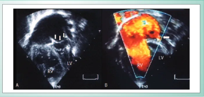
An 11-month-old male infant presented with intense dyspnea that required immediate mechanical respiratory assistance. The clinical examination was compatible with the diagnosis of a large atrial septal defect with a large left to right shunt. There was an intense systolic ejection murmur at the upper left sternal border and a splitted second heart sound, right ventricular overload on electrocardiogram and significant cardiomegaly due to dilation of right heart cavities. Echocardiographic images strongly suggested a diagnosis of cor triatriatum with a restrictive orifice communicating the two left atrial cavities and a large flow through the atrial septal defect, above the obstructive membrane (Fig. 1).
The correct diagnosis was established during surgery when we observed a total anomalous pulmonary venous drainage directly into the right atrium with a restrictive foramen ovale
and accentuated left-sided deviation of the atrial septum.
Fig. 1 - A) Image of the atrial septum deviated to the left, mimicking cor triatriatum (arrows). B) Color flow mapping of the right atrium and the restrictive foramen ovale mimicking respectively a large atrial septal defect and a small membrane orifice of the false cor triatriatum (arrow). LV: left ventricle; RV: right ventricle.
The atrial septum was enlarged and a patch directed all pulmonary veins to the left atrium. The postoperative period was uneventful, with normalization of the heart function.
The peculiarities of the echocardiographic images were better interpreted retrospectively. The atrial septum deviated to the left was parallel to the mitral valve, and mimicked an obstructive left atrial membrane and the foramen ovale
had also been wrongly construed as the orifice of this false membrane.
We think that this very unusual image is liable to misdiagnosis on echocardiogram, this being the reason why we decided to publish this report.
Potential Conflict of interest
No potential conflict of interest relevant to this article was reported.
Mailing Address: Edmar Atik •
Av. Enéas de Carvalho Aguiar, 44 – 05433-000 – São Paulo, SP, Brazil E-mail: [email protected]
Manuscript received May 3, 2006; revised manuscript received May 9, 2006; accepted May 9, 2006.