Frequency of Hypertension in Patients with Chronic Chagas’ Disease
and its Consequences on the Heart: A Clinical and Pathological Study
Cristina Brandt Friedrich Martin Gurgel and Eros Antonio de Almeida
Hospital e Maternidade Celso Pierrô da Pontifícia Universidade Católica de Campinas - Campinas, SP - Brazil
Summary
Background: Data from the literature on the frequency of concomitant Chagas’ disease and hypertension are controversial and, when available, do not mention the consequences of this concomitance in the natural history of either Chagas’ disease or of hypertension.
Objective: To assess the frequency of concomitant Chagas’ disease and hypertension and the clinical and anatomopathological consequences of this association.
Methods: The cases were selected from necropsies performed in the Department of Pathological Anatomy of Hospital e Maternidade Celso Pierrô of Pontifícia Universidade Católica de Campinas and divided into three groups: CH + SH group, of patients with Chagas’ disease plus hypertension; CH group, of patients with Chagas’ disease without hypertension; and SH group, of patients with hypertension without Chagas’ disease. The variables of gender, age, race, clinical forms of Chagas’ disease, and electrocardiographic and anatomopathological findings were statistically analyzed.
Results: In this assessment, a total of 101 (2.9%) cases of patients with Chagas’ disease was found, and 33 (32.7%) of them also had hypertension. A slight predominance of the male gender was observed; racial distribution and mean age were similar in the three groups. Severe hypertension was not frequently found among chagasic patients. When present, hypertension did not change the clinical and anatomopathological findings compatible with Chagas’ disease
Conclusion: The frequency of hypertension in chagasic patients was similar to that observed in the general population. Hypertension, when present in chagasic patients, occurred in those with a higher mean age. The concomitance of hypertension and Chagas’ disease did not change the natural history of either one of the two diseases. (Arq Bras Cardiol 2007;89(3):174-182)
Key words: Hypertension; Chagas cardiomyopathy; heart/physiopatology.
Mailing address: Cristina Brandt Friedrich Martin Gurgel •
Rua MMDC, 47/101 - 13025-130 - Campinas, SP - Brazil E-mail: [email protected]
Manuscript received July 07, 2006; revised manuscript received February 20, 2007; accepted March 13, 2007.
Introduction
Chagas’ disease is a chronic endemic zoonosis, usually transmitted to humans through the bite of a triatomine bug. It has a wide geographic distribution in the Americas, extending from central-western Mexico to far-southern Chile and Argentina1. The diagnosis in the chronic phase is based on the
presence of anti-Trypanosoma cruzi antibodies in the serum of infected individuals, and its clinical polymorphism is patent.
The disease may present itself in the following forms: indeterminate form, which is characterized by the absence of significant signs and symptoms of organic involvement; digestive form, which is secondary to the lesion of the myenteric plexus that results in motor incoordination and, in advanced cases, dilation of the affected organ (megacolon
and megaesophagus); and cardiac form, which is considered the most frequent in our setting.
Because of the different cardiac manifestations that result from varying extensions and degrees of inflammation, it is difficult to make a single clinical classification. Patients may have the myopathic form when signs and symptoms compatible with heart failure are present; the dromotropic form when cardiac conduction abnormalities are present; the bathmotropic form when automatism disturbances are present; or the combined form, when two or more clinical presentations are concomitant2. In the current literature,
these manifestations are presented in a simplified manner as congestive, arrhythmic and/or embolic manifestations.
Essential hypertension is asymptomatic in most of the cases, and was basically defined by the III Brazilian Consensus on Hypertension as the presence of systolic pressure levels equal to or higher than 140 mmHg and diastolic levels equal to or higher than 90 mmHg3. Its prevalence depends on the
specifically analyzed: 1) heart weight, to assess the presence of hypertrophy according to Fulton’s criterion9 which considers
increased weights those equal to or higher than 250 g; 2) analysis of the ventricular apex to verify the presence of apical lesion (ventricular apical recess); 3) analysis of the coronary arteries to verify atherosclerosis, which was quantified as mild – luminal stenosis equal to or lower than 50%; moderate – lesion ranging between 50% and 75%; and severe – stenosis higher than 75% (intimal alterations by the presence of fatty material without stenosis was called lipoidosis); 4) analysis of the presence of gross myocardial infarction, characterized by dense fibrosis, when previous, and by myomalacia, when recent; 5) analysis of gross intracardiac thrombosis; 6) presence of cerebral infarctions (ischemic or hemorrhagic); and 7) evaluation of the cause of death after analysis of the final clinical presentation and at necropsy, by the presence of bulbar compression due to cerebellar tonsil herniation, cardiac arrhythmia, shock of varying etiologies, respiratory failure, hypoglycemia, cachexia, and indeterminate, when the cause of death could not be established.
For the statistical analysis, the arithmetic mean and standard deviation were used for each of the quantitative variables that required this calculation10. For the inferential analysis,
the analysis of variance, Student’s t test, chi-square test, and Fischer’s exact test were used, considering p < 0.05 as the significance level11. Student’s t test was used for the comparison
between two means, and the ANOVA analysis of variance was used for the comparisons between three or more sample means. When a significant difference between the means was found, the Tukey-HSD test was used for multiple comparison of means, in order to locate the significant differences11,12.
The non-parametric chi-square test and Fischer’s exact test, which do not require the supposition of normality between the variables compared, were used for the analysis of the relation between quantitative variables13.
Results
From a total of 3,464 necropsies, 101 cases corresponded to patients with Chagas’ disease (2.9%). Nine cases were excluded for not meeting some of the inclusion criteria or for meeting the exclusion criteria. In 33 (32.7%) cases concomitant hypertension was found. In the CH and SH groups, 59 and 46 were included, respectively.
Of the total 138 cases studied, 58% were males and 42% were females, with no significant statistical difference between the groups. Also, no difference was observed in relation to race frequency. Statistical significance was found in relation to age in the three groups studied, with a significant difference between the group of patients with Chagas’ disease plus hypertension and the group of patients with Chagas’ disease without hypertension (Table 1).
No statistically significant difference was found in relation to the frequency of the different clinical forms between the two groups of patients with Chagas’ disease (Table 2).
Statistical difference was found in relation to the degree of hypertension among patients with Chagas’ disease, unlike among non-chagasic patients. In these two groups, predominance of mild hypertension among patients with made, and may be influenced by several variables such as
gender, age, obesity, race, alcohol use, environmental factors (diet, social conditions, psychological conflicts), and others. Cardiac hypertrophy secondary to high pressure levels is responsible for a higher frequency of severe arrhythmias, thus predisposing to myocardial infarction and the onset of heart failure4.
Few data on the association of Chagas’ disease with other chronic or acute diseases are available in the literature. Among them, there is controversy as to the frequency of concomitant trypanosomiasis and hypertension, as well as to the consequences of this association to the patient5-8.
The objective of the present study was to evaluate the clinical and pathological consequences of the coexistence of Chagas’ disease and hypertension, with special emphasis on heart diseases.
Methods
Medical records of patients undergoing necropsy, as well as their pathological data were studied. For the diagnosis of Chagas’ disease, in addition to a strongly positive epidemiology, at least two different positive serologic reactions performed in the living patient (complement fixation titer higher than 1/4, and/or indirect immunofluorescence titer equal to or higher than 1/40, and/or passive hemagglutination titer equal to or higher than 1/32, and/or a positive ELISA) were considered.
Hypertension, defined according to the criteria of the III Brazilian Consensus on Hypertension3, was classified
as: mild, when diastolic levels were between 90 mmHg and 99 mmHg; moderate, between 100 mmHg and 109 mmHg; and severe, when the levels were equal to or higher than 110 mmHg. Pressure levels were measured with a mercury sphygmomanometer available in wards, outpatient units, and urgency and emergency hospital units. Patients who had at least three measurements equal to or higher than those established as criteria for hypertension were included.
All cases with findings such as the presence of heart valve diseases, hypertrophic cardiomyopathy, amyloidosis and other deposition diseases that could influence the results, as well as the cases diagnosed as secondary hypertension, were excluded.
General data such as skin color, gender, age, clinical form of Chagas disease, final clinical presentation, and result of electrocardiography were collected from the medical records. For the skin color parameter, two variables were defined: white and non-white races.
To facilitate the analysis of the different variables, the cases were divided into three groups: CH + SH group, of patients with Chagas’ disease plus hypertension; CH group, of patients with Chagas’ disease without hypertension; and SH group, of patients with hypertension without Chagas’ disease. The CH and SH groups were considered controls for Chagas’ disease and hypertension. Cases from the SH group were selected by matching gender and age with the other groups, with the purpose of homogenizing the sample.
Chagas’ disease and of severe hypertension among the non-chagasic patients was also observed (Table 3).
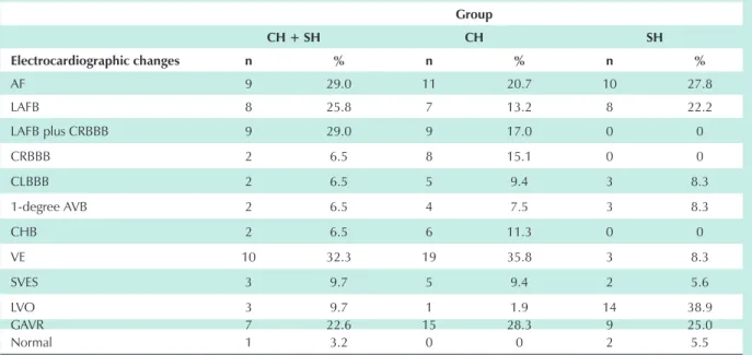
The main electrocardiographic changes are shown in Table 4.
No statistically significant difference was found regarding electrocardiographic changes between the groups studied, except for ventricular extrasystoles and for the presence of left ventricular overload. When one of the parameters studied was not present in a determined group, specifically in left anterior fascicular block with or without right bundle branch block, and in normal electrocardiograms, the other two groups were studied comparatively. However, no statistically significant difference was found in any of the cases.
The comparative analysis did not demonstrate a statistically significant difference in relation to heart weight between the CH + SH group (minimum weight of 250 g, maximum weight of 590 g, mean weight of 415.45 g, with standard deviation of 94.15) and the CH group (minimum weight of 155 g,
maximum weight of 850 g, mean weight of 443.81 g, with standard deviation of 155.53). Likewise, when the SH group (minimum weight of 150 g, maximum weight of 760 g, mean weight of 437.17 g, with standard deviation of 133.03) was added to the analysis, no significant difference was observed either.
The results of the study of coronary artery changes are shown in Table 5. Coronary study could not be performed in four cases of the CH + SH group (12.1%), in five cases of the CH group (8.5%), and in two cases of the SH group (4.3%). No statistically significant difference was found in relation to the degree of atherosclerosis for patients of the CH + SH and SH groups, but in the CH group a predominance of mild atherosclerosis was verified (Table 5).
Of all myocardial and cerebral infarctions observed, none was recent. Study of the brain tissue was not performed in eight cases of the CH group (13.6%), and in two cases of the SH group (4.3%). No statistically significant difference was found
Table 1 – Distribution of subjects according to gender, age and race
Group Gender Age (years) Race
Male Fem. Minimum Maximum Mean Stand. deviat. White Non-whitewhite
CH+ HAS 18 (54.5%) 15 (45.5%) 33 85 60.94 14.36 19 (57.6%) 14 (42.4%) CH 35 (59.3%) 24 (40.7%) 16 82 52.00 16.15 33 (55.9%) 26 (44.1%) SH 27 (58.7%) 19 (41.3%) 27 83 56.57 14.90 27 (58.7%) 19 (41.3)
Total 80 (58%) 58 (42%) 79 59
Statistical significance was found in relation to age in the three groups studied. Fem - female; CH + SH - patients with Chagas’ disease plus hypertension; CH - Chagas’ disease without hypertension, SH - hypertension without Chagas’ disease.
Table 2 – Clinical forms of hypertensive and non-hypertensive patients with Chagas’ disease
CH + SH Group CH Group
Clinical forms n % n %
Dromotropic + bathmotropic + myopathic 11 33.3 22 37.3
Dromotropic + myopathic 7 21.2 13 22.0
Dromotropic 4 12.1 5 8.5
Bathmotropic 0 4 6.8
Bahtmotropic + myopathic 3 9.1 5 8.5
Myopathic 2 6.1 2 3.4
Bathmotropic + dromotropic 2 6.1 2 3.4
Indeterminate 1 3.0 1 1.7
Neurovegetative without heart disease 1 3.0 0
Myopathic without electrocardiography 1 3.0 4 6.8 No myopathy, without electrocardiography 1 3.0 1 1.7
Total 33 100.0 59 100.0
Table 3 – Distribution of subjects in relation to the degree of hypertension
Degree of hypertension
Mild Moderate Severe Total
Group n % n % n % n %
CH + SH group 16 48.5 13 39.4 4 12.1 33 100.0
SH group 21 45.7 8 17.4 17 36.9 46 100.0
Total 37 46.8 21 26.6 21 26.6 79 100.0
Statistical difference was found between the degrees of hypertension only among patients with Chagas’ disease. CH + SH - patients with Chagas’ disease plus hypertension; SH - patients with hypertension without Chagas’ disease; n - number of patients.
Table 4 – Distribution of the electrocardiographic changes according to the group
Group
CH + SH CH SH
Electrocardiographic changes n % n % n %
AF 9 29.0 11 20.7 10 27.8
LAFB 8 25.8 7 13.2 8 22.2
LAFB plus CRBBB 9 29.0 9 17.0 0 0
CRBBB 2 6.5 8 15.1 0 0
CLBBB 2 6.5 5 9.4 3 8.3
1-degree AVB 2 6.5 4 7.5 3 8.3
CHB 2 6.5 6 11.3 0 0
VE 10 32.3 19 35.8 3 8.3
SVES 3 9.7 5 9.4 2 5.6
LVO 3 9.7 1 1.9 14 38.9
GAVR 7 22.6 15 28.3 9 25.0
Normal 1 3.2 0 0 2 5.5
Statistically significant difference was found only for the presence of ventricular extrasystoles and left ventricular overload. CH + SH - patients with Chagas’ disease plus hypertension; CH - patients with Chagas’ disease without hypertension; SH - patients with hypertension without Chagas’ disease; n - number of patients; AF - atrial fibrillation; LAFB - left anterior fascicular block; CRBBB - complete right bundle branch block; CLBBB - complete left bundle branch block; 1-degree AVB - first-degree atrioventricular block; CHB - complete heart block; VE - ventricular extrasystoles; SVES - supraventricular extrasystoles; LVO - left ventricular overload; GAVR - global alterations of ventricular repolarization.
Table 5 – Distribution of coronary artery changes according to the groups
Group
CH + SH CH SH
Coronary study n % n % n %
No changes 9 31.0 31 57.4 13 29.5
Lipoidosis 7 24.2 9 16.7 12 27.3
Mild atherosclerosis 5 17.2 10 18.5 8 18.2
Moderate atherosclerosis 4 13.8 3 5.5 3 6.8
Severe atherosclerosis 4 13.8 1 1.9 8 18.2
Total 29 54 54 44
Table 6 – Distribution of cardiovascular complications according to the groups
Group
CH + SH CH SH Total
Complications n % n % n % n %
Myocardial infarction 4 25.0 3 18.8 9 56.2 16 100.0
Stroke 10 31.3 7 21.9 15 46.8 32 100.0
No cerebral study 0 0 8 80.0 2 20.0 10 100.0
No complication 19 24.19 41 51.8 19 24.1 79 100.0 Myocardial infarction + stroke 0 0 0 0 1 100.0 1 100.0
Total 33 59 46 138
No statistically significant difference in relation to the number of infarctions alone. CH + SH - patients with Chagas’ disease plus hypertension; CH - patients with Chagas’ disease without hypertension; SH - patients with hypertension without Chagas’ disease; n - number of patients.
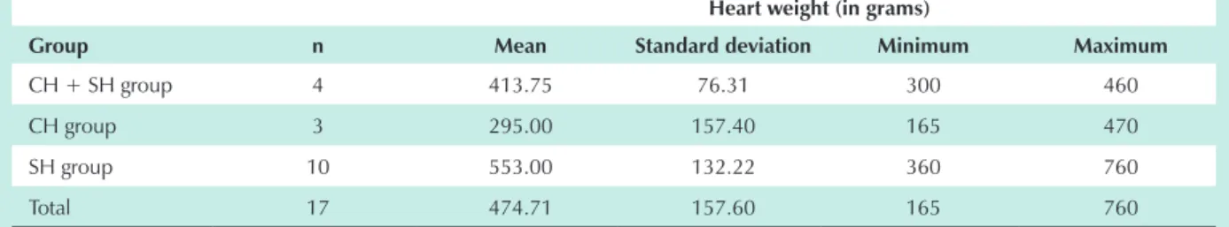
Table 7 - Distribution of subjects with myocardial infarction in the different groups according to heart weight
Heart weight (in grams)
Group n Mean Standard deviation Minimum Maximum
CH + SH group 4 413.75 76.31 300 460
CH group 3 295.00 157.40 165 470
SH group 10 553.00 132.22 360 760
Total 17 474.71 157.60 165 760
Statistically significant difference was found for the presence of myocardial infarction in relation to heart weight. n - number of patients; CH + SH - patients with Chagas’ disease plus hypertension; CH - patients with Chagas’ disease without hypertension; SH - patients with hypertension without Chagas’ disease.
in relation to the number of infarctions alone (Table 6). No statistically significant difference was found regarding age and gender in relation to cardiovascular complications in the CH + SH group, and neither in the CH and SH groups. A significant difference was observed when the presence of myocardial infarction and heart weight were analyzed. For the confirmatory analysis, the Tukey-HSD test showed that the statistical significance was found between heart weight of patients with Chagas’ disease without hypertension and between those of patients with hypertension alone (Table 7).
As regards the degree of hypertension and the complications in the subjects of the CH + SH and SH groups, no significant difference was found in the CH + SH group, as opposed to the SH group (Table 8).
From a total of 92 patients with Chagas’ disease, 57 (62%) had apical lesion, of which 18 (54.5%) were in the CH + SH and 39 (66.1%) in the CH groups; no statistically significant difference was found between the groups. However, the difference in the frequency of intracardiac thrombosis was significant between the group of hypertensive patients and that of patients with Chagas’ disease, whether hypertensive or not. In seven (100%) patients of the CH + SH group, thrombosis was located in the apical lesion, versus 11 cases (42.3%) of the
CH group. Of the total 37 patients who presented intracardiac thrombosis, four (10.8%) also had myocardial infarction and five (13.5%) had stroke. No statistically significant difference was found in the frequency of thrombosis in relation to heart weight in the CH + SH group, unlike in the CH group (mean heart weight of 489.23 g and standard deviation of 133.90 in individuals with thrombosis, and mean heart weight of 408.03 g and standard deviation of 163.84 in individuals without thrombosis), thus demonstrating that, in this specific group, patients with a heavier heart weight were those who presented a higher frequency of thrombosis.
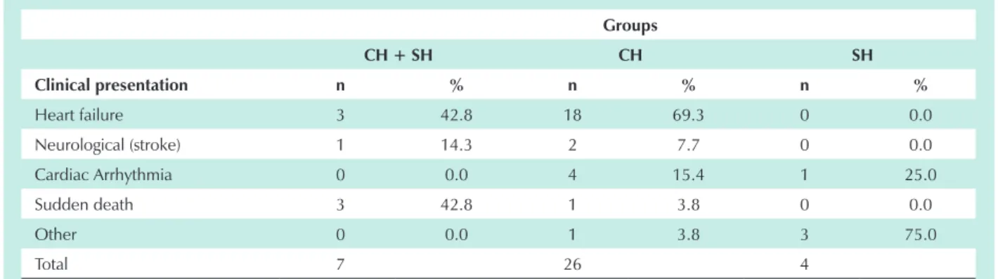
The final clinical presentation was compared in relation to intracardiac thrombosis. A statistically significant difference was found only for heart failure when patients with Chagas’ disease were compared (Table 9). Because of the small number of cases with atrial fibrilation, which is another recognized thrombogenic, an adequate statistical analysis was not possible to present.
Table 8 - Distribution of subjects of the CH + SH and SH groups according to the degree of hypertension and cardiovascular complications
Complications – CH + SH group
Degree of hypertension
Myocardial
infarction Stroke No complication Total
n % n % n % n %
Mild 2 12.5 5 31.3 9 56.2 16 100.0
Moderate 2 15.4 4 30.8 7 53.8 13 100.0
Severe 0 12.1 1 25.0 3 75.0 4 100.0
Total 4 10 30.3 19 57.6 33 100.0
Complications – SH group
Degree of Hypertension
Myocardial
infarction Stroke
No cerebral
study No complication MI + Stroke Total
n % n % n % n % n % n %
Mild 6 28.6 4 19.0 2 9.5 9 42.9 0 0.0 21 100.0 Moderate 0 0.0 1 12.5 0 0.0 7 87.5 0 0.0 8 100.0 Severe 3 17.6 10 58.9 0 0.0 3 17.6 1 5.9 17 100.0 Total 9 19.6 15 32.6 2 4.3 19 4.3 1 2.2 46 100.0
Statistically significant difference was found between the degree of hypertension and complications only in patients with hypertension without Chagas’ disease. CH + SH - patients with Chagas’ disease plus hypertension; n - number of patients; SH - patients with hypertension without Chagas’ disease; MI - myocardial infarction.
Table 9 – Clinical presentation of patients with intracardiac thrombosis in the different groups
Groups
CH + SH CH SH
Clinical presentation n % n % n %
Heart failure 3 42.8 18 69.3 0 0.0
Neurological (stroke) 1 14.3 2 7.7 0 0.0
Cardiac Arrhythmia 0 0.0 4 15.4 1 25.0
Sudden death 3 42.8 1 3.8 0 0.0
Other 0 0.0 1 3.8 3 75.0
Total 7 26 4
Statistically significant difference was found in the presence of heart failure. CH + SH - patients with Chagas’ disease plus hypertension; CH - patients with Chagas’ disease without hypertension; SH - patients with hypertension without Chagas’ disease; n - number of patients.
the causes of death were analyzed separately in each group, significant differences were found in the three groups, with the predominance of cardiac arrhythmia (Table 10).
Discussion
Studies on the concomitance of Chagas’ disease and hypertension are pertinent, given the recognized presence of neuronal impairment, especially of the parasympathetic system, in patients with Chagas’ disease. This alteration determines the unbalance of the autonomic nervous system, thus causing an increased sympathetic activity that can influence the genesis of hypertension, which would therefore
be secondary, so that there could be a pathophysiological connection between the two conditions.
No comprehensive study on the prevalence of hypertension is available in Brazil. Mean pressures in different regions of the country range from 16.1% to 37.1%14-21. The highest
percentage was found in the region of the city of Campinas, State of Sao Paulo, and was specially interesting because this region has peculiarities in relation to hypertension that make it different from the rest of the country.
Guariento et al6,7 (26.1%), and Gurgel et al23 (33.3%), and
are significantly different from those found in Argentina by Palmero et al5. These latter authors found lower pressure levels
in patients with Chagas’ disease in comparison to the general population, regardless of the presence of heart failure.
Andrade and Andrade24,25 studied the autonomous
nervous system in Chagas’ disease in laboratory animals and concluded that both the sympathetic and the parasympathetic systems are affected, with a sparse and quantitatively non-homogenous distribution, which may contribute to explain the discrepancy in the findings of hypertension or hypotension in patients with Chagas’ disease. The sympathetic and parasympathetic lesions found corroborate our own finding that the neurological mechanism may be responsible for the onset of hypertension; however, this mechanism may be not enough to determine hypertension in the patient with Chagas’ disease and hypertension. Although the method used in the present study does not allow for the conclusion of the existence or non-existence of a relationship between the autonomous nervous system and hypertension in patients with chronic Chagas’ disease due to the fact that the myenteric plexus and the sympathetic chain were not studied, we can assume that the evidences found in the literature tend to favor the non-participation of this mechanism as a single causal factor in the genesis of hypertension in these patients. The high frequency of hypertension found in our case series probably results from the multiple factors that contribute for the incidence of hypertension in the region studied. In this study, no statistically significant difference was observed for the differences reported for gender, thus also showing that the number of males and females were
matched in the sample studied. Therefore, this variable did not influence the others, since, according to the literature, both hypertension and Chagas’ disease may be more severe in male patients26,27.
No significant difference was found as regards race in the frequency of hypertension in the three groups studied. This finding shows that, although this variable is known to be capable of influencing the frequency of hypertension, it was not relevant in this study. A significant predominance of mild hypertension (48.5%) was found only in the group of patients with Chagas’ disease plus hypertension. This fact, associated with the age range, brings these study cases closer to those of essential hypertension, since hypertension resulting from catecholamine unbalance should perhaps be similar to that seen in pheochromocytoma. The fact that both systolic and diastolic levels are elevated also supports this hypothesis, because hypertension generated by adrenergic discharge, such as in emotional stress situations, is characterized by a predominance of the systolic component. This is difficult to prove in the present sample of the CH + SH group, since the severe myopathic Chagas’ heart disease may distort this result.
A significant difference was also found in the other degrees of hypertension, and severe hypertension was verified especially among non-chagasic patients (36.9%). This can be explained by the selection criteria of cases in this group of patients. For the evaluation of the anatomical and clinical observations, these cases were selected according to their main diagnosis, that is, the diagnosis that resulted in the individual’s death. Thus, hypertension would surely be more severe in these individuals, because it allowed changes that resulted in death. These types of study make it impossible to weigh the severity of pressure levels
Table 10 – Distribution of the subjects according to group and cause of death
Groups
CH + SH CH SH Total
Cause of death n % n % n % n %
Bulbar compression 1 7.7 3 23.1 9 62.2 13 100.0 Heart arrhythmia 23 27.1 41 48.2 21 24.7 85 100.0
Toxic shock 6 42.9 2 14.3 6 42.6 14 100.0
Cardiogenic shock 2 18.2 8 72.7 1 9.1 11 100.0 Hypovolemic shock 1 14.3 3 42.9 3 42.9 7 100.0
Indeterminate 0 0.0 1 50.0 1 50.0 2 100.0
Hypoglycemia 0 0.0 0 0.0 2 100.0 2 100.0
Cachexia 0 0.0 1 50.0 1 50.0 2 100.0
Neurogenic shock 0 0.0 0 0.0 1 100.0 1 100.0
Respiratory failure 0 0.0 0 0.0 1 100.0 1 100.0
Total 33 23.9 59 42.8 46 33.3 138 100.0
as a relevant factor in the classification of the different groups. The other items discussed refer to the probable influence of a disease on the natural course of another associated condition.
The concomitant presence of hypertension in chagasic patients did not change the expression of the clinical form of Chagas’ disease. The difference in the presentation of the clinical forms was not statistically significant in the CH + SH and CH groups, and the combined form (dromotropic, bathmotropic and myopathic forms combined) predominated both in the CH + SH group (33.3%) and in the CH group (37.3%). This was the expected result, since the sample studied was from necropsy, which made us suppose that the patients might have had more severe presentations. In this sense, it was not possible to confirm that concomitant hypertension was responsible for the change in the natural history of Chagas’ disease.
The electrographic assessment showed that ventricular extrasystoles were significantly more frequent in the CH + SH and CH groups, unlike in relation to supraventricular extrasystoles and atrial fibrillation, which had a similar frequency in the three groups. The high frequency of atrial fibrillation in patients with Chagas’ disease (29% in the hypertensive and 20.7% in the non-hypertensive patients) may be attributed to the severe clinical status of these patients. In patients with Chagas’ disease treated on an outpatient basis the frequency of this arrhythmia is of 1.8% to 2.5%28-30
and it is associated with a poor prognosis26, 31, which is also
attributed to the presence of complete left bundle branch block (CLBBB). In this study, no increased number of cases with CLBBB was observed among patients with Chagas’ disease plus hypertension, which can mean that the determinant mechanisms did not influence the course of Chagas’ disease when it was associated with hypertension.
Mean heart weight in the three groups was similar, however the CH group had a mean weight slightly higher in relation to the CH + SH group, with no statistically significant difference. We can conclude that the concomitance of the two diseases did not determine significant additional myocardial changes that could explain the increased heart weight in the CH + SH group patients. In the SH group, however, a higher number of myocardial infarctions was observed among those who had heavier heart weight.
In this study, in relation to coronary stenosis, no significant difference was found as regards the degree of atherosclerosis between the CH + SH and SH groups; however, in the CH group there was a significant predominance of mild atherosclerosis. Without the hypertension factor, the coronaries were significantly free from stenosis in 40 cases (74%). This shows that the hypertension factor had a decisive influence on the onset of atherosclerosis, both in patients with and without Chagas’ disease.
In relation to cardiovascular complications, no significant difference was found as regards the number of myocardial or cerebral infarctions, with no influence of the gender and age variables. The degree of hypertension was fundamental and statistically significant for the onset of these complications only in the SH group, with a higher frequency of complications in individuals with the severe form, unlike in patients with
Chagas’ disease plus hypertension. Incidentally, severe hypertension was not a common finding among patients with Chagas’ disease who presented some cardiovascular complication. The mechanism leading to myocardial infarction in patients with Chagas’ disease seems to be different from that observed in patients with hypertension, since it may result from embolic phenomena usually originating in the left ventricular apex32. The main etiology of stroke is also
embolism33, and in this study a high frequency was found in
the CH + SH and CH groups (30.3% and 11.9%, respectively), with no statistically significant difference between them. The lack of statistical significance in the number of myocardial and cerebral infarctions between the groups of patients with Chagas’ disease allowed for the conclusion that concomitance with hypertension, again, was not proven to influence the natural history of trypanosomiasis.
Chagasic apical lesions were present in the CH + SH and CH group individuals alike (54.5% and 66.1%, respectively), most of them located in the left ventricle. Therefore, there was no influence of hypertension on the progression of the apical lesion present in chagasic heart disease. Apical lesions should remain important for the diagnosis of chagasic heart disease in hypertensive patients.
Of the 92 cases of Chagas’ disease studied, 33 (35.9%) presented intracardiac thrombosis when compared with only 8.7% of patients with hypertension without Chagas’ disease. In 100% of the CH + SH group cases, thrombosis was located in the apical lesion. The same finding was observed in 42.3% of the CH group cases; in 100% of the SH group cases thrombosis was located in one of the atrial appendages. Statistical significance was found only in the CH group: the higher the heart weight, the higher the frequency of thrombosis. The final clinical presentation of patients with Chagas’ disease was congestive heart failure (63.6%) which was probably the most important factor for the higher frequency of intracardiac thrombosis, in addition to the presence of apical lesion. Heart failure was, therefore, a key factor for the onset of intracardiac thrombosis in patients with Chagas’ disease, especially in non-hypertensive patients. Atrial fibrilation was present mainly in the group of hypertensive patients, whether or not with Chagas‘ disease. Because of the small case series (three cases in the CH + SH group, four in the CH group, and two in the SH group), a proper statistical analysis could not be performed. In patients with Chagas’ disease, whether or not hypertensive, the most prevalent causes of death were cardiac causes: arrhythmia in the CH + SH group and cardiogenic shock in the CH group. However, in the SH group, 62.2% of the patients died secondarily to bulbar compression.
Conclusion
References
1. Fundação Nacional da Saúde. Situação atual e futuro da doença de Chagas no Brasil. [conferência]. Rio de Janeiro; 1997. (Memórias do Instituto Oswaldo Cruz on line).
2. Curti HJV, Sanches PCR, Bittencourt LAK, Carvalhal SS. Revisão da classificação anátomo-clínica da doença de Chagas. Arq Bras Cardiol. 1979; 33: 277-81.
3. III Consenso Brasileiro de Hipertensão Arterial. Rev Bras Cardiol. 1999; 1: 96-132.
4. Messerli FH, Amodeon C. Left ventricular hypertrophy as a risk factor. In: Frölich ED. Cardiology clinics. Philadelphia: W.B. Saunders; 1986. p. 137-44.
5. Palmero HA, Caiero TF, Iosa DJ. Effect of Chagas’ disease on arterial blood pressure. Am Heart J. 1979; 97: 38-42.
6. Guariento ME. Doença de Chagas e hipertensão arterial [dissertação]. Campinas: Unicamp; 1985.
7. Guariento ME, Ramos MC, Gontijo JAR, Carvalhal SS. Doença de Chagas e hipertensão arterial primária. Arq Bras Cardiol. 1993; 60: 71-5.
8. Guariento ME, Rocha RH, Freitas AG. Doença de Chagas e outras patologias associadas num serviço de referência. Rev Soc Bras Med Trop. 1994; 27 (supl II): 141-3.
9. Fulton RM, Hutchinson ME, Jones AM. Ventricular weight in cardiac hypertrophy. Br Heart J. 1952; 14: 413.
10. Werkema MCC. In: Ferramentas estatísticas para o gerenciamento de processos. Belo Horizonte: Fundação Cristiano Otonni, Escola de Engenharia – UFMG; 1995.
11. Levin J. Estatística aplicada em ciências humanas. São Paulo: Harper e Row do Brasil Ltda; 1978.
12. Drummond FB, Werkema MCC, Aguiar S. Análise de variância: comparação de várias situações. Belo Horizonte: Fundação Cristiano Otoni, Escola de Engenharia – UFMG, 1996.
13. Siegel S. Estatística não-paramétrica para as ciências do comportamento. São Paulo: Editora McGraw-Hill do Brasil Ltda; 1981.
14. Carvalho JJM, Silva NAS, Oliveira JM, Arguelles E, Silva JAF. Pressão arterial e grupos sociais: estudo epidemiológico. Arq Bras Cardiol. 1983; 40: 115-20.
15. Lolio CA. Prevalência da hipertensão arterial em Araraquara. Arq Bras Cardiol. 1990; 55: 167-73.
16. Ayres JEM. Prevalência da hipertensão arterial na cidade de Piracicaba. Arq Bras Cardiol. 1991; 57: 33-6.
17. Carneiro O, Jardim PCBV. Pressão arterial em tribo Xavante: comparação 15 anos depois. Arq Bras Cardiol. 1993; 61: 275-82.
18. Fuchs FD, Moreira LB, Moraes RS, Bredemeier M, Cardozo SC. Prevalência da
hipertensão arterial sistêmica e fatores associados na região urbana de Porto Alegre: estudo de base populacional. Arq Bras Cardiol. 1995; 63: 473-9.
19. Trindade IS, Heineck G, Machado J. Prevalência da hipertensão arterial sistêmica na população urbana de Passo Fundo. Arq Bras Cardiol. 1998; 71: 127-30.
20. Freitas OC, Carvalho FR, Neves JM, Veludo PK, Parreira RS, Gonçalves RM, et al. Prevalência da hipertensão arterial sistêmica na população urbana de Catanduva, SP. Arq Bras Cardiol. 2001; 77: 9-15.
21. Camilo DF, Volpini CCA, Zoldan CM, Cecchetti DFA, Oliveira EF, Oliveira FT, et al. Casos novos de hipertensão arterial, Distrito de Saúde Noroeste, Campinas, Estado de São Paulo. Hipertensão. 2001; 4 (supl): 32.
22. Fragata AA Fº. Doença de Chagas e hipertensão arterial sistêmica. Arteríola. 2001;3:43-8.
23. Gurgel CBFM, Miguel A Jr, Mendes CR, Zerbini CO, Carcioni TM. Freqüência da hipertensão arterial na doença de Chagas: estudo clínico retrospectivo. Arq Bras Cardiol. 2003; 81 (6): 541-4.
24. Andrade SG, Andrade ZA. Doença de Chagas e alterações neuronais no plexo de Auerbach (estudo experimental em camundongos). Rev Inst Med Trop São Paulo. 1966;8(5):219-24.
25. Andrade SG, Andrade ZA. Patologia da Doença de Chagas experimental de longa duração. Rev Inst Med Trop São Paulo. 1968; 10 (3): 180-7.
26. Mady C, Nacruth R. História natural da cardiopatia chagásica crônica: fatores prognósticos. Rev Soc Cardiol Estado de São Paulo. 1994; 4: 124-9.
27. Pereira Barretto AC, Arteaga E, Mady C, Ianni BM, Bellotti G, Pileggi F. Sexo masculino: fator prognóstico na Doença de Chagas. Arq Bras Cardiol. 1993; 60: 225-7.
28. Dias JCP, Kloetzel K. The prognostic value of the electrocardiographic features of chronic Chagas’ disease. Rev Inst Med Trop São Paulo. 1968; 10: 158-62.
29. Porto C. O eletrocardiograma na Doença de Chagas. Arq Bras Cardiol. 1964; 17: 313-46.
30. Garzon SAC, Lorga AM, Nicolau JC. Correlações entre alterações eletrocardiográficas e a fração de ejeção do VE em chagásicos crônicos considerando anormalidades isoladas por análise de regressão múltipla [abstract]. Arq Bras Cardiol. 1993; 61: 130.
31. Bestetti RB, Dalbo CMR, Arruda CA, Correa Filho D, Freitas OC. Predictors of sudden cardiac death for patients with Chagas’ disease: a hospital cohort study. Cardiology. 1996;87:481-7.
32. Morais CF, Higuchi ML, Lage S. Chagas’ heart disease and myocardial infarct. Incidence and report of four necropsy cases. Ann Trop Med Parasitol. 1989; 83: 207-14.
33. Guilhon F, Nasser W. Acidente vascular cerebral e cardiopatia chagásica. Rev Bras Neurol. 1989; 25 (2): 51-3.
Chagas’ disease did not change the natural history of either one of the diseases.
Potential Conflict of Interest
No potential conflict of interest relevant to this article was reported.
Sources of Funding
There were no external funding sources for this study.
Study Association