ABSTRACT
BACKGROUND AND OBJECTIVES: The convolutions gen-erated on the patient’s skin with the application of the elastic bandage reduce the pressure on the mechanoreceptors and thus, the nociceptive stimulus. The objective of this study was to com-pare the effect of the elastic bandage application with the appli-cation of the medical tape in myofascial pain in the region of the upper fibers of the trapezius muscle in teachers.
METHODS: Participants were assessed using the McGill-Melz-ack Pain questionnaire and the numeric pain rating scale, palpa-tion for the detecpalpa-tion of trigger points, goniometry for shoulder abduction and lateral neck flexion, and the upper trapezius mus-cle strength test. Participants were randomly divided into two groups. In the first moment, the participants of group A received an application of elastic bandage, with the “Y” technique, and those belonging to group B received the application of the same technique, however, using the medical tape. Both groups were reassessed after teaching class and after 24 hours. Two weeks lat-er, there was the inversion of the materials used.
RESULTS: The sample consisted of 16 teachers. Group A had a significant statistical pain reduction, according to the numeric pain rating scale, between the initial assessment and post-appli-cation at the first moment (p=0.00) and at the second moment (p=0.02). A similar result was found in group B, according to the numeric pain rating scale, both at the first moment (p=0.01) and at the second moment (p=0.03). In both groups, there was pain attenuation with no significance on the effect of the elastic bandage or the medical tape.
CONCLUSION: The application of elastic bandage has the same effect that the medical tape in reducing pain.
Keywords: Faculty, Myofascial pain syndromes, Pain, Physical therapy, Physical therapy modalities.
Comparison of the application of elastic bandage and medical tape in
pain reduction in primary and secondary teachers
Comparação da aplicação de bandagem elástica e esparadrapo na redução da dor em
professores de ensino fundamental e médio
Debora Mottin1, Cássio Preis1, Luiz Bertassoni Neto1
1. Pontifícia Universidade Católica do Paraná, Departamento de Fisioterapia, Curitiba, PR, Brasil.
Submitted on May 01, 2018.
Accepted for publication on September 17, 2018. Conflict of interests: none – Sponsoring sources: none.
Correspondence to:
Rua Zacarias de Paula Xavier, 221 – Centro 83414-160 Colombo, PR, Brasil. E-mail: [email protected]
© Sociedade Brasileira para o Estudo da Dor
RESUMO
JUSTIFICATIVA E OBJETIVOS: As circunvoluções geradas na pele do paciente com a aplicação da bandagem elástica reduzem a pressão dos mecanorreceptores e assim, o estímulo nociceptivo. O objetivo deste estudo foi comparar o efeito da aplicação da ban-dagem elástica com a aplicação de esparadrapo na dor miofascial na região das fibras superiores do músculo trapézio de professores. MÉTODOS: Os participantes foram avaliados através do ques-tionário da dor de McGill-Melzack e a escala numérica, palpa-ção para detecpalpa-ção de pontos-gatilho, goniometria de abdupalpa-ção do ombro e láteroflexão de cervical e teste de força no músculo tra-pézio superior. Aleatoriamente foram divididos em dois grupos. No primeiro momento, os participantes do grupo A, receberam a aplicação de bandagem elástica, com a técnica em “Y” e, os per-tencentes ao grupo B, receberam a aplicação da mesma técnica, entretanto com esparadrapo. Ambos os grupos foram reavaliados após ministrar aula e após 24 horas. Duas semanas após, ocorreu a inversão dos materiais utilizados.
RESULTADOS: A amostra foi constituída por 16 professores. O grupo A apresentou redução estatisticamente significativa da dor na escala numérica entre a avaliação inicial e pós-aplicação no primeiro momento (p=0,00) e no segundo momento (p=0,02). Resultado semelhante foi encontrado no grupo B na escala nu-mérica, tanto no primeiro momento (p=0,01) quanto no segundo momento (p=0,03). Em ambos os grupos a dor atenuou sem haver significância para o efeito da bandagem elástica ou esparadrapo. CONCLUSÃO: A aplicação de bandagem elástica apresenta o mesmo efeito da aplicação de esparadrapo para a redução do qua-dro álgico.
Descritores: Dor, Fisioterapia, Modalidades de fisioterapia, Pro-fessores, Síndromes da dor miofascial.
INTRODUCTION
The myofascial pain syndrome is a muscular condition caused by trigger points (TP), attributed to repetitive effort, maintenance of incorrect postures, musculoskeletal disorders, systemic diseas-es, sedentary lifestyle, and sleep disorders1,2.
Teachers constitute an occupational group with a high preva-lence of musculoskeletal pains. The causes are multifactorial. Among them are the intense physical effort, inadequate furni-ture, and long working hours. It is also associated with psycho-social factors, such as high level of stress, low psycho-social support, and professional satisfaction3,4.
The treatment of myofascial pain includes the Kinesiotaping® method, which uses an elastic bandage (EB)5,6. Originally devel-oped in Japan by Kase, it has become more and more popular7 over the years7.
The elastic bandage expands up to 140% of its natural size, thus matching the skin elasticity, with its thickness and weight also being comparable. The bandage is also hypoallergenic and water resistant. Equally important is that it does not contain drugs, and all the reported benefits come from the elasticity of the material8. The EB effects are not fully elucidated. This method proposes analge-sia, improvement of the muscle function via muscle tone regulation, to help the joint function, eliminate blockages in the bloodstream, and help lymphatic drainage. The studies show that it is effective when applied isolatedly. This fact can be associated with the placebo effect. When combined with the physical therapy, presumably the ideal method of treatment, there was no meaningful difference7,9-13. The combination of the EB expansion capacity and the appli-cation over the elongated muscle creates circumvolutions on the patient’ skin when it returns to the neutral position. These circumvolutions reduce the pressure on the mechanoreceptors located below the dermis, thus decreasing the nociceptive
stim-ulus14,15. These circumvolutions also increase the space between
the skin and the muscle, improving blood and lymphatic flows16. There is evidence that the pain is partially mediated by an en-dogenous mechanism of the brain, called the medial nociceptive system, which can contribute to the emotional component of pain. This system can be influenced by the patient’s expectations which, in turn, reduce the pain through the descendent inhibi-tion and release of opioids. These results suggest that a placebo has a true physiological effect17.
If the EB indeed acts inhibiting the descending pathway or an-other similar mechanism, it is possible that the application of medical tape provides enough stimulus to have a therapeutic ef-fect regarding the reduction of the pain14.
The purpose of this study was to compare the effect of the EB ap-plication with the medical tape apap-plication in myofascial pain in the region of the upper fibers of the trapezius muscle of teachers.
METHODS
Applied, experimental, qualitative, quantitative, and descriptive research conducted from March to June 2015, with teachers from four schools of the public and private education system, located in the metropolitan area of Curitiba.
Female teachers with TP and pain complaints in the region of the upper fibers of the trapezius muscle were included, aged from 20 to 50 years, who have been working for at least two years and at least 25 hours per week. The exclusion criteria were pregnan-cy, participants allergic to the medical tape, and with diseases in the EB application area. One participant who had supraspinatus muscle tendonitis and two above the age group were excluded. All volunteers were informed about the performed procedures and signed the Free and Informed Consent Form (FICT). The sample calculation was based on the assumption that for pop-ulations with more than 150 people the sample should be of 10%, and for populations with less than 150 people, 20%. Knowing that
there is an average of 80 teachers and that it is not the totality with the required characteristics, the minimum sample for the study was of 16 participants, who represent 20% of the population.
The participants were divided according to the order they were evaluated; the first evaluated was sent to group A, the second to group B, and so forth. The participants were identically evaluated during the breaks between classes. The personal information was collected (age, weight, height, marital status). In the patient’s his-tory, the main complaint was investigated, as well as information on the professional activity, and the history of pain using the Mc-Gill-Melzack Pain Questionnaire18 and the numeric scale (NS). The TP was identified by the palpation as a circumscribed point that is presented spontaneously or by acupressure, hypersensitivity, taut band, and referred pain. The Goniometer Pro Preview pro-gram was used to measure the range of motion (ROM) of the cer-vical lateral flexion, installed in a Samsung S5 Galaxy Smartphone. With the participant sitting, head in a neutral position, the device was positioned on the spinous process of the seventh cervical ver-tebra to establish the articular 0º and then the desired motion was requested. The mentioned program was also used to measure the motion of the shoulder abduction, with the participant sitting, with the upper limbs in an anatomical position, and the device was positioned in the distal and anterior region of the forearm. Finally, the strength test was applied in the superior trapezius muscle using the principles of the Oxford scale19.
In a first moment, group A received the EB application, accord-ing to the Application Manual8. The participants of group B, in this first moment, received the application of the same described technique, but with the medical tape. At the end of the class and on the following day, all the volunteers were reassessed according to the same procedures of the evaluation. Two weeks after the first application, group A received the application with medical tape, and group B the EB, in the same way as in the first moment, and they were reassessed. It is worth mentioning that the participants were not aware of which material was being applied, they were only informed that a tape was being placed in the region.
This study was approved by the Ethics Committee of the Pon-tifícia Universidade Católica do Paraná, under opinion number 987.607, and follows the Resolution 466/12 of the National Health Council.
Statistical analysis
RESULTS
The sample was composed of 16 participants. In the demographic data of the studied population, the statistical difference between the groups was only found in the weight (p=0.04). All the participants were right-handedness and presented a taut band or TP on that side. Table 1 shows that group A significantly reduced the pain (McGill and NS). In the post-significance test, the McGill variable showed a statistically significant difference between the initial evaluation and post-application (p=0.04) and between the initial evaluation and post-24h (p=0.03) in the first application, using the EB. In the second application, with the medical tape, there was a reduction of pain between the initial evaluation and post-24h (p=0.03).
The NS showed a statistically significant difference between the ini-tial evaluation and post-application (p=0.01) and between the iniini-tial evaluation and post-24h (p=0.01) in the first application (EB). In the second application, with the medical tape, there was a pain reduction between the initial evaluation and the post-application evaluation (p=0.04), and between the initial evaluation and after 24h (p=0.04). In the detection of the TP and the taut band tension in the three evaluation moments, the taut band tension decreased compared to the initial evaluation; however, the TP was present in the post-appli-cation and the post-24h evaluations in all the participants.
Another issue raised by the participants was the sensation that the pain increased in the member contralateral to the application. It is worth noting that two participants reported that the application of the medical tape was more effective, while the rest of the group did not express a preference.
In the comparison of the three evaluation moments in group B, there was also a statistically significant reduction of pain (McGill and NS) (Table 2). The McGill variable showed a statistically signifi-cant difference between the initial evaluation and post-24h (p=0.02) and between the post-application evaluation and post-24h (p=0.02) in the first care, using the medical tape. In the second application
with the EB, the difference was between the initial evaluation and the post-24h (p=0.03).
The NS showed a significant difference between the first evaluation and post-24h (p=0.01) in the first application (medical tape). The second application (EB) also showed a difference between the initial evaluation and the post-application (p=0.04), and between the ini-tial evaluation and after 24h (p=0.03).
A result similar to group A was found regarding the presence of TP and the taut band after the application. Regarding the participant’s perception, four of them stated that the medical tape provided a more effective and faster result. In contrast, two preferred the EB. Table 3 shows the mean of the differences of the comparison be-tween the evaluation moments for groups A and B. In the first application, where group A received EB and group B the medical tape, a statistically significant difference was found in the lateral in-clination of the cervical to the right, between the initial evaluation and post-application, and between the initial evaluation and after 24 hours. In the second application, no variable showed any difference. Group B, in the first application, presented more effective progress in the lateral inclination of the cervical to the right between the ini-tial evaluation and post-24h, and this variable receded in group A at the abovementioned moment.
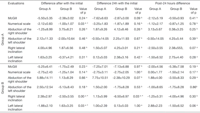
Table 4 shows the comparison between group A and group B taking the type of application into consideration, EB or medical tape. In the application of the EB, the variable that showed a statistically significant difference was the NS between the initial evaluation and post-application. In the medical tape application, the variables that presented a statistical difference was the goniometry of the lateral inclination of the cervical to the right between the initial evaluation and post-application, and between the initial and post-24h, and go-niometry of the lateral inclination of the cervical to the left between the initial evaluation and post-application.
One can note that both in the EB moment and the medical tape, group A showed a higher pain reduction (McGill and NS) than group B.
Table 1. Evaluation of pain and goniometry of group A (n=8)
First application
Initial evaluation Post-application evaluation Post-24 hours evaluation Value of p
McGill 10.50±6.21 8.75±6.88 3.38±3.34 0.00 a *
Numerical scale 5.50±1.41 4.25±1.49 2.75±1.98 0.01 b *
Goniometry
Right shoulder 162.75±14.06 163.88±11.00 160.38±9.02 0.83 b
Left shoulder 159.50±15.21 154.38±13.51 157.75±10.67 0.74 b
Right cervical 27.38±5.60 29.50±5.18 33.13±3.04 0.07 b
Left cervical 30.63±6.93 31.38±5.55 33.50±4.78 0.60 b
Second application
McGill 6.38±4.66 4.00±3.55 3.50±4.28 0.01 a *
Numerical scale 4.13±1.64 3.13±1.36 2.25±1.67 0.03 a *
Goniometry
Right shoulder 160.63±12.29 164.38±11.24 164.75±12.14 0.75 b
Left shoulder 155.50±14.07 153.50±15.30 157.75±16.46 0.27 a
Right cervical 30.38±5.45 32.25±7.05 34.63±4.21 0.34 b
Left cervical 32.63±5.15 32.25±8.01 35.00±5.68 0.65 b
Source: research data.
Table 2. Evaluation of pain and goniometry of group B (n=8)
First application
Initial evaluation Post-application evaluation Post-24 hours evaluation Value of p
McGill 10.50±6.21 8.75±6.88 3.38±3.34 0.00 a *
Numerical scale 5.50±1.41 4.25±1.49 2.75±1.98 0.01 b *
Goniometry
Right shoulder 162.75±14.06 163.88±11.00 160.38±9.02 0.83 b
Left shoulder 159.50±15.21 154.38±13.51 157.75±10.67 0.74 b
Right cervical 27.38±5.60 29.50±5.18 33.13±3.04 0.07 b
Left cervical 30.63±6.93 31.38±5.55 33.50±4.78 0.60 b
Second application
McGill 6.38±4.66 4.00±3.55 3.50±4.28 0.01 a *
Numerical scale 4.13±1.64 3.13±1.36 2.25±1.67 0.03 a *
Goniometry
Right shoulder 160.63±12.29 164.38±11.24 164.75±12.14 0.75 b
Left shoulder 155.50±14.07 153.50±15.30 157.75±16.46 0.27 a
Right cervical 30.38±5.45 32.25±7.05 34.63±4.21 0.34 b
Left cervical 32.63±5.15 32.25±8.01 35.00±5.68 0.65 b
Source: research data.
a Friedman; b ANOVA; * p<0.05.
Table 3. Intergroup Evaluation - comparison of the differences between groups A and B in the two moments of application
Evaluations Difference after with the initial Difference 24h with the initial Post-24 hours difference
Group A Group B Value of p
Group A Group B Value of p Group A Group B Value of p
First application
McGill -5.50±5.35 -1.75±2.49 0.13 a -7.625±6.63 -7.125±6.88 0.88 b -2.125±5.19 -5.38±7.58 0.15 a
Numerical scale
-2.12±0.83 -1.25±1.04 0.08 b -3.25±1.83 -2.75±2.05 0.62 b -1.125±2.17 -1.50±2.14 0.73 b
Abduction of the right shoulder
-1.25±8.99 1.13±8.29 0.59 b 1.88±9.26 -2.375±10.29 0.40 b 3.13±5.67 -3.5±8.33 0.08 b
Abduction of the left shoulder
2.12±11.33 -5.125±8.43 0.17 b -0.5±14.05 -1.75±8.28 0.83 b -0.5±14.05 -1.75±8.28 0.83 b
Right lateral inclination
4.00±4.96 -2.5±3.55 0.01 a * 1.50±5.07 -6.5±6.97 0.02 b * -2.5±3.55 -4.00±4.96 0.28a
Left lateral inclination
1.63±3.25 1.63±3.25 1.00 b 0.13±5.03 0.13±5.03 1.00 a -1.5±6.52 -1.50±6.52 1.00 a
Second application
McGill -5.25±6.41 -2.38±3.02 0.41a -7.25±7.01 -2.88±3.00 0.14 b -2.00±4.57 -0.50±0.93 0.87 a
Numerical scale
-2.75±2.43 -1.00±1.07 0.09 b -2.75±3.11 -1.88±1.89 0.51 b 0.00±1.77 -0.88±1.25 0.35 a
Abduction of the right shoulder
5.88±14.11 3.75±8.21 0.92 a 7.75±10.51 4.13±8.46 0.46 b 1.88±11.70 0.38±3.25 0.92 a
Abduction of the left shoulder
2.50±12.54 -2.00±10.64 0.45 b 1.50±11.66 2.25±11.93 0.90 b -1.00±8.65 4.25±5.44 0.17 b
Right lateral inclination
2.38±2.97 1.88±6.56 0.53 a 1.13±5.99 4.25±3.01 0.21 b -1.25±5.31 2.38±5.53 0.34 a
Left lateral inclination
-1.88±2.10 -0.38±4.21 0.63 a 1.00±2.39 2.38±3.16 0.34 b 2.88±2.23 2.75±4.40 0.94 b
Source: research data.
DISCUSSION
The results showed that the EB and the medical tape reduce the pain (McGill and NS) and there is no statistically significant dif-ference between them.
According to the theory proposed by Simons, the region around the TP is in an ischemic condition, resulting in lack of oxygen and glucose for the metabolism. After the TP relaxation, the blood flow to the tissue is activated, allowing the oxygen perfu-sion so that the skeletal muscle may recover the homeostasis20,21. The mechanism of action of the EB correlates to this theo-ry due to the fact the it lifts the subcutaneous space, which would lead to the increase of the circulation and the removal of the heat produced by the inflammation. And due to the re-duction of pressure on the nociceptors, the sensation of pain would diminish2.
The afferent stimulation, which is promoted both by the EB and the medical tape application, may be cited as another mechanism of action. According to the Gate Control Theory by Melzack and Wall, the velocity of the proprioceptive stimulus is higher, thus inhibiting the transmission of nociceptive signs at the spinal level in chronic musculoskeletal pain situations, leading to the attenu-ation of the pain experience11,22-24.
A similar result was found with the application of the EB in patients with latent TP in the sternocleidomastoid muscle, with a pain reduction and increase of the range of motion of the joint when compared to the group control, which did not receive care1.
To lift the subcutaneous space, improve the blood circulation, and consequently reduce pain, the BE must be applied with the elongated muscle to generate circumvolutions14. Howev-er, a study obtained a similar reduction in the intensity of pain and disability in patients treated according to the treat-ment manual of the Kinesiotaping Method and those who received the EB application without tension, and not creating circumvolutions15.
Once again, comparing the EB with and without tension, the statistically significant difference only existed up to the third day, indicating that the potential benefits of the ban-dage application are immediate. No differences were found between the groups regarding the measurement of pain and disability13.
These studies supported the results found since in the medical tape application, the circumvolutions were not generated, and both materials significantly reduced the pain (McGill and NS), referring this fact to the exteroceptive effect.
Regarding the range of motion of the joint, the muscle with the TP is stiff, with shortened sarcomeres, leading to motion restric-tion. When the myofascial pain is treated, the muscle function should increase, the muscle fibers relax, and, therefore, the re-duction of the muscle stiffness and the extension of the range of motion are expected25-27.
No statistically significant alteration was found in the range of motion in this research. This can be explained by the fact that the TP is present in all the participants after the application of the materials, although the pain sensation had decreased.
Table 4. Intergroup evaluation - comparison of the differences between groups A and B for the application of the elastic bandage and medical tape Evaluations Difference after with the initial Difference 24h with the initial Post-24 hours difference
Group A Group B Value of p
Group A Group B Value of p
Group A Group B Value of p
Gr
oup A
McGill -5.50±5.35 -2.38±3.02 0.24 a -7.62±6.63 -2.87±3.00 0.09 b -2.12±5.19 -0.50±0.93 0.41 b
Numerical scale -2.12±0.83 -1.00±1.07 0.03 b * -3.25±1.83 -1.87±1.89 0.16 b -1.12±2.17 -0.87±1.25 0.78 b
Abduction of the right shoulder
-1.25±8.99 3.75±8.21 0.26 b 1.87±9.26 4.12±8.46 0.26 b 3.13±5.67 0.38±3.25 0.25 b
Abduction of the left shoulder
2.12±11.33 -2.00±10.64 0.46 b -0.50±14.05 2.25±11.93 0.67 b -0.50±14.05 4.25±5.44 0.39 b
Right lateral inclination
4.00±4.96 1.87±6.56 0.48 b 1.50±5.07 4.25±3.01 0.21 b -2.50±3.55 2.38±553, 0.07 a
Left lateral inclination
1.63±3.25 -0.37±4.21 0.31 b 0.12±5.03 2.38±3.16 0.42 a -1.50±6.52 2.75±4.40 0.28 a
Gr
oup B
McGill -5.25±6.41 -1.75±2.49 0.23 a -7.25±7.01 -7.13±6.88 0.97 b -2.00±4.56 -5.38±7.58 0.19 a
Numerical scale -2.75±2.43 -1.25±1.04 0.14 b -2.75±3.11 -2.75±2.05 1.00 b 0.00±1.77 -1.50±2.14 0.17 a
Abduction of the right shoulder
5.88±14.11 1.13±8.29 0.66 a 7.75±10.51 -2.38±10.29 0.07 b 1.88±4.00 -3.50±8.33 0.29 a
Abduction of the left shoulder
2.50±12.54 -5.13±8.43 0.18 b 1.50±2.00 -1.75±8.28 0.53 b -1.00±8.65 -1.75±8.28 0.86b
Right lateral inclination
2.38±2.97 -2.50±3.55 0.00 a * 1.13±5.99 -6.50±6.97 0.03 b * -1.25±5.31 -4.00±4.96 0.30 b
Left lateral inclination
-1.88±2.10 1.63±3.25 0.03 a * 1.00±2.39 0.13±5.03 1.00 a 2.88±2.23 -1.50±6.52 0.06 a
Source: research data.
The study had limitations as to the size of the sample, and the use of an algometer is recommended for future researches to de-termine the pain sensation.
CONCLUSION
The results of this study have shown that the EB application had the same effect as the medical tape application for the reduction of the pain condition and increase of the range of motion. Future studies are necessary to clarify the EB clinical effects since the mechanism of action, according to the creator of the technique, are the circumvolutions. However, they were not generated with the application of the medical tape that showed positive results.
REFERENCES
1. Bae Y. Change the myofascial pain and range of motion of the temporomandibular joint following kinesio taping of latent myofascial trigger points in the sternocleido-mastoid muscle. J Phys Ther Sci. 2014;26(9):1321-4.
2. Wu WT, Hong CZ, Chou LW. The kinesio taping method for myofascial pain con-trol. Evid Based Complement Alternat Med. 2015;2015:950519.
3. Cardoso JP, Araújo TM, Carvalho FM, Oliveira NF, Reis EJ. [Psychosocial work-re-lated factors and Musculoskeletal pain among schoolteachers]. Cad Saude Publica. 2011;27(8):1498-506. Portuguese.
4. Erick PN, Smith DR. A systematic review of musculoskeletal disorders among school teachers. BMC Musculoskeletal Disord. 2011;12(260):1-11.
5. Jung-Ho L, Min-Sik Y, Bong-Jun K, Jin-Sang K. The effect of stabilization exercises combined with taping therapy on pain and function of patients with myofascial pain syndrome. J Phys Ther Sci. 2012;24(12):1283-7.
6. Luz Júnior MA, Sousa MV, Neves LA, Cezar AA, Costa, LO. Kinesio Taping® is not better than placebo in reducing pain and disability in patients with chronic non-speci-fic low back pain: a randomized controlled trial. Braz J Phys Ther. 2015;19(6):482-90. 7. González-Iglesias J, Fernández-de-Las-Peñas C, Cleland JA, Huijbregts P, Del Rosario
Gutierrez-Vega M. Short-term effects of cervical kinesio taping on pain and cervical range of motion in patients with acute whiplash injury: a randomized clinical trial. J Orthop Sports Phys Ther. 2009;39(7):515-21.
8. Sijmonsma J. TNM Manual da Banda Neuromuscular. Portugal: Aneid Press; 2000. 9. Taylor RL, O’Brien L, Brown T. A scoping review of the use of elastic therapeutic tape
for neck or upper extremity conditions. J Hand Ther. 2014;27(3):235-46. 10. Castro-Sánchez AM, Lara-Palomo IC, Matarán-Peñarrocha GA, Fernández-Sánchez
M, Sánchez-Labraca N, Arroyo-Morales M. Kinesio Taping reduces disability and pain slightly in chronic non-specific low back pain: a randomised trial. J Physiother.
2012;58(2):89-95.
11. Kachanathu SJ, Alenazi AM, Seif HE, Hafez AR, Alroumim MA. Comparison be-tween Kinesio Taping and a Traditional Physical Therapy program in treatment of nonspecific low back pain. J Phys Ther Sci. 2014;26(8):1185-8.
12. Paoloni M, Bernetti A, Fratocchi G, Mangone M, Parrinello L, Del Pilar Cooper M, et al. Kinesio Taping applied to lumbar muscles influences clinical and electromyo-graphic characteristics in chronic low back pain patients. Eur J Phys Rehabil Med. 2011;47(2):237-44.
13. Thelen MD, Dauber JA, Stoneman PD. The clinical efficacy of kinesiotape for shoul-der pain: a randomized, double-blinded, clinical trial. J Orthop Sports Phys Ther. 2008;38(7):389-95.
14. Montalvo AM, Cara EL, Myer GD. Effect of kinesiology taping on pain in individuals with musculoskeletal injuries: systematic review and meta-analysis. Phys Sportsmed. 2014;42(2):48-57.
15. Parreira Pdo C, Costa Lda C, Takahashi R, Hespanhol Junior LC, Luz Junior MA, Silva TM, et al. Kinesio taping to generate skin convolutions is not better than sham taping for people with chronic non-specific low back pain: a randomised trial. J Phys-iother. 2014;60(2):90-6.
16. Kalron A, Bar-Sela S. A systematic review of the effectiveness of Kinesio Taping--fact or fashion? Eur J Phys Rehabil Med. 2013;49(5):699-709.
17. Ossipov MH, Dussor GO, Porreca F. Central modulation of pain. J Clin Invest. 2010;120(11):3779-87.
18. Magee DJ. Avaliação musculoesquelética. 5aed. Barueri: Manole; 2010.
19. Kendall FP, McCreary EK, Provance PG, Rodgers M, Romani WI. Músculos: provas e funções. 5ª ed. São Paulo: Manole; 2007.
20. Moraska AF, Hickner RC, Kohrt WM, Brewer A. Changes in blood flow and cellular metabolism at a myofascial trigger point with trigger point release (ischemic compres-sion): a proof-of-principle pilot study. Arch Phys Med Rehabil. 2013;94(1):196-200. 21. Zhang Y, Ge HY, Yue SW, Kimura Y, Arendt-Nielsen L. Attenuated skin blood flow
response to nociceptive stimulation of latent myofascial trigger points. Arch Phys Med Rehabil. 2009;90(2):325-32.
22. Lim EC, Tay MG. Kinesio taping in musculoskeletal pain and disability that lasts for more than 4 weeks: is it time to peel off the tape and throw it out with the sweat? A systematic review with meta-analysis focused on pain and also methods of tape appli-cation. Br J Sports Med. 2015;49(24):1558-66.
23. Saavedra-Hernández M, Castro-Sánchez AM, Arroyo-Morales M, Cleland JA, Lara-Palomo IC, Fernández-de-las-Penas C. Short term effects of kinesiotaping versus cervical thrust manipulation in patients with mechanical neck pain: a randomized clinical trial. J Orthop Sports Phys Ther. 2012;42(8):724-30.
24. Artioli DP, Bertolini GR. Kinesio Taping: aplicação e seus resultados sobre a dor. Fi-sioter Pesq. 2014;21(1):94-9.
25. Ge HY, Arendt-Nielsen L. Latent myofascial trigger points. Curr Pain Headache Rep. 2011;15(5):386-92.
26. Lee SH, Chen CC, Lee CS, Lin TC, Chan RC. Effects of needle electrical intramus-cular stimulation on shoulder and cervical myofascial pain syndrome and microcircu-lation. J Chin Med Assoc. 2008;71(4):200-6.