Centro de Ciências da Saúde - Universidade Estadual de Londrina Mailing address: José Carlos dos Santos Guitti Rua Paranaguá, 934 86020-030 – Londrina, PR, Brazil
Objective – To determine the prevalence and other epi-demiological characteristics of congenital heart diseases.
Methods – A retrospective population based study of children who were born in Londrina, from January ’89 to December ’98 (80,262 live births). Diagnoses were confir-med through autopsy, surgery, catheterization, or echo-cardiography.
Results – A total of 441 patients was as certained what corresponds to a prevalence of 5.494:1,000 live bir-ths. Ventricular septal defect was the commonest lesion. A small number of transpositions of the great vessels and of left ventricular hypoplasia was observed. A high propation of ventricular septal defect (28.3%) and atrioventricular sep-tal defects (8.1%) occurred. Fifty-one (11.35%) affected children had syndromic diseases and 52 (12.01%) children had nonsyndromic anomalies.
Conclusion – The prevalence of congenital heart di-seases in Londrina is in accordance with that of other regi-ons of the globe. This prevalence also may reflect the reali-ty in the southern region of Brazil, because population characteristics are very similar in the 3 southernmost Bra-zilian states.
Key words: congenital heart diseases, epidemiology, Brazil.
Arq Bras Cardiol, volume 74 (nº 5), 400-404, 2000
José Carlos dos Santos Guitti
Londrina, PR - Brazil
Epidemiological Characteristics of Congenital Heart Diseases
in Londrina, Paraná South Brazil
“O Solomon! What wisdom is needed for that
physici-an who deals with a child physici-and a “pre-existing condition.”
Ron Louie, MD
Based on the pioneering studies by Abbott in the ‘20s, a large number of articles about the prevalence of congenital heart diseases in North America and Europe have already been published. In 1967, Caddell 1 pointed out that the actual
incidence and distribution of congenital heart diseases in tropical and subtropical regions were not known. The preva-lence of congenital heart diseases in Latin America remains unknown, because no information has been published about this population (source: MEDLINE, LILACS).
Londrina is the second most important city of Paraná State and the third greatest city in the southern region of the country. Its population is 426,607 inhabitants, 90% of whom live in the urban area. The major causes of death in the first year of life are perinatal diseases (47%), congenital anomalies (21%), and respiratory and infectious diseases (18%). Infant mortality is 14.48:1,000 live births, which, even though high, is far below the Brazilian rate, which is 40.0:1,000 live births (Brazilian Demographic Census, 1996). Approximately 3 out 4 inhabitants of Londrina live in the peripheral region of the city, in extreme poverty or in lower middle class conditions. In regard to the more indi-gent population, which lives mainly in the rural area, several factors account for the few visits that occur to the pediatric services, where the precocious detection of cardiovascular anomalies may cause a significant difference in the survival of children with lesions that may be treated, but that are le-thal if not diagnosed and treated in time 2.
Methods
This study comprised 441 children born in Londrina (rural and urban areas) from January ’89 to December ’98. The precise determination of the live-born population was performed through official demographic data (Fundação
Instituto Brasileiro de Geografia e Estatística – Brazilian
Data collected included the following: 1) all children wi-th congenital heart disease born in public hospitals or in wi-the hospital network accredited to SUS (Sistema Único de Saúde – Integrated Health System); 2) children suspected of having congenital heart disease, referred from the basic health units to the pediatric cardiology ambulatory service. All diagnoses were confirmed, corrected, or complemented, by echocardio-graphy, catheterization, surgical procedures, or autopsy. The cases of complete atrioventricular block were diagnosed by electrocardiography and echocardiography to exclude con-comitant structural defects.
Congenital heart disease has been defined as a struc-tural anomaly of the heart or the great vessels, which has a real or potential functional significance. This definition dif-fers from the classical definition by Mitchell et al 3 that
inclu-des only the gross malformations. Children with persis-tence of ductus arteriosus were only admitted if older than 10 days (normal weight at birth) or if older than 3 months when gestational age below 37 weeks. The cases of pulmo-nary atresia with ventricular septal defect were classified as tetralogy of Fallot.
As a considerable number of patients had more than one heart defect, a hierarchic system of classification was adopted, which allowed the inclusion of each patient in only one diagnostic category 4.
Those patients suspected of having genetic or chro-mosomal disorders underwent a genetic study, except those with a conclusive phenotype. In those cases with concomi-tant syndrome and nonsyndromic extracardiac anomaly, the most severe disease prevailed for effect of classification.
Results
Most of the patients had the diagnosis established in the first year of life (Table I).
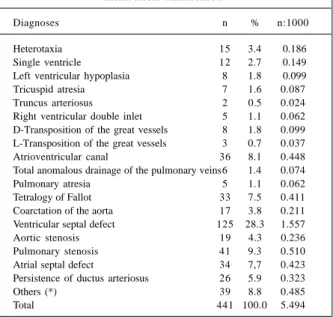
The calculated prevalence of congenital heart disea-ses was 5.494:1,000 live births. Lesions distribution and cor-responding frequencies are shown in Table II.
Seventy (17%) patients had 2 or more heart defects, which were classified according to an order of hierarchical importance, which takes the severity of the lesions into consideration. The defects most commonly found in association with other defects were ventricular septal de-fect, pulmonary stenosis, and atrioventricular canal.
Under the diagnosis of heterotaxia, 16 underlying defects were detected, which if considered within their own anatomical categories, some mild alterations in number, per-centage and prevalence would have been observed (single ventricle: 15, 3.4%, 0.186; tricuspid atresia: 8, 1.8%, 0.099; truncus arteriosus: 3, 0.7%, 0.037; D-transposition of the great vessels: 10, 2.26%, 0.124; L-transposition of the great vessels: 4, 0.9%, 0.049; atrioventricular canal: 39, 8.8%, 0.485; pulmonary atresia: 8, 1.8%, 0.099; tetralogy of Fallot: 34, 7.7%, 0.423; ventricular septal defect: 126, 28.5%, 1.569). Noncardiac anomalies were detected in 104 patients (24%): syndromic diseases in 51 (Table III) and nonsyndro-mic anomalies in 53 patients (Table IV).
The tendency to not perform palliative surgeries but to perform corrective ones preceded the onset of this study. Nevertheless, 52 (11.8%) patients underwent such proce-dures, because of the high risk that corrective surgeries represented to them at the time of the diagnosis. Percutane-ous procedures were performed in 29 (6.6%) patients and corrective surgeries in 154 (34.9%) patients. The total mor-tality rate was 10% (deaths after palliative procedures: 7; deaths after percutaneous procedures: 2; deaths after cor-rective surgeries: 14).
Fourteen defects evolved to spontaneous resolution, all confirmed by echocardiography (11 ventricular septal defects and 3 atrial septal defects).
Out of the 441 children studied, 346 (78%) were com-pletely followed up during the study period. In 35 cases,
Table I – Distribution of the patients according to age
Age n %
<1 month 81 18
1 – 3 months 102 23
4 – 6 months 69 16
7 – 12 months 97 22
1 year 55 12
2 – 4 years 24 5
5 – 9 years 13 3
Total 441 100
Table II – Diagnostic frequencies according to the hierarchical classification
Diagnoses n % n:1000
Heterotaxia 15 3.4 0.186
Single ventricle 12 2.7 0.149
Left ventricular hypoplasia 8 1.8 0.099
Tricuspid atresia 7 1.6 0.087
Truncus arteriosus 2 0.5 0.024
Right ventricular double inlet 5 1.1 0.062 D-Transposition of the great vessels 8 1.8 0.099 L-Transposition of the great vessels 3 0.7 0.037 Atrioventricular canal 36 8.1 0.448 Total anomalous drainage of the pulmonary veins6 1.4 0.074
Pulmonary atresia 5 1.1 0.062
Tetralogy of Fallot 33 7.5 0.411 Coarctation of the aorta 17 3.8 0.211 Ventricular septal defect 125 28.3 1.557
Aortic stenosis 19 4.3 0.236
Pulmonary stenosis 41 9.3 0.510
Atrial septal defect 34 7,7 0.423 Persistence of ductus arteriosus 26 5.9 0.323
Others (*) 39 8.8 0.485
Total 441 100.0 5.494
contact with the respective families was not possible. Twen-ty-six children withdrew during he follow-up because of psychosocial or cultural factors or both of these. Of those children not returning for follow-up, those who underwent corrective surgeries predominated. Therefore, late surgical mortality could not be determined.
Discussion
Not all congenital heart defects are evident at early childhood. Therefore, studies of prevalence in children above one year of age may obviously detect a greater num-ber of cases. On the other hand, the set of heart diseases seen in admission units of any large tertiary hospital may not represent the population as a whole.
Recognition and diagnosis of congenital heart
disea-ses may occur among children who were born or who lived, or both, in modern urban centers or close to them, where a special interest in the problem usually exists. In addition, these centers depend on resources necessary for complete diagnosis and appropriate treatment. The epidemiological study that most probably provides reliable results about the real incidence of congenital heart diseases is the one that comprises a great number of births and in which the inciden-ce of heart diseases is determined based on official popula-tion data 5.
In the last 40 years, several studies about the estima-ted index of congenital heart diseases in population groups were carried out, as were studies about the distribution of specific lesions. During this long period, significant diag-nostic and therapeutical developments occurred. Echocar-diography brought a notable improvement in diagnostic
ac-Table III – Association between syndromic disorders and diagnostic categories
Syndrome n % Diagnostic category n %
Trisomy 21 28 55 Atrioventricular canal 17 33
Ventricular septal defect 9 18 Persistence of ductus arteriosus 2 4
Congenital rubella 10 20 Ventricular septal defect 1 2
Pulmonary stenosis 3 6
Atrial septal defect 1 2 Persistence of ductus arteriosus 3 6
Others 2 4
Marfan 2 4 MVP + dilation of the aortic root 2 4
Trisomy13 1 2 Atrioventricular canal 1 2
Beckwith-Wiedeman 1 2 Pulmonary stenosis 1 2
Pierre Robin 1 2 Tetralogy of Fallot 1 2
Cornelia de Lange 1 2 Ventricular septal defect 1 2
Holt-Oram 1 2 Atrial septal defect 1 2
Coffin-Lowry 1 2 Atrial septal defect 1 2
Not defined 2 4 Aortic stenosis 2 4
CHARGE association 2 4 Tricuspid atresia 1 2
Ventricular septal defect 1 2
VACTERL association 1 2 Single ventricle 1 2
Total 51 100 51 100
MVP- mitral valve prolapse.
Table IV - Extracardiac anomalies and diagnostic categories
Diagnostic category Extracardiac anomalies
n % mild moderate severe
Heterotaxia - - 1 1 2
Single ventricle - - 1 1 2
Tricuspid atresia 1 1 - 2 4
RV double outflow tract - 1 1 2 4
Transposition of the great vessels 1 3 - 4 8
Atrioventricular canal 1 4 2 7 13
Pulmonary atresia 2 - - 2 4
Tetralogy of Fallot 3 1 1 5 9
Coarctation of the aorta - 1 - 1 2
Ventricular septal defect 6 8 2 16 30
Aortic stenosis 1 - - 1 2
Pulmonary stenosis 1 1 - 2 4
Atrial septal defect 1 1 - 2 4
Others 5 2 - 7 13
Total 21 23 9 53 100
curacy, which previously was only obtained through inva-sive methods. Due to their own experience, Tubman et al 6
concluded that Doppler echocardiography should be con-sidered the gold standard for detecting congenital heart di-seases.
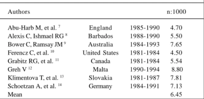
The relation between the number of children with con-genital heart disease and that of the live-born infants was 5.494:1,000. The mean obtained based on 8 significant stu-dies performed in the last 2 decades in different regions with diverse ethnicities was 6.450:1,000 (Table V).
If this mean could be applied to the reality of Londrina, 77 cases of congenital heart disease would not have been detected among the 80,262 live-born infants in the period studied. Such increase would correspond to 0.956:1,000 live-born infants. Considering that 20% of the children with congenital heart disease die in the first year of life and that 5% of the remaining die every year in the 4 subsequent years 15, we may determine that at least 53 children with
un-recognized congenital heart diseases would be living in the community. The studies shown in Table V were selected after the echocardiographic assessment had been included as an essential criterion for the diagnosis of congenital heart diseases. The indices obtained, however, varied from 4.50 to 8.80 per 1,000 live-born infants. Methodological differences, mainly the comprehensiveness of the diagnos-tic approach and the strictness in the criteria of case admis-sion, may explain these significant discrepancies.
Ferencz et al 10 have admitted that very restrictive
crite-ria may constitute biases when evaluating the real preva-lence of congenital heart diseases. Hoffman and Christi-anson 16 and also Lorenzo et al 17 have defended a similar
concept based on a study carried out in Spain in a populati-on of 38,674 students, with ages ranging from 4 to 12 years, all of them without a previous diagnosis of congenital heart disease. Those authors have detected an incidence of con-genital heart disease of 2.3:1,000 and have cited several stu-dies carried out in other regions with similar results.
Our study comprised all children with congenital heart disease born in Londrina from 1989 to 1998. The adoption of this criterion allowed the inclusion in the study of 92 (21%) patients older than one year. If the admission criterion had been more strict, considering only the children under one year of age, the prevalence would have been reduced to 4.348:1,000 live-born infants, which is very similar to the
Table V - Prevalence (n: 1.000 live births) in different regions in the last 20 years
Authors n:1000
Abu-Harb M, et al. 7 England 1985-1990 4.70 Alexis C, Ishmael RG 8 Barbados 1988-1990 5.50 Bower C, Ramsay JM 9 Australia 1984-1993 7.65 Ferencz C, et al. 10 United States 1981-1984 4.50 Grabitz RG, et al. 11 Canada 1981-1984 5.54 Greh V 12 Malta 1990-1994 8.80 Klimentova T, et al. 13 Slovakia 1981-1987 7.81 Schoetzan A, et al. 14 Germany 1984-1991 7.13
Mean 6.45
mean value of 4.185:1,000 obtained considering data from 3 of the most important studies carried out in North America, which adopted this criterion 4,10,11. Therefore, our
methodo-logy is in accordance with the one that Ferencz et al 10
poin-ted out in the Baltimore-Washington Infant Study, which states that the systematic assessment of cohort studies is of great value for establishing the prevalence of congenital heart diseases using an admission criterion that includes children at school age. On the other hand, if only the conge-nital heart diseases diagnosed through invasive methods in children in the first year of life (222 cases) had been consi-dered in our case series, the prevalence would have been 2.765:1,000, which is very close to the index reported in the Report of the New England Infant Cardiac Program 4.
In our study, the frequencies obtained for transposi-tion of the great vessels and left ventricular hypoplasia were significantly smaller than the ones expected, probably because of not diagnosing all cases. If the actual prevalence of these defects was equal to that obtained in other studies, 8 cases of transposition of the great vessels and 7 of left ventricular hypoplasia would not have been detected. Ado-pting this hypothesis, it would be possible to admit that su-ch patients, considering the severity of the lesions, would have died a few hours or days after birth, before the diagno-sis could be made. Abu-Harb et al 7 have shown that the
diagnosis of congenital heart disease may pass unnoticed in 30% of infants during the first weeks of life. According to data gathered by these authors, about 200 children die an-nually in Great Britain due to unrecognized congenital heart disease. They conclude that the occurrence of these cases may be even greater in countries where access to more spe-cialized services is more difficult. When the deaths resulting from unrecognized congenital heart diseases are not consi-dered, a low prevalence of complex heart diseases is obtai-ned, which masks the actual scenario. Therefore, it is neces-sary to assure a more efficient diagnostic approach through training programs. These programs, according to the same authors, should involve neonatal and primary health care staff, aiming to increase the recognition of congenital heart diseases in newborn and small infants, making the early treatment and reduction of infant mortality possible. The cur-rent trend, however, is directed toward intrauterine diagnosis using fetal echocardiography. This method, however, is still unavailable to the population of most of our urban centers, because of its costs and the lack of skilled technicians.
Congenital cardiovascular malformations are frequen-tly associated with other anomalies. According to Good-ship et al 18, the chromosomal defect that most commonly
ac-companies congenital heart disease is the 21trisomy. Mitchell et al 3 have found in their series that 30.1% of all
children with congenital heart disease also had extracardiac malformations, with a high mortality rate, calling attention to Down’s syndrome that was present in 14% of their case series. Similar data have been reported by other authors 4, 6,9,19.
The most frequent condition was the trisomy 21, which was detected in 28 patients, corresponding to 54.9% of the syn-dromic patients and to 8.02% of all patients with congenital heart disease. The second most frequent syndrome was congenital rubella, affecting 10 patients. The vaccine agai-nst rubella has been available in the public health system of Paraná state since 1996, but it was only in 1998 that the first state program of vaccination for women in childbearing age was carried out.
Nonsyndromic extracardiac anomalies, similar to the criterion used by Fyler 4, were arbitrarily classified as
fol-lows: a) severe – those representing a significant threat to life and that can only be treated in a supportive way; b) mo-derate – responsible for a variable degree of physical inca-pacity and that can be treated in a partial corrective way; c) mild – with a small repercussion in the well-being of the patients, without the risk of death and that can be comple-tely corrected. The combination of any extracardiac anoma-ly with congenital heart disease, however, may interfere in a significant way in the prognosis of both diseases and, the-refore, the adopted classification has only a didactic merit.
Spontaneous occlusion of some congenital defects has already been reported 4, 20.
In our case series, the spontaneous occlusion of the defect occurred in 11 patients with ventricular septal de-fect and in 3 with atrial septal dede-fect. No patient with per-sistence of ductus arteriosus had a spontaneous occlu-sion of the defect, and, therefore, all patients with that type of defect were systematically referred for surgical repair. As our study has a longitudinal characteristic and the fol-low-up of patients with atrial or ventricular septal defect is mainly clinical most of the time, we expect to detect the oc-currence of other spontaneous occlusions in the follo-wing years.
Special attention was directed at those patients not re-turning to follow-up. Many patients belonging to the most needy layers of the population come from families living on the periphery of the city and working in the rural area. These families move from one place to the other according to the availability of work. Most of them have a low level of educa-tion, lacking appropriate understanding of the real conditi-on of the patient, as Allen et al 2 have already reported.
1. Caddell JC. The pattern of congenital heart disease in Yoruba children of Western Nigeria. Am Heart J 1967; 73: 431-2.
2. Allen HD, Taubert KA, Deckelbaum RJ, et al. Poverty and cardiac disease in chil-dren. Am J Dis Child 1991; 145: 550-3.
3. Mitchell MD, Korones SB, Berendes HW. Congenital heart disease in 56,109 births. Circulation 1971; 43: 323-32.
4. Fyler DC. Report of the New England Infant Cardiac Program. Pediatrics 1980; 65(suppl): 375-461.
5. Keith JD. Prevalence, Incidence and Epidemiology. In: Keith JD, Rowe & Vlad. Heart Disease in Infancy and Childhood. 3rd ed. New York: Mac Millan Publi-shing Co. Inc., 1979: 3-4.
6. Tubman TRJ, Shields MD, Craig BG, Mulholland HC, Nevin NC. Congenital heart disease in Down’s syndrome: two year prospective early screening study. Br Med J 1991; 302: 1425-7.
7. Abu-Harb M, Hey E, Wren C. Death in infancy from unrecognised congenital heart disease. Arch Dis Child 1994; 71: 3-7.
8. Alexis CA, Ishmael RG. The incidence of congenital heart disease in Barbados. West Indian med j 1992; 41:44.
9. Bower C, Ramsay JM. Congenital heart disease: a 10 year cohort. J Pediatrics Child Health 1994; 30: 414-18.
10. Ferencz C, Rubin JD, McCarter RJ, et al. Congenital heart disease: prevalence at li-vebirth. The Baltimore-Washington Infant Study. Am J Epidemiol 1985; 121: 31-6. 11. Grabitz RG, Joffres MR, Collins-Nakai RL. Congenital heart disease: incidence
in the first year of life. Am J Epidemiol 1988; 128: 381-8.
References
12. Greh V. Spectrum of congenital heart disease in Malta. An excess of lesions causing right ventricular outflow tract obstruction in a population-based study. Eur Heart J 1998; 521-5.
13. Klimentova T, Cernay T, Bircak J, Raisova A. Congenital heart defects in children born 1981-1987 in Bratislava District and surrouding countryside. Ceskolo-venska Pediatric 1990; 45:327-30
14. Shoetzan A, van Sauten F, Sauer U, Irl C. Cardiovascular abnormalities in Bavaria 1984-1991. Zeitschrift fur Kardiologie 1997;86:496-504.
15. Cabo JM. Problemas sociales del niño cardiopata. In: Sanchez PA. Car-diologia Pediatrica. Clinica y Cirugia. Barcelona: Salvat Ed., 1986: 1294-300.
16. Hoffman JIE, Christianson R. Congenital heart disease in a cohort of 19,502 bir-ths with long-term follow-up. Am J Cardiol 1978; 42: 641-7.
17. Lorenzo JG, Terol I, Quintana ME, Bautista JM, Plaza L. Prevalencia de anomalías congénitas cardiacas en una población de 38.674 escolares. Rev Esp Cardiol 1985; 38: 46-9.
18. Goodship J, Cross I, Liling J, Wren C. A population study of chromosome 22 q11 delections in infancy. Arch Dis Child 1998; 79: 348-51.
19. Ferencz C, Neill CA, Boughman JA, Rubin JD, Brenner JI, Perry LW. Congenital cardiovascular malformations associated with chromosome abnormalities: An epidemiologic study. J Pediatrics 1989; 114: 79-86.