569
DOI: 10.1590/0004-282X20150066
ARTICLE
Neurodegenerative changes in the brainstem
and olfactory bulb in people older than 50
years old: a descriptive study
Alterações neurodegenerativas no tronco cerebral e bulbo olfatório em indivíduos acima
de 50 anos: um estudo descritivo
Francine Hehn de Oliveira, Edson Rodrigues Neto, Mariana Kumaira Fonseca, André Silvestre Reitz da Costa, Marcio Aloisio Bezerra Cavalcanti Rockenbach, Renata dos Santos Padilha, Liana Lisboa Fernandez, Arlete Hilbig
he elderly population in Brazil has increased signiicantly over the past several decades1, comprising 8.6% of the coun
-try’s total population in 2010 and is estimated that they will account 25% of the Brazilian population in 20502. In the elder
-ly population, there has been an increase in the frequency of neurodegenerative diseases (NDs)3, which, from the patholog
-ic point of view, are characterized by neuronal loss in speci-ic regions associated with the presence of intra- or extracellular
protein deposits. hese pathologic changes increase in inten -sity and frequency with the evolution of disease, afecting dif -ferent brain regions in a stereotypical manner4.
Alzheimer’s disease (AD) is the most prevalent ND3,4. Early
clinical detection of AD is uncommon during the early years3,
and its clinical diagnosis is always based on probability. he deinitive diagnosis depends on an anatomopathological exam of the post-mortem brain, conducted out through the
Universidade Federal de Ciências da Saúde de Porto Alegre, Faculdade de Medicina, Departamento de Patologia, Porto Alegre RS, Brazil.
Correspondence: Francine Hehn de Oliveira; Laboratório de Patologia da UFCSPA; Rua Sarmento Leite, 245/Anexo II; 90050-170 Porto Alegre RS, Brasil; E-mail: [email protected]
Conflict of interest: There is no conflict of interest to declare.
Support: Fundação de Amparo à Pesquisa do Rio Grande do Sul (FAPERGS - Rio Grande do Sul Research Support Foundation).
This study was submitted to the Postgraduate Program in Pathology of the Universidade Federal de Ciências da Saúde de Porto Alegre as a requirement for the Master’s degree and was conducted in the Department of Forensic Medicine of Porto Alegre and in the Pathology Laboratory of UFCSPA.
Received 11 April 2014; Received in final form 22 February 2015; Accepted 16 March 2015.
ABSTRACT
With the increase in life expectancy in Brazil, concerns have grown about the most prevalent diseases in elderly people. Among these diseases are neurodegenerative diseases, such as Alzheimer’s and Parkinson’s diseases. Protein deposits related to the development of these diseases can pre-date the symptomatic phases by years. The tau protein is particularly interesting: it might be found in the brainstem and olfactory bulb long before it reaches the limbic cortex, at which point symptoms occur. Of the 14 brains collected in this study, the tau protein was found in the brainstems of 10 (71.42%) and in olfactory bulbs of 3 out 11. Of the 7 individuals who had a final diagnosis of Alzheimer’s disease (AD), 6 presented tau deposits in some region of the brainstem. Our data support the idea of the presence of tau protein in the brainstem and olfactory bulb in the earliest stages of AD.
Keywords: neurodegenerative diseases, Alzheimer, Parkinson, tau protein, brainstem.
RESUMO
Com o aumento da expectativa de vida no Brasil e no mundo, crescem as preocupações com as doenças mais prevalentes entre os idosos, dentre elas as doenças neurodegenerativas (DN) como a doença de Alzheimer (DA) e a doença de Parkinson (DP). Sabe-se que os depósitos proteicos relacionados com o desenvolvimento destas doenças podem preceder a fase sintomática em anos. A proteína tau é de particular interesse, uma vez que parece ser encontrada no tronco encefálico e bulbo olfatório muito antes de atingir o córtex límbico, quando ocorrem os primeiros sintomas. Dos 14 encéfalos coletados neste estudo, a proteína tau foi encontrada, no tronco encefálico, em 10 (71,42%) e no bulbo olfatório em 3 de 11. Dos 7 indivíduos que tiveram diagnóstico final de DA, todos apresentavam depósitos de tau em alguma região do tronco encefálico. Nossos dados estão de acordo com a literatura mais recente, que tem confirmado a presença de proteína tau no tronco encefálico e bulbo olfatório nos estágios mais precoces da DA.
570 Arq Neuropsiquiatr 2015;73(7):569-577
identiication of selective neuronal loss in speciic areas and through the detection, by an immunoperoxidase reaction, of intracellular deposits of hyperphosphorylated tau protein and extracellular deposits of β-amyloid protein4,5,6.
here is evidence that the pathologic processes of AD begin approximately 10 years before cognitive deicits emerge. AD is understood as a continuum, from early asymptomatic neu -rodegenerative stages until dementia. he deposition of tau protein is present from the beginning through the late stag -es of the disease and cannot be reversed6,7. he tau aggregates
accumulate in the neurons, as pretangles (pNFTs), neurites (NTs) and tangles (NFTs)6,8. he process of intraneuronal dis
-ease progresses in a topographically systematic manner and can require a very long period, almost an entire life, to reach its full extent6,8. According to Braak et al.9, the presence of tau
deposits is classiied in six stages, from I to VI, beginning with the transentorhinal region (stage I) and progressing until stage VI with severe pathology of the isocortex. Late phases of the disease cause recognizable symptoms and have been correlat -ed with the clinical manifestations of AD6,10,11. However, studies
of these phases have been laden with diiculties, due to the large number of altered nerve cells. In the early phases of the disease, the circumstances are clearer, and individuals without clinical symptoms can present pathologic changes associated with AD, such as the presence of abnormally phosphorylated tau protein in some nerve cells or even in a single neuron12.
β-amyloid deposits in senile or neuritic plaques are also hallmarks of the pathogenesis of AD9. According to hal et al., there are ive phases of β-amyloid deposits. In phase 1, the
isocortex is involved; in phase 2, the hippocampus and the en -torhinal cortex; in phase 3, the striatum and the diencephalic nucleus; in phase 4, various brainstem nuclei; and, inally, in phase 5, the cerebellum and the additional brainstem nuclei13,14.
Parkinson’s disease (PD) is the second most common ND after AD, considered a multisystem disorder with variegat -ed non-motor deicits and neurological symptoms, including impaired olfaction, sleep disorders, gastrointestinal and uri -nary abnormalities and cardiovascular dysfunction, in addi -tion to other symptoms and signs such as pain, depression and mood disorders15. PD has a prevalence of 0.5 to 1% among
individuals 65 to 69 years old, reaching 1 to 3% in individu -als 80 years old and older4. Sporadic PD (90% of cases) is the
most frequent Lewy body disease. It is progressive and clini -cally manifests after the pathological stages have advanced. he anatomopathological diagnosis, both in preclinical and clinical phases, is accomplished through the identiication of selective neuronal loss in the substantia nigra pars compac -ta and many other neuronal systems besides the presence of Lewy bodies (LB) e Lewy neuritis (LN)16. Damage to speciic
subnuclei of the substantia nigra is frequently considered the most important hallmark of PD16. he Lewy pathology can be
found in necropsies of individuals without clinical manifes -tations of the motor symptoms of PD (incidental Lewy pa -thology)17,18. Currently, Braak’s classiication16 is used for the
pathologic staging of the disease, and it ranges from I to VI, beginning with the involvement of the medulla oblongata and the posterior pons in stage I through stage VI, with de -posits in the neocortex16.
he present study is the initial phase of a human brain bank study of neurodegenerative diseases conducted in our Institution and intend to describe the presence of pathologi -cal changes related to NDs in the brainstem (midbrain, pons and medulla oblongata) and olfactory bulb (OB) in a sample of patients from south of Brazil.
METHOD
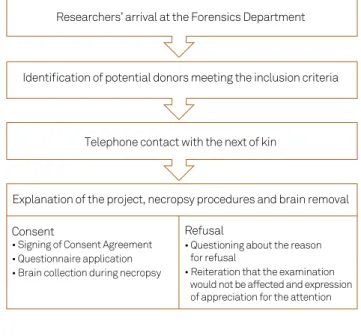
his was a descriptive study, in which the samples were collected by convenience and with the informed consent of irst-degree relatives. Fourteen brains were collected from individuals 50 years old or older who underwent death ver -iication in the Forensics Department (FD) of Porto Alegre. Individuals who were the victims of violent deaths, whose rel -atives did not provide informed consent and who had acute neurological events as the cause of death were excluded.
he donors’ clinical information was obtained after death through the next of kin, using a questionnaire to evaluate cognitive decline (IQCODE)19 and a customized form to eval
-uate motor and sensory changes. For the IQCODE, the cut-of point for dementia was a score cut-of 3.27 or greater, as sug -gested by Sanchez and colleagues19. After removal, the brains
were ixed in a 10% formalin solution for four weeks and were weighed, sectioned, and macroscopically evaluated, in ac -cordance with the international protocol (Figure 1). After the macroscopic examination, in addition to the speciic areas described in the paragraph below, the frontal, parietal, tem -poral and occipital cortices, hippocampus, amygdala, basal ganglia and cerebellum were sampled for histopathological evaluation, were stained with hematoxylin-eosin (HE) and were submitted to immuno-histochemical reactions (IHC).
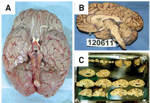
Figure 1. Case 3 (MSA patient) - Basal view of the brain after removal (A) and sagital section after four weeks in formalin 10%, showing severe atrophy of brainstem and cerebellum (B); standardized sections after fixation in formalin (C).
A
B
571
Francine Hehn de Oliveira et al. Neurodegeneration in the brainstem and olfactory bulb
For this study, the following specific regions of in -terest were selected: the midbrain (substantia nigra at the level of the emergence of the oculomotor nerve), pons (section including segment of the locus ceruleus) and medulla oblongata (at the level of the dorsal mo -tor nucleus of the vagus). Besides these regions, we also evaluate the olfactory bulb, which was submitted to full examination and paraffin embedded in craniocaudal ori -entation. These regions were also evaluated by routine HE staining and IHC.
The immuno-histochemical technique was performed in compliance with the routine protocols of the Laboratory of Pathology at UFCSPA. After deparaffination, the sam -ples were immersed in 3% hydrogen peroxide and 10% methanol for 15 minutes to inhibit endogenous peroxi -dase activity. Then, they were boiled in citrate (10 mM, pH 6.0) and/or treated with 1% formic acid to recover the antigen. After washing with PBS, they were incubat -ed with common horse serum for 1 hour and then with primary antibody at 4°C in a wet chamber overnight. The following primary antibodies were used: anti-Aβ (human monoclonal mouse antibody, DAKOCYTOMATION, clone 6F/3D, code M0872), 1:60 dilution, following 3 minutes of incubation in 1% formic acid; anti-phosphorylated tau (monoclonal mouse antibody, INNOGENETICS, clone AT-8, code 90206), 1:500 dilution, following 10 minutes of incubation in citrate; and anti-alpha-synuclein (mouse monoclonal antibody, NOVOCASTRA, clone KM51, code ASYN-L), 1:200 dilution, following 4 minutes of incubation in 1% formic acid and 20 minutes of incubation in citrate. After overnight incubation in primary antibodies, the slides were washed three times in PBS and were incubated in DAKO secondary polymer for 40 minutes, and horse -radish peroxidase for 30 minutes and, finally, DAB. All of the slides were counterstained with hematoxylin for 10 seconds. All of the procedures were performed with nega -tive and posi-tive controls.
The slides were studied by light microscopy for his -topathological diagnosis, aiming to identify possible changes in the morphological and anatomical struc -tures. The presence of protein inclusions on IHC against specific antibodies was semi-quantitatively classified as follows: absent, 1+ (1 to 4 positive cells/10 HPF), 2+ (5 to 9 positive cells/10 HPF) and 3+ (more than 10 posi -tive cells/10 HPF).
he data collected from each case were compared with the corresponding values on the IQCODE, with the evalua -tion form for motor changes and with clinical informa-tion.
he study was approved by the ethics committees at both of the involved institutions (CEP – UFCSPA; and CEP – DML Porto Alegre).
he data are presented in a descriptive manner, and to compare the mean ages between the demented and non-demented patients, Fisher’s test was performed.
RESULTS
Sample description
he average age of the individuals in our sample was 72.07 years old (± 15.79). When the subjects were categorized in clinically demented (D) and non-demented (NoD) categories, based on the IQCODE, the average ages were 72.87 years old and 71.16 years old, respectively. No statistically signiicant diference was found between the groups regarding age. he demented in -dividuals corresponded to 57% of the sample, and their average IQCODE score was 3.72. he non-demented individuals’ average IQCODE score was 2.74, and there was a statistically signiicant diference between the two groups for this variable (p = 0.016, Fisher’s test), as expected. Regarding the questionnaire about motor changes, only one patient presented light tremor and ri -gidity. A detailed sample description is provided in Table 1.
Macroscopic findings
Six of the 14 analyzed brains presented macroscopic changes, as detailed in Table 2. We highlighted the presence of mild frontal atrophy with expansion of the frontal horn of the lateral ventricles as the most frequent inding ( found in 3 cases). Moreover, brain number 10 presented temporal atrophy with volumetric reduction of the hippocampus. Brain number 3 was from a patient with clinical diagnosis of
Table 1. Clinical data of patients.
Demented (D) Non demented (ND)
n (%) 8 (57.14) 6 (42.86)
Age (years) 72.87 71.16
Gender
Female 4 (50%) 3 (50%)
IQCODE Mean (SD) 3.72 (0.6) 2.74 (0.7) IQCODE: Informant Questionnaire on Cognitive Decline in the Elderly; SD: Standard Deviation.
Consent
• Signing of Consent Agreement • Questionnaire application • Brain collection during necropsy
Refusal
• Questioning about the reason for refusal
• Reiteration that the examination would not be affected and expression
of appreciation for the attention
Researchers’ arrival at the Forensics Department
Identification of potential donors meeting the inclusion criteria
Telephone contact with the next of kin
Explanation of the project, necropsy procedures and brain removal
572
Ar
q Neur
opsiquia
tr 2015;
73(7):569-577
Table 2. Description of 14 analyzed brains sample.
Case Age of Death Gender IQCODE Cause of Death Microscopic Findings β-amyloid Tau α-synuclein Pathological Diagnostic DP SP NTs pNFTs NFTs Gr LB NTs PNIs CGIs
1 91 F 3.00 Pneumonia Undefined
M - - 2+ 1+ - 2+ - - - - Tau deposit
P - - 2+ - 1+ 1+ - - -
-MO - - 1+ 1+ - 1+ - - -
-OB NA NA NA NA NA NA NA NA NA NA
2 79 M 3.15 Cardiac Undefined
M Tamponade - - 1+ 1+ 1+ - - - Tau deposit
P - - -
-MO - - -
-OB - - -
-3 59 F 4.52 Septic shock Cerebellar atrophy MSA
M BG atrophy - - - 3+ 3+
P - - - 3+ 3+
MO - - - 3+ 3+
OB - - - 3+ 3+
4 56 M 3.00 Undetermined NORMAL
M - - 1+ - 1+ 1+ - - -
-P - - -
-MO - - -
-OB NA NA NA NA NA NA NA NA NA NA
Frontal atrophy
5 93 F 3,33 Myocardial Increase TH Alzheimer Pathology
M Infarction - - 3+ 1+ 3+ 1+ - - - - Braak III/IV
P - - 2+ 1+ 3+ - - - -
-MO - - 1+ 1+ 1+ - - - -
-OB NA NA NA NA NA NA NA NA NA NA
Alzheimer Pathology
6 68 F 3,27 Undetermined Braak I/II
M - - 1+ - - -
-P - - -
-MO - - -
-OB Metastatic - - - Alzheimer Pathology
carcinoma of the cervix Braak III/IV
573
F
rancine Hehn de Oliv
eir
a e
t al
. Neur
odeg
ener
a
tion in the br
ains
tem and ol
fac
tory bulb
Continuation
7 89 F 3,00 Multiorgan
M Failure - - 1+ - - -
-P - - - Isolate β-amyloid
MO - - 1+ - - - deposit
OB - - -
-8 49 M 3,00 Undetermined
M - - -
-P - - - Alzheimer Pathology
MO - - - Braak I/II
OB - - -
-9 54 M 3.86 Pneumonia Alzheimer Pathology
M - - - Braak V/VI
P - - -
-MO - - -
-OB Frontal and - - -
-Temporal atrophy
10 62 M 4.77 Acute Alzheimer Pathology
M Pulmonar 1+ 1+ 3+ 3+ 3+ 2+ - - - - Braak III/IV
P edema - - 2+ 2+ 3+ 1+ - - -
-MO Frontal atrophy - - 2+ 1+ 2+ 2+ - - -
-OB Increase of LV - - 3+ 1+ 1+ - - - -
-Alzheimer Pathology
11 62 M 3.38 Liver Braak V/VI
M Cirrhosis Frontal atrophy - - 2+ 1+ 2+ 1+ - - -
-P Increase of LV - - 2+ 2+ 1+ 1+ - - -
-MO - - 1+ - - -
-OB - - -
-Undefined
12 81 F 3.36 Undetermined Tau deposit
M - - 2+ 1+ 2+ 1+ - - -
-P - - 1+ - 1+ - - - -
-MO - - - - 1+ - - - -
-OB - - 2+ 1+ 1+ 1+ - - -
574 Arq Neuropsiquiatr 2015;73(7):569-577
multiple system atrophy (MSA) and showed severe cerebellar atrophy, predominantly in the white matter, putamen, pons basis and cerebellar peduncle, as well as slight paleness of the substantia nigra (Figure 2).
Microscopic findings
Hematoxylin-eosin (HE)
In case 1, we found an old 0.3 cm infarction in the pons. In the case of the clinic diagnosis of MSA (case 3), microscopy conirmed the atrophic areas described in the macroscopic exam, in addition to neuronal loss and areas of demyelin -ation, mainly in the pons and cerebellar white matter.
Another common inding, observed in 11 of 14 brains, was the presence of corpora amylacea, ranging from rare in some cases to numerous in others, predominantly localized in the subpial and periventricular regions. his observation did not appear to be related to the patient’s age or to the presence of protein deposits associated with the ND, because they were present both in younger and older patients, as well as in pa -tients with and without protein deposits.
In 11 of 14 cases, the OB was available, and no histological alterations were identiied.
Immuno-histochemical findings
Histopathological diagnosis and classification of AD according to Braak and Braak9
In addition to the protein evaluation in the four areas pro -posed in this study, we evaluated the same proteins in the areas previously described by Braak et al.12, including the hip
-pocampus and the entorhinal, frontal, parietal, and occipital cortices, to establish anatomopathological diagnoses.
Of the 10 patients who presented tau protein depos -its in some location of the brainstem, 8 also had tau in the cortical areas in association with β-amyloid protein deposits, characterizing Alzheimer-type pathology. Of these 8 patients, 3 met the criteria for stage V/VI of Braak’s Classiication for AD and were also considered dement -ed patients according to the IQCODE evaluation; 3 were classiied as stage III/IV and 2 as stage I/II. In the case in which the IQCODE score was less than 3.27 (case 7), the
Continua tion A lzheimer P a thol og y 13 62 F 3. 00 Unde ter mined Br aak V /VI M -P P arie to-occipital inf ar c tion -MO -OB -14 17 M 3.28 Car diac M Tamponade -2+ 1+ 1+ -P -2+ 1+ 2+ -MO -OB -3+ 3+ 1+ -DP : diff use plaque; SP : senil e plaque; NT s: neurit es; pNFT s: pr e-tangl es; NFT: tangl es; Gr: gr anul es; LB: L e wy Bodies; PNIs: P erineur onal Nucl ear Inclusions; CGIs: Cyt
oplasmic Glial Inclusions;
M: Midbr ain; P : P ons; MO: medulla obl onga ta; OB: ol fac tory bulb; BG: basal ganglia; NA: no t a vailabl e; MS A: multipl e s ys tem a tr oph y; TH: t empor al hor n; L V: la ter al v entricl e .
Figure 2. Case 3 - Anterior view of the brainstem showing pons basis atrophy (A); sagital section also shows pons basis atrophy and severe atrophy of the cerebellum (B).
575
Francine Hehn de Oliveira et al. Neurodegeneration in the brainstem and olfactory bulb
individual was classiied with Alzheimer-type pathology, stage V/VI, without clinical correlation.
he other 3 cases presented only tau deposits in corti -cal neurons without beta-amyloid. In all of these cases, the IQCODE score was less than 3.27 (cases 1, 2 and 13).
Beta-amyloid protein (Tables 2 and 3)
Only 1 of the 14 brains (case 10) presented two beta-amyloid deposits in the brainstem, in the form of difuse and senile plaque (Figure 3) localized in the mesencephalic-diencephalic transition of the midbrain. Considering that this case had also beta-amyloid deposits in isocortex, hippocampus, ento -rhinal, cortex and striatum, we could classify it as stage IV, according to hal. et al.13 classiication.
Phosphorylated tau protein (Tables 2 and 3)
he data on the distribution and quantity of tau protein deposits are detailed in Table 2. Ten of 14 brains present -ed several tau deposits in one of the four areas evaluat-ed. In these cases, the most common forms of protein depos -its were pretangles (pNFT) (Figure 4A) and dystrophic neu -rites (DN) (Figure 4B) and, less frequently, tangles (NFT) (Figure 4C) and granules (Figure 4D). Of the cases in which the OB was available for analysis (n = 11), three presented difuse and strong positivity to tau antibody, under the form of dystrophic neuritis (DN), mainly in the external plexi -form layer (Figure 5).
Alpha-synuclein protein (Tables 2 and 3)
Deposits of α-synuclein were only found in one case, in the individual with a clinical diagnosis of probable MSA in a distribution compatible with the disease, thus conirming
Table 3. Brainstem location of protein deposits.
Case Beta-amyloid Tau Alfa-synuclein
Midbrain 1/14 10/14 1/14
Substantia Nigra 1/14 10/14 1/14
Periaqueductal gray matter 0 4/14 1/14
Pons 0 6/14 1/14
Locus ceruleus 6/14 1/14
Reticular formation 4/14 1/14
Medulla Oblongata 0 5/14 1/14
Dorsal motor nucleus of the vagus 4/14 1/14
Hypoglossal nucleus 3/14 1/14
Total number of brainstem segments: 14.
the diagnosis. In this case, α-synuclein deposits in the form of cytoplasmic glial inclusions (CGIs) (Figure 6A) and perineu -ronal nuclear inclusions (PNIs) (Figure 6B) were identiied and mainly observed in the white matter of the cerebellum (Figure 6C), in the pons, medulla oblongata, and midbrain. Many CGIs and PNIs were also identiied in the putamen, and several were identiied in the cortex.
DISCUSSION
We interviewed 30 relatives and 16 of them (53.3%) did not consent to the donation. he main reasons ofered were disagreement with the necropsy procedure that the subject would undergo (25%; n = 4), philosophical or religious is -sues (12.5%; n = 2), objections from other relatives (12.5%; n = 2) and consideration for the deceased person’s wishes ex -pressed when alive (12.5%; n = 2). Most commonly, no reason for refusal was given (31.2%; n = 5)20.
Figure 5. Case 14 - Pretangles and dystrophic neurites (A) and tangles (B) in the olfactory bulb (Tau, 400X).
A
B
20 µm
B
Figure 3. Case 10 - senile plaques of β-amyloid deposit in the
midbrain (A and B) (β-amyloid, 600X).
A
20 µm
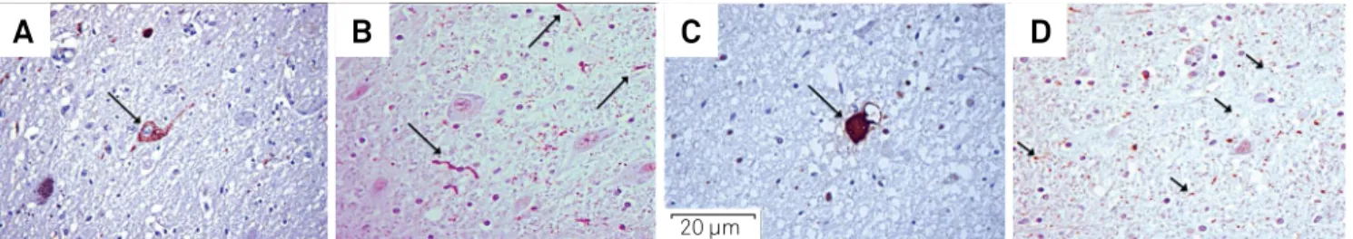
Figure 4. Cases 5 and 10 - pretangles in the medulla oblongata (A); dystrophic neurites in the midbrain (B); tangle in the pons (C) and granules in the pons (C) (Tau, 400X).
A
B
C
D
576 Arq Neuropsiquiatr 2015;73(7):569-577
To assess mental status of the patients, we used IQCODE as a screening method and we believe that despite this questionnaire does not deine the etiology of dementia, it has demonstrated utility as a good tracking instrument for cognitive changes and has been strongly correlated with anatomopathological indings on deceased people.
We categorized the patients into two groups: non-demented (NoD) and demented (D). We found that the same number of men and women were afected, which could be explained by the small sample size. Another noteworthy inding was that the majority of our sample (57%) was classi -ied, according to the IQCODE, in the demented group. It is possible that families who sufer from the diiculties of this disease are more amenable to donation.
Although we found a signiicant number of individuals (n = 11) with a protein deposit in at least one brainstem segment or in the OB, when we compared demented to non-demented cases, our data did not show statistically signiicant diferences regarding deposits. Researchers have attempted to ind clinical signs of early ND and these changes could be the result of protein deposits in the brainstem and OB, because, according to more recent studies, these segments are afected long before the corti -cal areas21. No association was found between protein deposits
in the brainstem and clinical changes as related by the next of kin through the IQCODE and the motor alteration questionnaire.
Some authors have demonstrated that the brainstem nu -clei present NFTs before the transentorhinal region. According to Grinberg et al.22, in all of the cases in which the transento
-rhinal region presented NFTs, they were also found in the dor -sal raphe nucleus (DRN). Moreover, 22% of the individuals who did not present NFTs in the cortical regions (Braak 0) exhib -ited NFTs in the DRN, suggesting that the brainstem was af -fected by AD before the supratentorial regions22. Our indings
agreed, in general, with this idea. Of the 8 cases in which we found Alzheimer-type pathology, seven (87.5%) presented tau protein deposits in some brainstem segment or in the OB, and in 10% of the cases (1/10) in which we found tau deposits in the brainstem, this protein was not found in cortical areas, a inding that could be related to the early stages of AD.
It has been well documented that olfactory dysfunction (hyposmia or anosmia) is a very common characteristic of various NDs, occurring particularly in patients with AD and PD23. In many cases, an olfactory deicit is an early
event in these diseases, and it can precede the typical clini -cal manifestations by many years. Deposits of α-synuclein, tau and β-amyloid proteins were present in many ND patients’ OBs and olfactory epithelia24, and it has been sug
-gested that the OB could be the irst neuronal structure in which these deposits appear during NDs23. We found NFTs
and NTs in the OBs of three of the 11 individuals (27,2%) whose OBs were available for analysis (cases 10, 12 and 14). Mundiñano et al. found NFTs in 42.9% of the OBs of pa -tients with conirmed diagnoses of AD, exactly the same percentage that we found23.
We found no cases of sporadic PD. Regarding α-synuclein deposits, only the case that had a clinical diagnosis of MSA presented this protein in all the segments of the brainstem, as well as in the OB. Alpha-synuclein is the main component of CGIs in the MSA25.
Only one case was positive to beta-amyloid deposits in the brainstem, more speciically in the midbrain, and this individual had stage V/VI AD, according to Braak’s classiica -tion. According to hal et al.13, we could classify this case in
phase 4, which include various brainstem nuclei.
his study revealed interesting indings regarding protein deposits in the brainstems and OBs of people with and with -out dementia, but due to the very small sample size, better interpretation of the results was precluded. For the irst time, a research group reproduces these indings in a population from the south of Brazil.
Acknowledgements
he authors would like to thank the Department of Forensic Medicine of the General Institute of Medical Forensic Analyses of Rio Grande do Sul and the staf of the pathology laboratory for their support.
Figure 6. Case 3 - Cytoplasmic glial inclusions in the pons (A); perinuclear neuronal inclusion in the midbrain (B) and cytoplasmic glial inclusions in the cerebellar white matter (C) (α-synuclein, 600X).
A
B
C
577
Francine Hehn de Oliveira et al. Neurodegeneration in the brainstem and olfactory bulb
References
1. Carvalho J, Garcia A. [The aging process in the Brazilian population: a demographic approach]. Cad Saúde Pública. 2003;19(3):725-33. Portuguese. http://dx.doi.org/10.1590/S0102-311X2003000300005 2. Fagundes SD, Silva MT, Silva MFR, Pereira MG. Prevalence of dementia
among elderly Brazilians: a systematic review. Sao Paulo Med J. 2011;129(1):46-50. http://dx.doi.org/10.1590/S1516-31802011000100009 3. Prado M, Caramelli P, Ferreira ST, Cammarota M, Izquierdo I.
Envelhecimento e memória: foco na doença de Alzheimer. Revista USP. 2007;75:42-9.
4. Nussbaum RL, Ellis CE. Alzheimer’s disease and Parkinson’s disease. N Engl J Med. 2003;348(14):1356-64. http://dx.doi.org/10.1056/NEJM2003ra020003
5. Braak H, Braak E. Demonstration of amyloid deposits and neurofibrillary changes in whole brain sections. Brain Pathol. 1991;1(3):213-6. http://dx.doi.org/10.1111/j.1750-3639.1991.tb00661.x 6. Braak H, Del Tredici K. Alzheimer’s disease: pathogenesis
and prevention. Alzheimers Dement. 2012;8(3):227-33. http://dx.doi.org/10.1016/j.jalz.2012.01.011
7. Braak H, Del Tredici K. Alzheimer’s pathogenesis: is there neuron-to-neuron propagation? Acta Neuropathol. 2011;121(5):589-95. http://dx.doi.org/10.1007/s00401-011-0825-z 8. Bancher C, Brunner C, Lassmann H, Budka H, Jellinger K,
Wiche G et al. Accumulation of abnormally phosphorylated tau precedes the formation of neurofibrillary tangles in Alzheimer’s disease. Brain Res. 1989;477(1-2):90-9. http://dx.doi.org/10.1016/0006-8993(89)91396-6 9. Braak H, Braak E. Neuropathological stageing of
Alzheimer-related changes. Acta Neuropathol. 1991;82(4):239-59. http://dx.doi.org/10.1007/BF00308809
10. Nelson PT, Jicha GA, Schmitt FA, Liu H, Davis DG, Mendiondo MS et al. Clinicopathologic correlations in a large Alzheimer disease center autopsy cohort: neuritic plaques and neurofibrillary tangles “do count” when staging disease severity. J Neuropathol Exp Neurol. 2007;66(12):1136-46. http://dx.doi.org/10.1097/nen.0b013e31815c5efb
11. Sabbagh MN, Cooper K, DeLange J, Stoehr JD, Thind K, Lahti T et al. Functional, global and cognitive decline correlates to accumulation of Alzheimer’s pathology in MCI and AD. Curr Alzheimer Res. 2010;7(4):280-6. http://dx.doi.org/10.2174/156720510791162340 12. Braak H, Del Tredici K. The pathological process underlying
Alzheimer’s disease in individuals under thirty. Acta Neuropathol. 2011;121(2):171-81. http://dx.doi.org/10.1007/s00401-010-0789-4 13. Thal DR, Rüb U, Orantes M, Brask H. Phases of A
beta-deposition in the human brain and its relevance for the development of AD. Neurology. 2002;58(12):1791-800. http://dx.doi.org/10.1212/WNL.58.12.1791
14. Montine TJ, Phelps CH, Beach TG, Bifio EH, Cairns NK, Dickson DW et al. National Institute on Aging-Alzheimer’s Association guidelines for the neuropathologic assessment of Alzheimer’s
disease: a practical approach. Acta Neuropathol. 2012;123(1):1-11. http://dx.doi.org/10.1007/s00401-011-0910-3
15. Ferrer I, Martinez A, Blanco R, Dalfó E, Carmona M. Neuropathology of sporadic Parkinson disease before the appearance of
parkinsonism: preclinical Parkinson disease. J Neural Transm. 2011;118(5):821-39. http://dx.doi.org/10.1007/s00702-010-0482-8 16. Braak H, Del Tredici K, Rüb U, Vos RA, Jansen Steur EN,
Braak E. Staging of brain pathology related to sporadic Parkinson’s disease. Neurobiol Aging. 2003;24(2):197-211. http://dx.doi.org/10.1016/S0197-4580(02)00065-9
17. Braak H, Vos RA, Bohl J, Del Tredici K. Gastric alpha-synuclein immunoreactive inclusions in Meissner’s and Auerbach’s plexuses in cases staged for Parkinson’s disease-related brain pathology. Neurosci Lett. 2006;396(1):67-72. http://dx.doi.org/10.1016/j.neulet.2005.11.012
18. Duyckaerts C, Dickson D. Neuropathology of Alzheimer’s Disease and its variants. In: Dickson D, Weller R, editors. Neurodegeneration: the molecular pathology of dementia and movement disorders. 2nd ed. Hoboken: Wiley-Blackwell; 2011. p. 62-91.
19. Sanchez MA, Lourenco RA. Informant Questionnaire on Cognitive Decline in the Elderly (IQCODE): cross-cultural adaptation for use in Brazil. Cad Saúde Pública. 2009;25(7):1455-65. Portuguese. http://dx.doi.org/10.1590/S0102-311X2009000700003 20. Fonseca MK, Rodrigues Neto E, Costa AS, Rockembach
MA, Padilha RS, Fernandez LL et al. Assessing families’ and patients’ attitudes toward brain donation for research purposes in a Brazilian population sample. Cell Tissue Bank. 2014. http://dx.doi.org/10.1007/s10561-014-9465-6
21. Grinberg LT, Rueb U, Heinsen H. Brainstem: neglected locus in neurodegenerative diseases. Front Neurol. 2011;2:42. http://dx.doi.org/10.3389/fneur.2011.00042
22. Grinberg LT, Rüb U, Ferretti RE, Nitrini R, Farfel JM, Polichiso L et al. The dorsal raphe nucleus shows phospho-tau neurofibrillary changes before the transentorhinal region in Alzheimer’s disease: a precocious onset? Neuropathol Appl Neurobiol. 2009;35(4):406-16. http://dx.doi.org/10.1111/j.1365-2990.2008.00997.x
23. Mundiñano IC, Caballero MC, Ordóñez C, Hernandez M, DiCaudo C, Marcilla I et al. Increased dopaminergic cells and protein aggregates in the olfactory bulb of patients with neurodegenerative disorders. Acta Neuropathol. 2011;122(1):61-74. http://dx.doi.org/10.1007/s00401-011-0830-2
24. Bloch A, Probst A, Bissig H, Adams H, Tolnay M et al. Alpha-synuclein pathology of the spinal and peripheral autonomic nervous system in neurologically unimpaired elderly subjects. Neuropathol Appl Neurobiol. 2006;32(3):284-95. http://dx.doi.org/10.1111/j.1365-2990.2006.00727.x