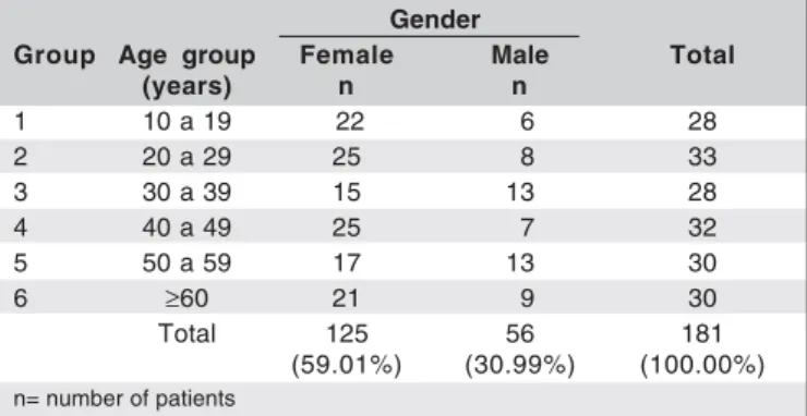
Arq. Bras. Oftalmol. vol.69 número5
Texto
Imagem
Documentos relacionados
The probability of attending school four our group of interest in this region increased by 6.5 percentage points after the expansion of the Bolsa Família program in 2007 and
Abstract: As in ancient architecture of Greece and Rome there was an interconnection between picturesque and monumental forms of arts, in antique period in the architecture
Ao Dr Oliver Duenisch pelos contatos feitos e orientação de língua estrangeira Ao Dr Agenor Maccari pela ajuda na viabilização da área do experimento de campo Ao Dr Rudi Arno
Ousasse apontar algumas hipóteses para a solução desse problema público a partir do exposto dos autores usados como base para fundamentação teórica, da análise dos dados
The false checks observed in 45.1 % of the stu- died chubs may be due to the small scale growth rates observed from August to October, which can be related with the Jack
Managers involved residents in the process of creating the new image of the city of Porto: It is clear that the participation of a resident designer in Porto gave a
(1984) analisando povoamentos de Eucalyptus saligna, aos 8 e 9 anos de idade, respectivamente, verificaram que em média 85% da biomassa aérea encontrava- se no fuste (madeira
didático e resolva as listas de exercícios (disponíveis no Classroom) referentes às obras de Carlos Drummond de Andrade, João Guimarães Rosa, Machado de Assis,
