Blood Pressure Measurement During Aerobic Exercise: Subsidies for
Cardiac Rehabilitation
Emanuel Couto Furtado
1, Plínio dos Santos Ramos
1, Claudio Gil Soares de Araújo
1,2Programa de Pós-Graduação em Educação Física da Universidade Gama Filho1; Clínica de Medicina do Exercício – CLINIMEX2, Rio de Janeiro,
RJ - Brazil
Summary
Background: Institutional documents recommend that hemodynamic variables – heart rate (HR) and systolic (SAP) and diastolic arterial pressure (DAP) – be routinely controlled at the aerobic part of supervised exercise sessions for coronary disease patients.
Objectives: a) to determine the pattern and reproducibility of the blood pressure (BP) throughout 5 minutes of physical exercise at constant and moderate intensity; and b) to compare the BP measurement obtained with digital and conventional device during the exercise.
Methods: Thirty adult individuals of both sexes (65± yrs) were assessed for 5 minutes during lower-limb cycle ergometry and the BP was measured every 2 minutes, between the 3rd and the 3th minutes, using a Tango digital sphygmomanometer (Suntech, USA) and in the 4th minute, using a mercury column sphygmomanometer. Seven days later, at similar time of the day, six individuals had the test repeated to evaluate reproducibility.
Results: Whereas the DAP did not vary throughout the exercise (p> 0.05), SAP increased from the 3rd to 7th minute (46±4. versus 58±4.5 mmHg, p<0.05) and thereafter remained practically constant. The digital and conventional measurements showed a strong correlation – r = 0.83 for SAP and 0.84 for DAP – with no differences for SAP (63±4.5 versus 62±4.3 mm Hg; p>0.05) and a small difference for DAP (72±2.4 versus 78±2.3 mm Hg; p<0.05).
Conclusion: For exercises of moderate and constant intensity in a cycle ergometer with a 5-minute duration, BP measurements must be carried out from the 7th minute on. The digital measurements with the Tango equipment and those obtained with the conventional mercury-column sphygmomanometer were, for clinical purposes, very similar and reproducible. (Arq Bras Cardiol 2009;93():42-48)
Key Words: Blood pressure; exercise; rehabilitation.
Mailing address: Claudio Gil S. de Araújo •
Rua Siqueira Campos, 93/101, Copacabana, 22031-070, Rio de Janeiro, RJ - Brazil
E-mail: [email protected], [email protected]
Manuscript received July 30, 2008; revised manuscript received October 2nd,
2008, accepted October 15, 2008.
Introduction
There is a consensus that regular physical exercises are an important component in the primary and secondary prevention of coronary disease1. Institutional documents2,3, have suggested that the aerobic component of the physical exercise must be prescribed regarding the weekly frequency and the duration and intensity of the exercise session. Although the rate of adverse cardiovascular events is relatively low during the supervised exercise sessions4, it seems appropriate to maintain a more accurate and individualized control of the aerobic prescription in patients with more severe cardiopathies5.
It is known that the heart rate (HR) increases rapidly within the first seconds of a dynamic exercise, primarily due to the inhibition of the cardiac vagal action6, which progressively increases with
time, especially when the effort intensity is ≥ 80% of the load in which the anaerobic threshold was detected7. However, although the blood pressure (BP) measurement is recommended and frequently performed during supervised exercise sessions, little is known about its behavior during a 10-20 minute exercise at constant intensity.
The objectives of this study were: a) to determine the BP behavior throughout 15 minutes of constant and moderate intensity physical exercise; and b) to compare and determine the reliability of the BP measurements during exercise in a lower-limb cycle ergometer obtained with a precise digital device as well as with a conventional one.
Methods
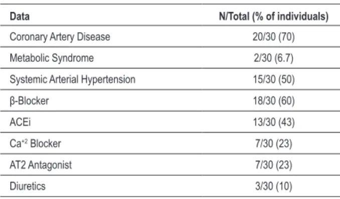
SampleA total of 30 adult individuals (28 men) were selected by convenience, according to the following inclusion criteria: a) to have regularly attended a Supervised Exercise Program (SEP) for at least three months; b) to have completed a minimum of 30 sessions; c) to perform, during the aerobic part of the session, uninterrupted 15-minute constant exercising, with a previously established load based on the results of the maximum cardiopulmonary exercise testing, sporadically adjusted to maintain the individual within the target heart rate zone in a lower-limb cycle ergometer; d) to exhibit sinus rhythm with no more than three extrasystoles per minute. The main clinical conditions and medication use are shown in Table 1.
Experimental protocol
Initially, the procedures were explained and a free and informed consent form was obtained from all participants. All the individuals had been previously submitted to a maximum cardiopulmonary exercise testing in a lower-limb cycle ergometer, along with the measurement and analysis of the expired gases, in which the data necessary for the individualized prescription of aerobic exercise was obtained.
After the heart rate monitor (Polar®, Finland) was placed on the left wrist and weight was measured, the pre-exercise BP and HR were obtained through an Omron professional digital sphygmomanometer, model XML-907 (Omron, USA)9, under the direct supervision of the physician. After this procedure, if the values obtained did not exceed the limits
Table 1 - Clinical conditions and anti-hypertensive drugs used by the individuals
Data N/Total (% of individuals)
Coronary Artery Disease 20/30 (70)
Metabolic Syndrome 2/30 (6.7)
Systemic Arterial Hypertension 15/30 (50)
β-Blocker 18/30 (60)
ACEi 13/30 (43)
Ca+2 Blocker 7/30 (23)
AT2 Antagonist 7/30 (23)
Diuretics 3/30 (10)
ACEi: angiotensina-converting enzyme inhibitor; Ca+2 Blocker; Calcium
channel blocker; AT2: Angiotensin II Antagonist; Obs. - Some individuals used more than one type of anti-hypertensive medication, which explains the sum of percentages > 100%.
of variation that were usually observed, the individual was positioned on a Cateye lower-limb cycle ergometer, model EC-1600 (Cateye, Japan) and then, the cuff of the Suntech digital sphygmomanometer, model Tango (Suntech, USA) was placed on the participant’s right arm and three thoracic electrodes were used to obtain the CC5 position. The digital sphygmomanometer was then configured to automatically measure BP in the 3rd, 5th, 9th, 11th and 13th minutes of the exercise, automatically inflating the cuff up to 30 mmHg above the maximum systolic arterial pressure reached in previous sessions under the same exercise conditions. The deflation rate was set at 8 mmHg/s, corresponding to approximately 35 seconds to inflate and deflate the cuff at each measurement. After the last measurement performed by the equipment, in the 13th minute, the cuff was removed and a conventional mercury-column sphygmomanometer was positioned on the same limb, and another examiner, who had not participated in the procedure so far, obtained the BP measurement.
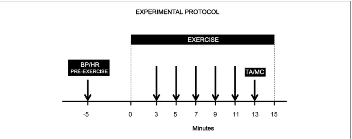
The individual’s arm was positioned at the level of the heart and supported by the examiner. The experimental protocol is shown in Figure 1. In order to standardize the measurement technique, the assessed individuals were advised to position the right arm on the supine position, with the hand resting on the cycle ergometer handlebar anytime the Tango™ sphygmomanometer started to inflate. The measurement obtained with the conventional sphygmomanometer is standardized in our research group and it is carried out by inflating the cuff up to around 30 mmHg above the individual’s previously known SAP and, thereafter, deflating the cuff at a rate of 2 to 4 mmHg/s. According to the characteristics of the cycle ergometer used in the study, the load progressively increases in the first three minutes until it reaches the established load, which is then maintained up to 15th minute of exercise. The load in the cycle ergometer was individually established according to the maximum cardiopulmonary exercise testing and the previous SEP sessions, in order to reach the proposed target heart rate zone for aerobic training. Subsequently, six individuals (20% of the sample selected for the study) were reassessed, with a one-week interval between the two assessments, at a similar time of the day and under the same conditions of regular medication use.
Statistical analysis
Figure 1 - Experimental protocol. The irst arrow represents the measurement of BP and HR before the start of the exercise; the following arrows represent the moments
in which the BP was measured by the digital sphygmomanometer and the last arrow represents the moment when the digital Tango sphygmomanometer (TA) was removed and the BP measurement was carried out with the conventional mercury-column sphygmomanometer (MC).
exercise. The statistical calculations were carried out with the GraphPad Prism program version 5 (GraphPad, USA) and the level of significance was set at 5%.
Results
Demographic characteristics
The age of the 30 assessed individuals was 65 ± 11 years, ranging from 46 to 86 years; height was 170 ± 6.5 cm and weight was 79 ± 6.5 kg. A total of 70% of the sample presented coronary artery disease; 10 individuals had been submitted to myocardial revascularization and 50% of the individuals had been diagnosed with and received treatment for systemic arterial hypertension. As for the use of anti-hypertensive drugs, it was observed that ß-blockers were the most often used ones, followed by angiotensin-converting enzyme inhibitors (ACEI), respectively, in 57 and 43% of the sample. The systolic (SAP) and diastolic (DAP) arterial pressure at rest were, respectively: 125 ± 14, 68 ± 8 mm Hg and HR was 65 ± 10 bpm.
Bp and HR during exercise
The upper part of Figure 2 shows that HR increases throughout the 15 minutes of exercise with a constant load at the cycle ergometer, initially faster (p < 0.05) and subsequently, more gradually, so that the values differed when compared to those obtained in the 3rd minute, 95 ± 2.4 bpm, reaching, respectively, in the 7th, 9th, 11th and 13th minutes, the values of 102 ± 2.7 bpm, 104 ± 2.9 bpm, 105 ± 3.2 bpm and 107 ± 3.2 bpm. Figure 1 also shows the SAP increase up to the 7th minute of exercise (p < 0.05), with values then remaining stable (p > 0.05), especially between the 9th and the 13th minutes of exercise, when they were virtually identical. The DAP presented a stable behavior throughout the exercise and there was no difference among the values obtained between the 3rd and 13th minutes (p > 0.05).
Digital and conventional BP measurements during the exercise
The other measurement obtained at the end of the exercise session allowed the comparison between the digital and the conventional BP measurements: similar values were observed for SAP - 163 ± 4.5 versus 162 ± 4.3 mm Hg (p > 0.05) and slightly distinct ones were observed for DAP - 72 ± 2.4 versus
78 ± 2.3 mm Hg (p > 0.05). The diagrams of dispersion in the two SAP and DAP measurements are shown in Figures 3 and 4, with very similar coefficients of correlation of, respectively, 0.83 and 0.84. The difference between the SAP measurements carried out with the digital sphygmomanometer and the conventional one exceeded 10 mmHg in 13 individuals (more than 10% in 5 cases); regarding DAP, the same difference was found in nine individuals.
Reassessment
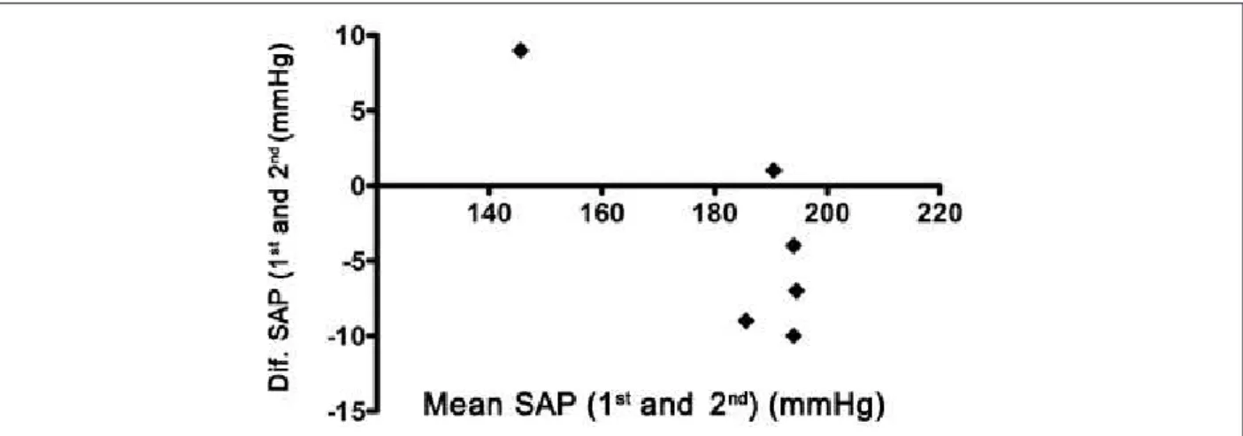
The protocol repetition in six individuals showed very similar values regarding the several measurements obtained throughout the 15-minute exercise session. The SAP (p = 0.31) and DAP (p = 0.53) values in the 13th minute not only did not differ, but also showed significant correlations – r = 0.99 – (p < 0.05) for SAP and 0.65 for DAP (p > 0.05). The similarity of the results can be better observed in the respective Bland-Altman diagrams (Figures 5 and 6).
Discussion
Figure 4 - Correlation between the diastolic arterial pressure measurements
carried out by the digital sphygmomanometer and the conventional, mercury-column sphygmomanometer. Coeficient of correlation - r = 0.84.
Figure 3 - Correlation between the systolic arterial pressure measurements
carried out by the digital sphygmomanometer and the conventional, mercury-column sphygmomanometer in the last minutes of a constant exercise. Coeficient of correlation - r = 0.83.
measurement during an aerobic exercise – of constant and moderate intensity, with a duration ≥ 10 minutes – as well as about threshold values, such as the moment when these measurements must be carried out, in either these documents or in those in which the Brazilian Society of Cardiology standardizes the procedures for supervised exercise sessions and cardiac rehabilitation programs12.
Several authors13-15 have studied the behavior of cardiovascular variables during prolonged, moderate-intensity exercise and, in particular, the concept of cardiovascular drift is worth mentioning, originally described by Saltin et al16 in 1964, which corresponds to the slight, but systematic HR increase with the exercise. According to Araújo7, during an exercise carried out in a cycle ergometer with an intensity of 80% of the individual anaerobic threshold by young and healthy adults, there would be an increase of approximately 0.6 bpm for each minute of exercise from the 10th minute on. The result obtained in the present study, a mean variation of 3 beats between the 9th and the 13th minutes of exercise, is quite similar to that obtained by Araújo7. This HR increase would be the result of a decrease in the systolic volume and would aim at minimizing a reduction in the cardiac output and would be influenced by the sympathetic activity, by the body temperature and by the redistribution of the peripheral blood flow, which could result in a slight SAP decrease after 15 minutes of exercise.
Interestingly, although in most situations of supervised exercise for patients with cardiopathies, the constant exercise has a more typical duration of 10 to 30 minutes, the behavior of the BP in this situation is virtually unknown.
If, on the one hand, the measurement of the HR during the exercise is much more simple, as it can be obtained non-invasively and continuously, with high reliability and low cost, the same is not true for the BP. The invasive and
Figure 2 - SAP and HR behavior during the constant-intensity aerobic
Figure 5 - Bland-Altman diagram representing the SAP difference between the irst and the second assessments, carried out with a digital sphygmomanometer in the
13th minute of constant aerobic exercise.
Figure 6 -Bland-Altman diagram representing the DAP difference between the irst and the second assessments, carried out with a digital sphygmomanometer in the
13th minute of constant aerobic exercise.
complex high-quality measurement does not apply, however, to the daily situations of supervised exercise or cardiac rehabilitation programs. Equipment that allows the continuous and noninvasive measurement of the BP - beat by beat – are not practical, in addition to being high-cost and subject to interference and technical difficulties that prevent their routine use in situations of prolonged exercise. Therefore, in clinical practice the measurement of BP during the exercise is carried out discontinuously (in general, only a single measurement for a constant and moderate-intensity exercise, with a duration between 10 and 30 minutes), using conventional sphygmomanometers and the auscultation technique carried out by a professional specifically trained for this measurement under exercise conditions. Considering the specific difficulties of the mechanical aneroid sphygmomanometer for exercise situations, most services that measure BP during exercise use the mercury-column sphygmomanometers.
In the last years, the use of the mercury-column sphygmomanometer has been the object of criticism due to its possible environmental implications, notably in European countries17, although in Brazil, this matter has not been more
broadly discussed. Equipment with digital technology has been launched in the market for some decades, primarily intended for the nonmedical population; however, more recently, the manufacturers have proposed to meet higher quality standards, with a degree of reliability that is compatible with the full clinical use18. Nevertheless, this is still a debatable question and far from being consensual, with many considering the century-old and classic measurement method as the “gold-standard” of BP measurement19-21.
attaining quite favorable results in individuals submitted to a treadmill exercise test with modified Bruce protocol. This piece of equipment uses an additional resource to reduce or eliminate artifacts or background noise that impair or prevent the BP measurement in exercise conditions. Obtaining valid BP measurements through this digital sphygmomanometer during exercise became possible due to an ingenious process of sound selection obtained by the amplification of Korotkoff sounds by a built-in microphone in the cuff, through the simultaneous obtaining of an electrocardiogram (ECG) through electrodes positioned on the individual’s chest, allowing the equipment to select which sounds would be true or caused by artifacts and irrelevant background noise, resulting in valid and reliable measurements.
At least in the context of the present study, lower-limb cycle ergometry, the BP measurements obtained with the digital and conventional sphygmomanometers did not differ and showed to be strongly associated.
In a small percentage of cases (< 20%), the differences were > 10 mmHg, without defining a uniform pattern of under or overestimation of one of the sphygmomanometers that could have some clinical relevance. Additionally, the digital measurements also showed to be quite reproducible when they were repeated after a one-week interval. However, it is worth mentioning that, in some rare cases (< 5% of the individuals), it was not possible to obtain BP measurements with this digital equipment. That occurred with an individual that used a pace-maker and another that was extremely obese, probably due to difficulties in the identification of the R-wave of the ECG. On the other hand, rare isolated extrasystoles did not seem to significantly influence the obtaining of the digital BP measurements. The digital measurement seems to be also extremely reproducible in repeated and sequential measurements of the same individual and the small individual differences do not seem to be clinically relevant.
The present study did not aim at evaluating or comparing operational advantages and the economical viability of routinely incorporating the digital equipment used in SEP or in exercise test situations.
The comparative results between the digital and the conventional measurements of DAP were somewhat different from those observed for SAP. The DAP measurement by the digital sphygmomanometer was around 6 mm Hg lower than the value obtained by an experienced examiner using a conventional mercury-column sphygmomanometer. It is known that the accurate criterion for the DAP measurement has always been a challenge, as, in opposition to the start of a clear and repetitive sound that characterizes the first phase of Korotkoff sounds when SAP is measured, there is no consensus if the DAP better corresponds to the muffling and change in the sound pattern or to its complete disappearance22, the latter occasionally absent in exercise conditions.
Considering this last aspect, the standardization of the DAP measurement during exercise by our group is carried out by the muffling or change in the sound pattern, perhaps justifying the significant difference, although of little clinical relevance, of 6 mmHg in the DAP measurement, which does not vary
throughout the moderate and constant intensity exercise with a 15-minute duration. In accordance with this discussion, the reproducibility of the DAP measurement was lower than that of the SAP.
Considering the protocol and the present study sample, some limitations must be considered: a) it is not possible to guarantee that in situations of moderate and constant intensity exercise with a duration > 15 minutes, the DAP and SAP will remain indefinitely constant from the 7th minute of exercise on; b) it is possible that in some environments with unfavorable climate conditions, with higher levels of temperatures and humidity, capable of inducing some degree of dehydration, the BP behavior can be different; c) the digital sphygmomanometer measurements were obtained in exercises performed on a cycle ergometer and the BP behavior during exercise on a treadmill or other types of equipment were not studied; and d) it is possible that distinct clinical responses be obtained in hypertensive patients without adequate clinical control.
As previously mentioned, the literature is very limited in the description of the BP behavior during constant exercises with a duration of 10-20 minutes, a more frequent form of exercise session, in comparison with the extensive description of the pressure response to submaximal exercise.
The use of a high-technology digital sphygmomanometer, which was previously validated for exercise conditions, in the present study allowed us to effectively control the possibility of intra-examiner variations in the measurements of SAP and DAP, an essential condition to obtain the study results.
This is an original approach and allows us to consider that the occasional variations obtained in the SAP and DAP measurements during exercise would be solely due to the physiological responses, without any influence of the physician-examiner. Considering that, the present study brings an important combination for the body of knowledge, mainly in the cardiac rehabilitation area and that of supervised exercise programs, supporting the fact that the BP measurements during exercise in a lower-limb cycle ergometer with constant load, can be performed, without any impairment, from the 7th minute of exercise on, considering that the measurement will not undergo significant alterations until the end of the exercise.
Based on this information, we intend to contribute to the improvement of the quality of information and a better clinical safety, especially for those individuals that need a more stringent follow-up of their hemodynamic variables during the exercise session.
Conclusions
References
1. Fraker TD Jr, Fihn SD, Gibbons RJ, Abrams J, Chatterjee K, Daley J, et al. 2007 chronic angina focused update of the ACC/AHA 2002 Guidelines for the management of patients with chronic stable angina: a report of the American College of Cardiology/American Heart Association Task Force on Practice Guidelines Writing Group to develop the focused update of the 2002 Guidelines for the management of patients with chronic stable angina. Circulation. 2007; 116 (23): 2762-72.
2. Graham I, Atar D, Borch-Johnsen K, Boysen G, Burell G, Cifkova R, et al. European guidelines on cardiovascular disease prevention in clinical practice: executive summary. Fourth Joint Task Force of the European Society of Cardiology and other societies on cardiovascular disease prevention in clinical practice constituted by representatives of nine societies and by invited experts. Eur J Cardiovasc Prev Rehabil. 2007; 14 (Suppl 2): E1-40. 3. Nelson ME, Rejeski WJ, Blair SN, Duncan PW, Judge JO, King AC. Physical
activity and public health in older adults: recommendation from the American College of Sports Medicine and the American Heart Association. Circulation. 2007; 116 (9): 1094-105.
4. Thomas RJ, King M, Lui K, Oldridge N, Pina IL, Spertus J, American Association of Cardiovascular and Pulmonary Rehabilitation ACC/AHA 2007 performance measures on cardiac rehabilitation for referral to and delivery of cardiac rehabilitation/secondary prevention services. Circulation. 2007; 116 (14): 1611-42.
5. ACSM’s Guidelines for exercise testing and prescription. 7th ed. Baltimore: Lippincot Willians & Wilkins; 2005.
6. Araújo CG, Nobrega AC, Castro CL. Heart rate responses to deep breathing and 4-seconds of exercise before and after pharmacological blockade with atropine and propranolol. Clin Auton Res. 1992; 2 (1): 35-40.
7. Araújo CG. Resposta cardiorrespiratória a um exercício submáximo prolongado. Arq Bras Cardiol. 1983; 41 (1): 37-45.
8. Cameron JD, Stevenson I, Reed E, McGrath BP, Dart AM, Kingwell BA. Accuracy of automated auscultatory blood pressure measurement during supine exercise and treadmill stress electrocardiogram-testing. Blood Press Monit. 2004; 9 (5): 269-75.
9. Mattioli GM, Teixeira FP, Castro CL, Araújo CG. Frequência cardíaca e pressão arterial em repouso: variação de 10 dias em participantes de um programa de exercício supervisionado. Rev SOCERJ. 2006; 19 (5): 404-8.
10. Pescatello L, Franklin B, Fagard R, Farquhar W, Kelley G, Ray C. American College of Sports Medicine. Exercise and hypertension: position stand. Med Sci Sports Exerc. 2004; 36 (3): 533-53.
11. Sociedade Brasileira de Cardiologia. Diretriz brasileira de reabilitação cardíaca. Arq Bras Cardiol. 2005; 84 (5): 431-40.
12. Araújo CG, Carvalho T, Castro CL, Costa RV, Moraes RS, Oliveira Filho JA, et al. Normatização de equipamentos e técnicas de reabilitação cardiovascular supervisionada. Arq Bras Cardiol. 2004; 83 (5): 448-52.
13. Lafrenz A, Wingo J, Ganio M, Cureton K. Effect of ambient temperature on cardiovascular drift and maximal oxygen uptake. Med Sci Sports Exerc. 2008; 40 (6): 1065-71.
14. Dawson E, Shave R, Whyte G, Ball D, Selmer C, Jans O, et al. Preload maintenance and the levt ventricular response to prolonged exercise in men. Exp Physiol. 2006; 92 (2): 383-90.
15. Coyle E, González-Alonso J. Cardiovascular drift during prolonged exercise: new perspectives. Exerc Sport Sci Rev. 2001; 29 (2): 88-92.
16. Saltin B, Stenberg J. Circulatory response to prolonged severe exercise. J Appl Physiol. 1964; 19: 833-8.
17. O’Brien E. Replace the mercury sphygnomanometer. BMJ. 2000; 320: 815-6. 18. El Assaad MA, Topouchian JA, Darne BM, Asmar RG. Validation of the Omron
HEM-907 device for blood pressure measurement. Blood Press Monit. 2002; 7 (4): 237-41.
19. Turner M, Speechly C, Bignell N. Sphygmomanometer calibration--why, how and how often? Aust Fam Physician. 2007; 36 (10): 834-8.
20. Jones D, Frohlich E, Grim C, Grim E, Taubert K. Mercury sphygmomanometers should not be abandoned: an advisory statement from the Council for High Blood Pressure Research, American Heart Association. Hypertension. 2001; 37: 185-6.
21. Varughese GI, Lip GY. Goodbye mercury? Blood pressure measurement and its future. J R Soc Med. 2005; 98 (3): 89-90.
22. Perloff D, Grim C, Flack J, Frohlich E, Hill M, McDonald M, et al. Human blood pressure determination by sphygmomanometry. Circulation. 1993; 88 (5 Pt 1): 2460-70.
Acknowledgments
Partial financial support from CNPq: Dr. Claudio Gil Soares de Araújo is a level-1A research productivity fellow and Emanuel Couto and Plínio Ramos are Master’s Degree fellows.
Potential Conflict of Interest
No potential conflict of interest relevant to this article was reported.
Sources of Funding
This study was partially funded by CNPq.
Study Association